.webp&w=384&q=75)

Loading, please wait...
Loading, please wait...

Trigeminal neuralgia causes excruciating, lancinating facial pain that severely impairs patient quality of life and functional independence. Clinicians identify neurovascular conflict at the cisternal segment of the fifth cranial nerve as the primary etiology in classic cases. High-resolution magnetic resonance imaging serves as the diagnostic gold standard for evaluating anatomical relationships around the root entry zone. However, qualitative visual review frequently suffers from inter-observer subjectivity, subtle anatomical nuances, and diagnostic variation. Fortunately, recent breakthroughs in trigeminal neuralgia AI present reliable computational strategies to automate nerve segmentation and detect offending neurovascular compression.
Trigeminal neuralgia represents one of the most painful conditions encountered in neurosurgical and neurological practice. Patients experience paroxysmal, unilateral electric-shock sensations triggered by innocuous stimuli such as talking, chewing, or brushing teeth. Pathologically, focal vascular compression against the root entry zone precipitates localized demyelination and aberrant ephaptic axonal transmission. Therefore, neurosurgeons prioritize microvascular decompression to physically separate the aberrant vascular loop from the vulnerable nerve trunk.
Nevertheless, identifying discrete neurovascular conflict poses formidable challenges for diagnostic radiologists and clinicians. Standard constructive interference in steady-state sequences offer sharp contrast, yet tortuous vessels often obscure the cisternal segment. In addition, small transverse pontine veins or subtle arterial branches produce faint signals that mimic normal cisternal architecture. Consequently, manual tracing demands extensive radiological expertise, consumes excessive clinical time, and exhibits noticeable inter-observer variability across different neuroimaging centers. When clinicians misinterpret subtle vascular compression, patients frequently endure protracted pharmacological trials with escalating adverse effects. Furthermore, missed neurovascular compression delays curative interventions like microvascular decompression. Thus, modern neurosurgical practice urgently requires automated, reproducible diagnostic workflows to standardize preoperative neurovascular evaluation and improve diagnostic confidence.
Recent developments in computer vision and artificial intelligence have dramatically transformed neuroimaging diagnostics. Deep learning networks, particularly convolutional architectures like U-Net and its modern variants, demonstrate remarkable aptitude for 3D volumetric segmentation. Specifically, these models ingest high-resolution constructive interference sequences and accurately differentiate neural pathways from adjacent arterial and venous structures. In addition, machine learning pipelines extract high-dimensional quantitative spatial features that surpass qualitative visual inspections.
Moreover, automated pipelines eliminate the subjectivity inherent in manual tracing routines. Deep learning algorithms rapidly process volumetric data, tracing the trigeminal pathway from the brainstem to Meckel's cave within seconds. Simultaneously, these tools quantify critical surgical metrics, including the surface area of vascular contact and the distance from the brainstem to the compression site. As a result, clinicians receive objective spatial coordinates and three-dimensional anatomical reconstructions without human fatigue or bias. Furthermore, automated segmentation bridges the gap between tertiary academic medical centers and resource-constrained community clinics. By democratizing expert-level image analysis, trigeminal neuralgia AI establishes a consistent benchmark for recognizing complex posterior fossa neurovascular conflicts.
To evaluate the true clinical value of these algorithms, investigators conducted a rigorous systematic review and meta-analysis across major medical databases. The pooled analysis examined five comprehensive studies comprising 577 patients with clinically confirmed trigeminal neuralgia. Researchers extracted core performance metrics, validating models against expert radiological annotations, clinical diagnoses, and intraoperative microvascular surgical confirmations.
Remarkably, the pooled analysis revealed an exceptional specificity of 92% across all evaluated deep learning frameworks. This elevated specificity indicates that the models rarely produce false-positive detections, thereby protecting patients from unnecessary invasive procedures. Meanwhile, the pooled sensitivity reached 71%, demonstrating dependable ability to capture genuine neurovascular conflict. Furthermore, the diagnostic odds ratio reached an impressive 26.71, highlighting robust overall discriminatory performance. The area under the summary receiver operating characteristic curve achieved 0.91, which confirms outstanding diagnostic accuracy. Similarly, the positive diagnostic likelihood ratio was 8.5, whereas the negative diagnostic likelihood ratio was 0.32. Therefore, these quantitative findings provide compelling statistical evidence that artificial intelligence reliably supports clinical assessment in complex craniofacial pain.
Qualitative evaluation of cranial nerve compression has historically generated substantial debate among neuroradiologists and skull base surgeons. Neurovascular contact without clinical symptoms occurs frequently in asymptomatic individuals, which complicates radiographic interpretations. Therefore, differentiating benign anatomical contact from true pathological compression with axonal distortion requires rigorous objective criteria.
Fortunately, deep learning models analyze subtle multiplanar voxel interactions that human observers might overlook. For example, neural networks evaluate subtle vessel impaction, focal nerve deviation, and cross-sectional thinning at the cisternal root entry zone. Furthermore, algorithmic models integrate high-resolution structural MRI with diffusion tensor imaging and MR angiography to assess microstructural integrity. Consequently, this multisequence synergy enables precise mapping of the nerve-vessel interface. In addition, automated algorithms maintain stable diagnostic accuracy irrespective of the imaging vendor, magnetic field strength, or institutional protocol variations. By standardizing quantitative criteria, deep learning reduces institutional diagnostic discrepancies. Hence, multidisciplinary neurovascular boards can make therapeutic recommendations backed by verifiable quantitative datasets rather than subjective visual impressions.
Microvascular decompression offers durable, long-term pain relief for patients suffering from medically refractory classic trigeminal neuralgia. During this delicate microsurgical procedure, neurosurgeons navigate the narrow cerebellopontine angle to mobilize impinging vessels away from the root entry zone. However, unexpected intraoperative anatomical configurations, such as hidden venous tributaries or multi-vessel compressions, elevate the risk of surgical complications.
Consequently, precise preoperative anatomical knowledge directly determines surgical success and minimizes neurological morbidity. Automated artificial intelligence reconstructions furnish neurosurgeons with patient-specific three-dimensional visual models prior to entering the operating room. These computational roadmaps illustrate the precise trajectory of the superior cerebellar artery, anterior inferior cerebellar artery, and petrosal vein. Furthermore, knowing the exact distance of the conflict from the pontine surface optimizes craniotomy placement and cerebellar retraction vectors. As a result, neurosurgeons can execute targeted, minimally invasive skull base approaches with reduced operative time. In addition, accurate preoperative identification of non-compressive etiologies prevents futile surgical explorations. Therefore, artificial intelligence provides indispensable preoperative guidance that translates into superior surgical outcomes and lower recurrence rates.
Although current evidence demonstrates substantial clinical promise, several technological hurdles remain before widespread clinical implementation occurs. First, the pooled sensitivity of 71% highlights that algorithms still miss approximately three in ten clinically meaningful compressions. Therefore, future model iterations must incorporate advanced architectures that distinguish small offending veins from adjacent arachnoid membranes.
Moreover, most available training datasets originate from single-center retrospective cohorts with modest sample sizes. To ensure broad clinical generalizability, researchers must validate these algorithms on diverse multi-institutional datasets from varied imaging platforms. In addition, integrating automated segmentation directly into standard picture archiving and communication systems will streamline daily neurological workflows. Clinicians should view artificial intelligence as a supportive diagnostic adjunct rather than an autonomous decision-maker. As prospective clinical trials validate these tools in real-time practice, deep learning will transform trigeminal neuralgia management. Ultimately, the synergy between algorithmic precision and clinical acumen will elevate patient care and diagnostic certainty worldwide.
Trigeminal neuralgia AI enhances MRI interpretation by automating three-dimensional segmentation of the trigeminal nerve and adjacent cerebellopontine vasculature. These deep learning algorithms objectively quantify critical anatomical features, including contact surface area and compression distance from the brainstem, thereby eliminating subjective observer variability and accelerating preoperative radiological reviews.
In this systematic review and meta-analysis of 577 patients, artificial intelligence models achieved a pooled specificity of 92% and a sensitivity of 71%. Additionally, the algorithms demonstrated an impressive diagnostic odds ratio of 26.71 and an overall area under the receiver operating characteristic curve of 0.91.
No, artificial intelligence cannot replace experienced neurosurgeons or radiologists in surgical decision-making. Instead, deep learning models serve as powerful clinical decision-support tools that automate complex segmentation, provide reliable quantitative measurements, and assist surgical teams in refining operative strategies while human experts retain final clinical responsibility.
Disclaimer: This content is for informational and educational purposes only. It should not be used as a substitute for professional medical advice, diagnosis, or treatment. Always seek the advice of your physician or other qualified health provider with any questions you may have regarding a medical condition. Refer to the latest local and national guidelines for clinical practice.
References

Read summarized clinical updates, watch expert medical content, and earn CME certifications right from your smartphone.
Open your smartphone camera to scan and install.





A meta-analysis demonstrates that trigeminal neuralgia AI models achieve 92% specificity and an AUC of 0.91 for neurovascular conflict detection on MRI, streamlining surgical decision-making for microvascular decompression.
Today

A recent Journal of Clinical Oncology study demonstrates that industry payments to oncologists directly sway prescribing behavior. Oncologists who received marketing transfers favored promoted therapies over non-promoted alternatives, highlighting critical ethical concerns for evidence-based cancer practice.
Today

A recent qualitative study examines how college students seek digital sexual health information under persistent uncertainty. Discover the concept of functional trust, the clinical hazards of search abandonment, and evidence-based approaches clinicians can adopt to guide youth toward verified reproductive healthcare.
Today
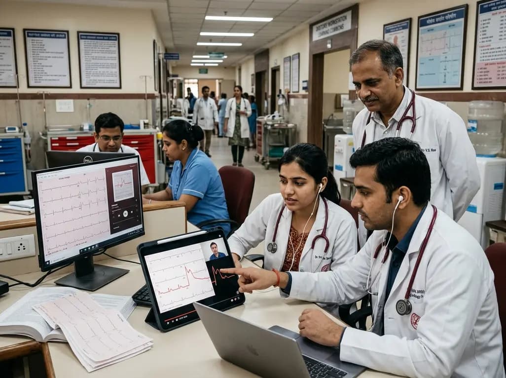
A randomized controlled trial demonstrates that curated FOAMed videos and podcasts produce superior gains in high-risk ECG interpretation and acute coronary syndrome clinical decision-making compared with print-based materials among interprofessional emergency care teams.
Today
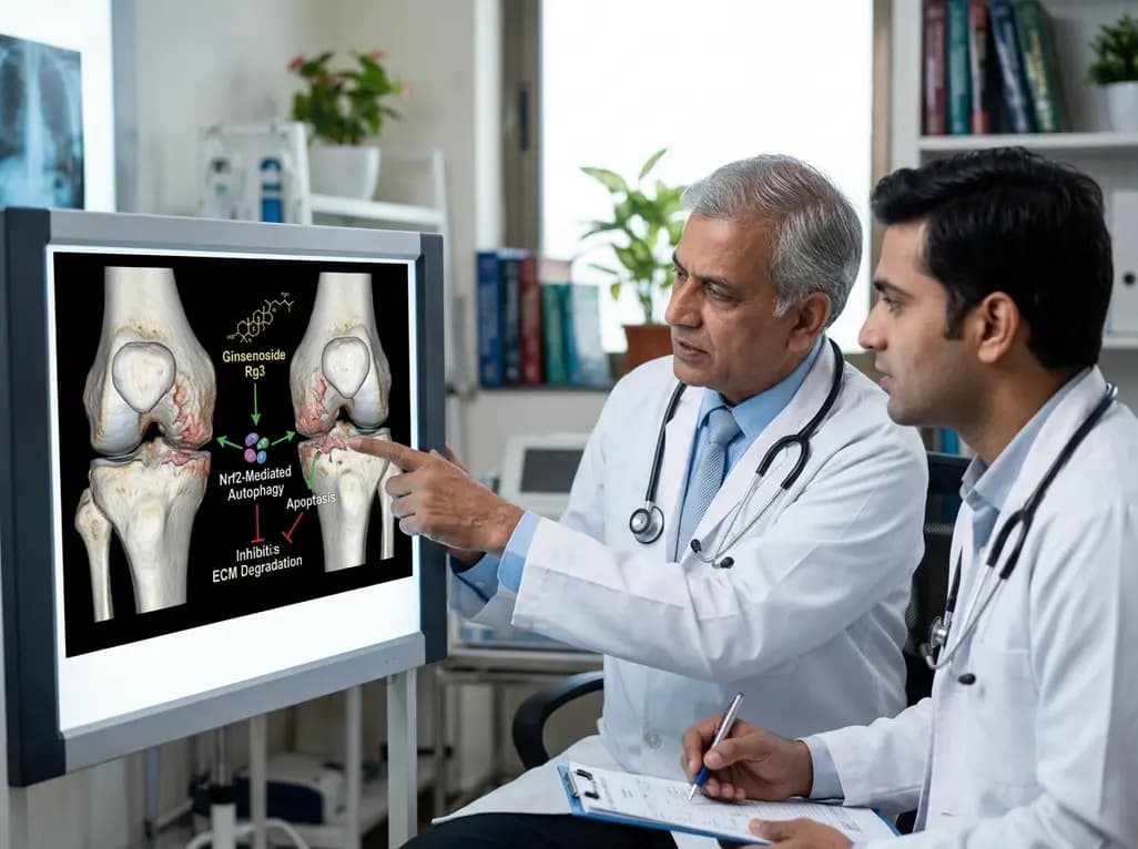
A study reveals that Ginsenoside Rg3 attenuates osteoarthritis progression by modulating the Nrf2-mediated autophagy pathway, reducing chondrocyte apoptosis, and curbing extracellular matrix degradation, offering promising chondroprotective potential.
Today

A cross-sectional study demonstrates that lower pediatric motor competence strongly correlates with adverse metabolic syndrome markers, including central adiposity, elevated triglycerides, and low HDL cholesterol, emphasizing early neuromuscular interventions in youth with overweight and obesity.
Today
