.webp&w=384&q=75)

Loading, please wait...
Loading, please wait...
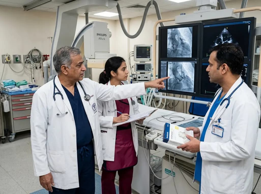
Adult congenital heart disease presents complex anatomical and hemodynamic challenges that demand innovative structural interventions. A ruptured sinus of Valsalva aneurysm represents an uncommon yet life-threatening complication in patients with surgically corrected congenital anomalies. When this rare lesion emerges decades after complete repair of tetralogy of Fallot, clinical decision-making becomes exceptionally intricate. Recently, interventional cardiologists described a groundbreaking approach that successfully eliminated a significant left-to-right shunt while restoring pulmonary valve competence in a single percutaneous procedure. This technique highlights how modern transcatheter solutions can overcome severe anatomical distortions in high-risk adult surgical candidates.
Sinus of Valsalva aneurysms arise primarily from congenital weakness at the junction between the aortic media and the fibrous annulus. Over several decades, systemic arterial pressures cause progressive thinning and progressive outward ballooning of the affected sinus wall. Eventually, sudden or subacute perforation occurs, which generates an acute or progressive left-to-right shunt into adjacent low-pressure cardiac chambers. In patients with repaired tetralogy of Fallot, previous surgical patches, altered geometry of the right ventricular outflow tract, and chronic hemodynamic stress amplify the vulnerability of the right coronary sinus. Consequently, a ruptured aneurysm in this setting frequently drains directly into the main pulmonary artery or the right ventricle. This abnormal communication precipitates rapid volume overload, progressive chamber dilation, and acute-on-chronic heart failure.
A 53-year-old male with a history of complete tetralogy of Fallot repair during infancy presented with rapidly progressive New York Heart Association functional class IV heart failure. Clinicians promptly initiated a comprehensive diagnostic workup to determine the precise etiology of his decompensation. Advanced cardiac computed tomography revealed a large ruptured sinus of Valsalva originating from the right aortic sinus, measuring 28.1 by 33.5 mm and communicating directly with the main pulmonary artery. Furthermore, invasive cardiac catheterization demonstrated a substantial left-to-right shunt with a pulmonary-to-systemic flow ratio of 2.6 to 1. Fortunately, the patient maintained normal pulmonary vascular resistance despite marked volume overload. Transthoracic and transesophageal echocardiography confirmed coexisting severe pulmonary regurgitation, which compounded the right ventricular volume overload.
Traditional management of an acute sinus rupture requires emergent open-heart surgical reconstruction with patch closure and valve repair. However, previous sternotomies, extensive mediastinal adhesions, and advanced heart failure made redo cardiac surgery prohibitively hazardous for this patient. Therefore, the multidisciplinary structural heart team devised a novel transcatheter approach using a self-expanding 25-mm Harmony pulmonary valve. By deploying this specialized covered transcatheter device across the native right ventricular outflow tract, operators aimed to cover the fistulous communication completely while simultaneously treating the pulmonary regurgitation. Precise angiographic and echocardiographic guidance ensured optimal positioning of the valve frame against the main pulmonary artery wall, thereby sealing the defect without obstructing coronary blood flow or compromising adjacent cardiac structures.
The transcatheter intervention achieved immediate and complete technical success without procedural complications. Post-deployment angiography confirmed full occlusion of the ruptured aneurysm with zero residual left-to-right shunting. In addition, the prosthetic valve established excellent competence, drastically reducing pulmonary regurgitation from severe to trace levels. Right ventricular systolic and diastolic pressures declined substantially within hours of device deployment, signaling immediate hemodynamic offloading. Following the procedure, the patient experienced rapid symptomatic recovery, and his functional status markedly improved from class IV to class II heart failure. Serial follow-up imaging corroborated stable device anchoring, intact coronary arterial perfusion, and sustained right ventricular remodeling.
This landmark case provides several crucial learning points for cardiologists managing adult congenital heart disease. First, clinicians must maintain a high index of suspicion for aortocardiac fistulas or sinus aneurysms when patients with repaired congenital defects deteriorate unexpectedly. Second, multimodal imaging combining cardiac computed tomography, echocardiography, and invasive hemodynamics remains indispensable for mapping intricate spatial relationships. Finally, transcatheter pulmonary valve technology offers a viable therapeutic alternative for selected patients who face prohibitive operative mortality. As transcatheter device designs and delivery systems continue to evolve, transcatheter therapies will increasingly provide life-saving alternatives to conventional open redo surgeries in complex structural heart disease.
Patients typically present with acute or subacute onset of dyspnea, fatigue, chest pain, and signs of congestive heart failure. Physical examination frequently reveals a continuous, loud cardiac murmur heard along the left sternal border. When the rupture produces a large left-to-right shunt, patients rapidly deteriorate into decompensated heart failure requiring urgent intervention.
Conventional open repair entails significant operative risk in patients with prior infant sternotomies, complex scar tissue, and severe cardiac dysfunction. A transcatheter valve prosthesis simultaneously occludes the aneurysm orifice along the pulmonary artery wall and corrects concomitant pulmonary regurgitation, effectively treating two major hemodynamic lesions through a single minimally invasive femoral access.
Comprehensive evaluation requires multidetector cardiac computed tomography angiography to assess three-dimensional anatomy, defect diameter, and coronary artery proximity. In addition, transesophageal echocardiography evaluates valve competence, while invasive right and left heart catheterization accurately quantifies shunt magnitude, cardiac output, and pulmonary vascular resistance.
Disclaimer: This content is for informational and educational purposes only. It is not intended to be a substitute for professional medical advice, diagnosis, or treatment. Always seek the advice of your physician or other qualified health provider with any questions you may have regarding a medical condition. Refer to the latest local and national guidelines for clinical practice.
References

Read summarized clinical updates, watch expert medical content, and earn CME certifications right from your smartphone.
Open your smartphone camera to scan and install.





A 53-year-old adult with repaired tetralogy of Fallot presented in severe heart failure caused by a ruptured sinus of Valsalva aneurysm. Clinicians successfully treated the defect and concomitant severe pulmonary regurgitation using a transcatheter 25-mm Harmony valve, avoiding high-risk redo surgery.
4 weeks back

A systematic review of 41 preclinical studies reveals that baicalin dampens systemic inflammation, inhibits NF-κB and NLRP3 pathways, and improves organ preservation in sepsis models. However, substantial study heterogeneity and a complete lack of human clinical trials highlight the need for further translational research.
Today
Discover the crucial distinctions between mesonephric and mesonephric-like proliferations of the female genital tract. Learn the key histomorphologic, immunohistochemical, and molecular differences essential for accurate diagnosis and clinical management.
Today

A large UK Biobank study reveals that plasma proteomic signatures capture preclinical organ damage and improve multiorgan risk prediction across early CKM syndrome stages 0 to 2 beyond traditional PREVENT clinical models.
Today
A narrative review investigates whether the articularis genus muscle functions as an independent anatomical entity or blends with the vastus intermedius. We evaluate its morphology, role in retracting the suprapatellar bursa, and direct clinical significance in anterior knee pain and arthroplasty.
Today

A Bayesian multilevel meta-analysis reveals that aerobic training combined with moderate carbohydrate restriction modestly lowers HbA1c in type 2 diabetes. However, sparse data and very low certainty leave incremental benefits over exercise or diet alone unproven, highlighting the need for individualized care.
Today
