.webp&w=384&q=75)

Loading, please wait...
Loading, please wait...
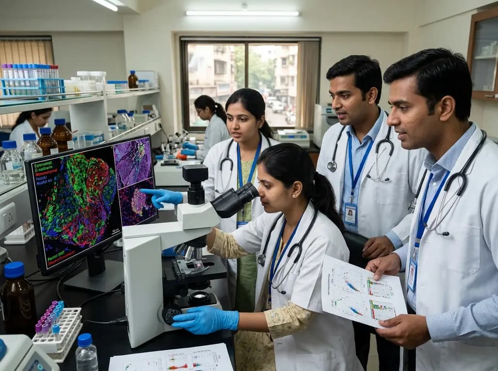
Oral squamous cell carcinoma represents one of the most formidable oncological challenges globally, particularly across the Indian subcontinent where tobacco, betel quid, and areca nut habits remain prevalent. Clinicians frequently encounter aggressive disease marked by rapid local invasion, extensive cervical nodal dissemination, and frequent treatment failure. Recent translational investigations highlight that TP53 mutations in OSCC function not merely as passive genomic scars, but as active biochemical engines driving extreme phenotypic plasticity. While traditional paradigms viewed malignant progression as a binary transition from epithelial to mesenchymal states, emerging evidence reveals a far more nuanced intermediate continuum. Specifically, malignant oral keratinocytes leverage a hybrid cellular program known as partial epithelial-mesenchymal transition. In this intermediate state, neoplastic cells retain critical intercellular cohesive elements while simultaneously acquiring strong migratory mesenchymal characteristics. Consequently, understanding the direct regulatory link between mutant p53 signaling and these intermediate cellular phenotypes offers vital diagnostic precision and unveils actionable therapeutic targets for high-risk oral malignancies.
Historically, pathologists recognized TP53 as the primary guardian of genomic stability, coordinating cell-cycle arrest, DNA repair, and programmed apoptotic demise upon cellular stress. However, in oral cavity carcinomas, somatic alterations within this gene occur in 60% to 80% of clinical cases. Neoplastic clones routinely display remarkable intratumoral genetic divergence across distinct geographic zones of the same lesion. To capture this complexity, investigators utilized multi-regional whole-exome sequencing spanning dozens of discrete tumor micro-domains alongside matched regional lymph node metastases. Most alterations localized directly within the conserved DNA-binding domain, predominantly presenting as missense substitutions. Rather than causing complete peptide absence, these structural alterations consistently provoked severe intracellular accumulation of abnormal p53 protein throughout both nuclear and cytoplasmic compartments. Importantly, spatial sequencing proved that these driver mutations arise early in clonal evolution and endure intact during regional nodal dissemination. Therefore, mutant p53 establishes a persistent genomic trunk that orchestrates diverse phenotypic programs across distinct subclonal branches, thereby amplifying overall intratumoral heterogeneity.
To characterize how genomic divergence impacts cellular architecture, researchers executed advanced multiplexed immunofluorescence combined with digital cellular spatial analysis. Traditional histology often overlooks fine gradients within complex microenvironments. In contrast, spatially resolved single-cell image profiling uncovered profound structural reorganization specifically within TP53-mutant tumor sectors. Unaltered wild-type oral tumors maintained robust membranous E-cadherin expression with minimal mesenchymal protein presence. Conversely, mutant tissue architectures exhibited marked E-cadherin suppression coupled with strong vimentin induction. Most strikingly, digital segmentation identified an enriched subpopulation of pan-cytokeratin and vimentin dual-positive hybrid cells. Rather than adopting an entirely spindle-like mesenchymal identity, these neoplastic cells preserve epithelial adhesion while activating mesenchymal motility machines. Notably, spatial distribution maps demonstrated that these partial EMT cells concentrate heavily along the invasive tumor front directly abutting the reactive stroma. Consequently, this localized spatial clustering explains why mutant tumors invade surrounding healthy tissues as cohesive, collective migration streams rather than isolated individual cells.
Beyond observational pathology, rigorous in vitro functional assays illuminate how specific mutant proteins direct cellular reprogramming. Investigators transiently transfected immortalized oral epithelial lines with the prevalent human hotspot mutation p53R248Q. This specific missense substitution severely disrupts essential protein-DNA contact surfaces. Remarkably, introducing this single genetic alteration rapidly precipitated noticeable morphological transitions. Cultured epithelial cells loosened their rigid polygonal contacts and assumed elongated, dynamic migratory shapes. Quantitative reverse-transcription polymerase chain reaction assays verified robust upregulation of core mesenchymal transcript factors alongside dramatic suppression of classic epithelial markers. Furthermore, cellular migration and wound-healing assays demonstrated markedly enhanced locomotive capacity in mutant-transfected keratinocytes compared to parental controls. Mechanistically, mutant p53 proteins engage in distinct gain-of-function oncogenic interactions, hijacking alternative transcription factor networks and promoting autocrine growth signaling loops. Thus, p53R248Q operates as a direct molecular switch that destabilizes epithelial fidelity and activates intermediate mesenchymal plasticity programs.
The clinical ramification of partial EMT driven by mutated p53 is profound for surgical and medical oncologists. Neoplastic cells suspended in a hybrid state exhibit elevated resistance to standard platinum-based chemotherapy and conventional ionizing radiation. Moreover, these plastic cells possess elevated self-renewal features characteristic of cancer stem cells. Multi-regional profiling confirms that identical mutant alleles persist between primary tumor cores, infiltrating fronts, and metastatic cervical lymph nodes. Therefore, primary tumors harboring high proportions of hybrid cells demonstrate an elevated propensity for early occult lymphatic spread and extranodal extension. During routine histopathological evaluation, deep invasive margins often display detached cohesive clusters exhibiting high budding indices. These histological patterns reflect active partial EMT underway at the leading edge. Consequently, identifying mutant p53 accumulation along with dual cytokeratin-vimentin expression can help pathologists detect high-risk disease variants early. Such granular spatial profiling promises to refine surgical margin assessment and guide decisions regarding elective neck dissections.
Translating these cellular breakthroughs into tangible patient outcomes requires innovative targeted approaches tailored to overcome tumor plasticity. Currently, standard systemic cytotoxic regimens yield limited survival improvements in refractory, recurrent oral cancer. However, targeting the p53-EMT signaling axis provides compelling therapeutic avenues. First, small-molecule refolding compounds, such as APR-246, aim to restore wild-type conformational binding to structurally altered p53 variants. By re-establishing normal transcription functions, these agents can re-sensitize malignant cells to apoptotic triggers. Second, interrupting downstream transcriptional regulators of partial EMT, including AXL, TGF-beta, and focal adhesion kinases, can effectively reverse cellular plasticity. Reverting invasive hybrid cells into differentiated, non-motile epithelial states restores susceptibility to localized radiation and immune surveillance. Furthermore, because hybrid EMT phenotypes intimately correlate with an immunosuppressive microenvironment, combining plasticity inhibitors with immune checkpoint blockade represents a rational clinical strategy. Ultimately, integrating spatial molecular diagnostics will allow oncologists to tailor targeted combinations for patients facing aggressive, TP53-driven oral malignancies.
A partial epithelial-mesenchymal transition state describes a plastic cellular condition where neoplastic cells express both epithelial and mesenchymal markers simultaneously. Instead of converting entirely into spindle-like mesenchymal cells, malignant keratinocytes retain intercellular adhesion proteins like cytokeratin while gaining motility factors such as vimentin. Consequently, these hybrid cells migrate collectively, withstand environmental shear stress, and drive aggressive local invasion alongside elevated metastatic dissemination.
Missense mutations within the DNA-binding domain of TP53 cause defective p53 peptides to accumulate heavily within nuclear and cytoplasmic compartments. Instead of undergoing normal rapid degradation, these stable mutant proteins engage non-canonical transcription factors, inhibit protective p63 and p73 family members, and activate downstream oncogenic cascades. Through these aberrant protein-protein interactions, mutant p53 directly transactivates mesenchymal gene networks, promotes stemness, and enhances therapeutic resistance.
Conventional bulk tissue profiling obscures regional clonal divergence and masks critical cellular behaviors occurring across specific tissue microdomains. In contrast, spatially resolved techniques examine the invasive tumor-stromal interface where partial EMT cells predominantly concentrate. Therefore, spatial mapping allows pathologists to identify aggressive subclonal cell clusters at surgical resection margins, predicting microscopic residual disease, local recurrence risks, and early occult regional lymph node metastases far more accurately.
Disclaimer: This content is for informational and educational purposes only... Refer to the latest local and national guidelines for clinical practice.
References
Ibrahim RA et al. TP53 mutations orchestrate partial EMT states in oral squamous cell carcinoma: spatially resolved and in vitro preliminary insights into tumour plasticity. J Pathol. 2026 Sep 30. doi: 10.1002/path.70123. PMID: 42812045.
Ma L, Zhang Y, Liu W, et al. The role of TP53 mutations in oral cancer: molecular mechanisms and prognostic implications. Adv Cancer Res. 2026;162:145-178.
Wang C, Liu X, Chen Z, et al. Partial-EMT in oral squamous cell carcinoma: molecular circuitry and clinical translation. Int J Oral Sci. 2026;18(1):12-25.
de Oliveira LR, Ribeiro-Silva A, Zucoloto S, et al. Functionally impactful TP53 mutations are associated with increased risk of extranodal extension in clinically advanced oral squamous cell carcinoma. Cancer. 2020;126(20):4500-4510.

Read summarized clinical updates, watch expert medical content, and earn CME certifications right from your smartphone.
Open your smartphone camera to scan and install.





A landmark study reveals that TP53 mutations in oral squamous cell carcinoma fuel partial epithelial-mesenchymal transition and tumor plasticity. Using multi-regional sequencing and spatial multiplexed immunofluorescence, researchers uncover key mechanisms driving invasive phenotypes and lymph node metastasis.
Today

Data from the 2026 Workforce Health Index reveals that 38.7% of urban employees and dependents exhibit abnormal glycemic control. Emerging at increasingly younger ages, this metabolic shift intersects with severe vitamin deficiencies, highlighting the urgent need for early workplace screenings and targeted clinical care.
Today

The Maharashtra Food and Drug Administration has enforced stringent compliance orders under food safety laws. The directive prohibits commercial promotions, infant formula samples, gifts, and clinician sponsorships while mandating explicit breastfeeding notices and rapid adverse event reporting across healthcare facilities.
Today

Haryana Chief Minister Nayab Singh Saini has rolled out landmark performance-linked financial incentives for public sector clinicians and allied staff. The comprehensive reform introduces free intravitreal diabetic retinopathy management, flexible empanelment for private specialists, and digital infrastructure upgrades.
Today

A cross-sectional study reveals a strong positive correlation between problematic internet use and kinesiophobia in adults aged 18-45 with non-specific low back pain, highlighting how digital overuse reinforces movement fear and complicates musculoskeletal recovery.
Today

A recent coronary CT angiography study demonstrates that diabetic patients exhibit accelerated plaque burden, impaired CT-FFR, and elevated perivascular fat attenuation index values, reflecting heightened vascular inflammation that worsens across adverse metabolic phenotypes.
Today
