.webp&w=384&q=75)

Loading, please wait...
Loading, please wait...

Orthognathic correction of severe dentofacial deformities demands rigorous evaluation of craniomandibular biomechanics. In clinical maxillofacial practice, surgeons frequently observe TMJ morphological changes following surgical repositioning of asymmetric jaws. Mandibular prognathism accompanied by facial asymmetry presents complex mechanical challenges to the articular eminence and mandibular condyle. Consequently, understanding these early adaptive responses helps clinicians optimize surgical stability, safeguard joint health, and refine multidisciplinary treatment protocols.
Patients presenting with mandibular prognathism and concurrent facial asymmetry exhibit distinctive baseline joint characteristics. Due to chronic unilateral chewing patterns and rotational skeletal discrepancies, the deviated and non-deviated sides endure vastly divergent biomechanical loads. Clinicians regularly observe marked structural disparities between contralateral temporomandibular joints before any orthodontic or surgical intervention begins. Specifically, the non-deviated side typically experiences unique functional loading that alters the articular fossa and condylar head over prolonged periods. Furthermore, the posterior slope of the articular eminence displays notable differences in inclination when comparing both sides. Preoperative multi-sectional computed tomography imaging clearly delineates these complex adaptive osseous configurations. Understanding this pre-existing structural asymmetry enables reconstructive surgeons to anticipate how realignment movements might impact joint stability. In addition, recognizing baseline remodeling prevents clinicians from misinterpreting established osseous morphology as acute progressive pathology. Therefore, comprehensive three-dimensional imaging remains indispensable before initiating presurgical orthodontic decompensation. Maxillofacial teams utilize these detailed diagnostic findings to design patient-specific surgical vectors that minimize harmful articular torsion. Consequently, precise anatomical baseline mapping ensures predictable skeletal alignment while safeguarding long-term joint function.
Surgeons tailor surgical interventions to the severity of the dentofacial deformity. In patients with isolated mandibular discrepancies, teams often execute bilateral sagittal split osteotomy alone. Conversely, severe three-dimensional discrepancies with canted occlusal planes require a combined approach consisting of Le Fort I maxillary osteotomy and mandibular ramus osteotomy. Both approaches reposition the tooth-bearing segments while simultaneously shifting the proximal condylar segments into new relationships. However, bimaxillary surgery introduces three-dimensional alterations across the midface and skull base that redistribute masticatory stresses differently than isolated mandibular surgery. Maxillofacial surgeons must carefully control the proximal segment during internal fixation to prevent adverse condylar rotation. Furthermore, rigid internal fixation with miniplates or bicortical screws provides initial stability, but it locks the condyle into a predetermined position within the glenoid fossa. As a result, the articular disc and surrounding fibrocartilage must adapt rapidly to this revised anatomical configuration during early healing. Clinicians continuously evaluate these mechanical dynamics to prevent condylar displacement, postoperative malocclusion, or progressive joint resorption. Accordingly, selecting the appropriate surgical technique directly influences the trajectory of postoperative articular remodeling.
Computed tomography reconstructed imaging at six months postoperatively reveals fascinating patterns of osseous adaptation. Research indicates that the posterior slope of the articular eminence remains significantly steeper on the non-deviated side compared to the deviated side both preoperatively and after surgery. This persistence demonstrates that intrinsic cranial base anatomy maintains its fundamental contours despite radical mandibular repositioning. Nevertheless, the functional repositioning triggers localized adaptive remodeling along the articular surface. Chondrocytes and subchondral bone cells respond dynamically to altered vectors of joint loading. Moreover, the condylar head often exhibits subtle surface flattening or cortical bone thickening as it establishes equilibrium within the fossa. Surgeons note that these early changes represent physiologic accommodation rather than destructive osteoarthritis when patient occlusion remains stable. Additionally, the non-deviated and deviated condyles demonstrate asymmetric remodeling rates due to uneven muscular pull during rehabilitation. By tracking these changes at the six-month mark, clinicians verify whether the joint tissues have successfully accommodated the new mandibular posture. Consequently, six-month postoperative tomographic scans provide an exceptional window for evaluating craniomandibular stability and detecting early osseous maladaptation.
Evaluating early morphological adaptations provides vital insights into long-term occlusal stability. When the surgical procedure successfully balances bilateral articular loading, patients generally enjoy improved masticatory efficiency and reduced myofascial discomfort. In contrast, improper positioning of the proximal segments can generate excessive compressive stress against the posterior slope of the eminence. Such unmitigated pressure frequently causes condylar resorption, joint pain, and skeletal relapse. Maxillofacial specialists must therefore combine meticulous intraoperative positioning with structured postoperative monitoring. Furthermore, clinicians in Indian maxillofacial centers emphasize phased physical rehabilitation to encourage coordinated neuromuscular adaptation. Early physiotherapy guides symmetrical jaw translation and prevents uncoordinated lateral excursions that strain healing joint tissues. Similarly, close collaboration between the orthodontist and the maxillofacial surgeon guarantees timely detection of minor occlusal interferences. Eliminating these prematurities through post-surgical orthodontics protects the remodeling condyle from chronic microtrauma. Ultimately, integrating radiographic assessments with clinical functional evaluations enables surgeons to maintain long-term skeletal harmony and protect temporomandibular well-being. Accordingly, treating clinicians establish proactive follow-up protocols to identify subtle morphological variations before they trigger functional compromise.
Modern three-dimensional imaging has revolutionized the diagnostic workup for orthognathic surgery. While conventional multidetector computed tomography delivers outstanding contrast for cortical bone morphology, low-dose cone-beam computed tomography now provides comparable precision with reduced radiation exposure. Clinicians routinely use these volumetric scans to perform multi-sectional reconstructions and voxel-based superimpositions. Consequently, surgical teams can quantify sub-millimeter modifications in articular eminence inclination, condylar volume, and joint space dimensions over serial intervals. Moreover, artificial intelligence algorithms now assist radiologists by automatically segmenting TMJ structures and identifying subtle osteolytic changes. Looking ahead, integrating three-dimensional bony models with dynamic magnetic resonance imaging will elucidate the interactions between soft tissue disc displacement and hard tissue remodeling. Furthermore, prospective multicenter registries across diverse patient populations will clarify how ethnic facial variations influence joint biomechanics. By embracing these innovative diagnostic technologies, surgical teams can enhance predictive treatment planning and minimize surgical complications. Therefore, continuous technological integration will redefine the standards of care for patients with asymmetric dentofacial deformities.
Patients with asymmetric mandibular prognathism routinely develop unequal biomechanical loading patterns across their jaw joints over many years. Because the non-deviated side frequently bears greater compressive forces during mastication and unilateral chewing, the local bone undergoes functional remodeling. Consequently, the posterior slope of the articular eminence becomes significantly steeper on the non-deviated side to accommodate these altered functional demands. This structural adaptation often persists even after corrective orthognathic surgery.
Orthognathic osteotomies alter condylar position and redistribute stress across the temporomandibular joint. If surgeons improperly rotate or compress the proximal segment during rigid internal fixation, excessive mechanical load concentrates on the condylar head. Consequently, this abnormal mechanical stress can compromise subchondral vascularity, provoking osteoclast activation and progressive bone resorption. However, precise preoperative digital planning, passive segment positioning, and regular radiographic monitoring effectively minimize this adverse complication during postoperative recovery.
Multi-sectional computed tomography provides exquisite three-dimensional visualization of osseous architecture that conventional panoramic radiographs cannot match. Clinicians utilize postoperative tomographic images to detect early condylar displacement, assess articular eminence inclination, and quantify bone remodeling patterns. Furthermore, advanced digital superimposition enables surgical teams to compare preoperative and postoperative anatomy with high precision. Therefore, volumetric imaging at six months postoperatively serves as a crucial diagnostic tool to verify joint stability and guide orthodontic finishing.
Disclaimer: This content is for informational and educational purposes only. It does not constitute medical advice and must not replace professional clinical judgment, diagnosis, or treatment. Always consult a qualified specialist for clinical decision-making. Neither the author nor the publisher assumes any liability for actions taken based on this publication. Refer to the latest local and national guidelines for clinical practice.
References

Read summarized clinical updates, watch expert medical content, and earn CME certifications right from your smartphone.
Open your smartphone camera to scan and install.





A prospective CT evaluation examines early postoperative morphological alterations in the temporomandibular joint among patients with mandibular prognathism and facial asymmetry, highlighting articular eminence adaptation and condylar remodeling after bimaxillary and mandibular ramus osteotomies.
Today
Modern renal nutrition is shifting toward additive-aware dietary guidance. This paradigm differentiates between intrinsic nutrients in whole foods and hyper-bioavailable inorganic additives, offering practical strategies to mitigate additive stacking and toxicity without restricting nutritious whole foods.
Today

A multiparametric MRI approach combining quantitative T2 shading, apparent diffusion coefficient (ADC) measurements, and morphological signs provides superior diagnostic accuracy over subjective evaluation to differentiate ovarian endometriomas from benign hemorrhagic cysts.
Today
A landmark case report reveals a donor heart beating for a century in a 63-year-old recipient, challenging conventional age limits in cardiac transplantation and offering critical lessons on organ longevity, donor selection, and long-term graft survival.
Today
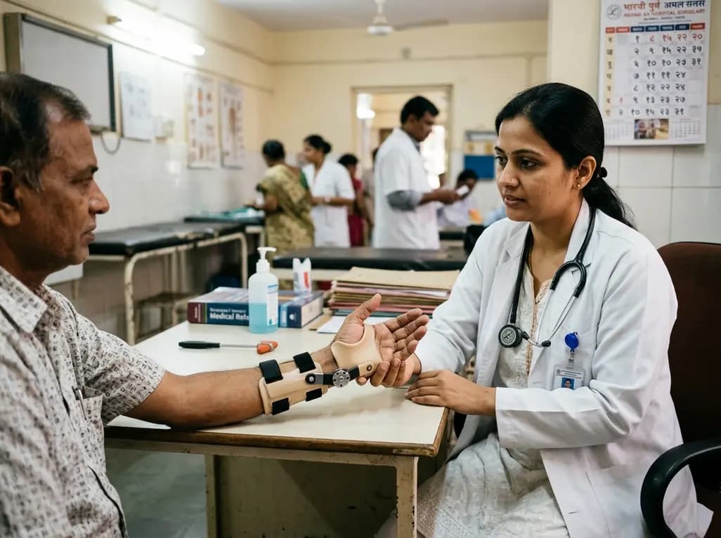
A new randomized crossover trial reveals that a hinged ulnar gutter orthosis significantly improves hand function and weight-bearing tolerance compared to standard volar splints in patients with degenerative triangular fibrocartilage complex lesions, supporting functionally permissive orthotic designs.
Today

New research reveals that M1 macrophage-derived exosomes aggravate diabetic nephropathy by transferring WTAP to stabilize S1PR2 mRNA. Silencing WTAP in these vesicles attenuates endothelial injury and renal fibrosis, pointing toward innovative nanomedicine therapies.
Yesterday
