.webp&w=384&q=75)

Loading, please wait...
Loading, please wait...
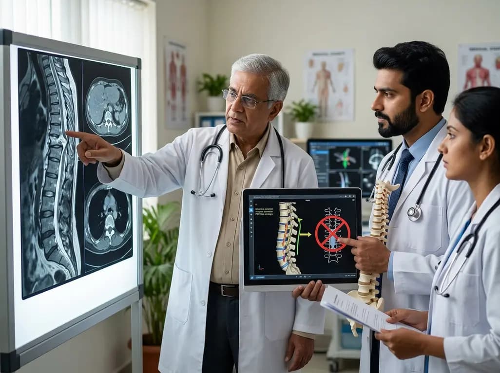
Managing thoracic ossification of the posterior longitudinal ligament (T-OPLL) represents one of the most significant challenges in modern spinal surgery, particularly when treating nonambulatory patients. Specifically, the thoracic OPLL surgical strategy must balance the need for adequate decompression with the high risk of iatrogenic injury to a vulnerable spinal cord. In the Indian clinical context, where late presentations of myelopathy are common, selecting an approach that minimizes surgical trauma while maximizing neurological recovery is essential. Traditional methods often involve aggressive circumferential decompression, which, although effective, is associated with high morbidity. Recent evidence now suggests that a more selective, intraoperatively guided approach may offer a safer alternative for these high-risk individuals.
Consequently, clinicians are increasingly exploring the posterior decompression with fusion (PDF)-first strategy. This approach focuses on initial posterior stabilization and indirect decompression, reserving more invasive ventral work for cases where it is strictly necessary. By utilizing real-time intraoperative data, surgeons can tailor the procedure to the specific physiological response of the spinal cord. This paradigm shift moves away from a one-size-fits-all aggressive resection toward a customized intervention. Such a strategy is particularly relevant for the "beak-type" morphology, where the ossified mass creates a localized, high-pressure point on the ventral cord that is notoriously difficult to manage without causing further damage.
Thoracic OPLL is categorized into several morphological types, with the "beak-type" being the most clinically aggressive. This variant is characterized by a localized, sharp protrusion of bone that directly impales the ventral aspect of the spinal cord. Unlike the "continuous" or "mixed" types, which spread the pressure over multiple segments, the beak-type focuses mechanical stress on a single point. This focal compression frequently results in severe neurological deficits, often rendering patients nonambulatory before they seek surgical consultation. Furthermore, the thoracic spine’s natural kyphosis and limited blood supply to the spinal cord make any surgical manipulation in this region exceptionally precarious.
Moreover, the radiographic presence of a beak-type lesion often prompts surgeons to consider circumferential decompression (CD) as the primary option. CD involves removing the ossified mass from both posterior and anterior/lateral directions, which is a technically demanding and high-risk maneuver. However, the inherent risks of CD, including massive blood loss and a high incidence of cerebrospinal fluid (CSF) leaks, necessitate a re-evaluation of its routine use. Understanding that not every beak-type lesion requires direct ventral resection is a cornerstone of the modern thoracic OPLL surgical strategy. By analyzing the mechanics of how the cord shifts after posterior decompression, surgeons can often achieve satisfactory outcomes without the morbidity associated with ventral ossification removal.
The posterior decompression with fusion-first (PDF-first) strategy is built on the principle of minimizing initial surgical trauma. In this protocol, all patients undergo a standard posterior decompression combined with instrumented fusion and dekyphosis. The rationale is that posterior decompression relieves the dorsal pressure, while instrumented fusion stabilizes the segment and corrects the kyphotic alignment. This correction often leads to an "indirect decompression," where the spinal cord shifts away from the ventral beak-type lesion. In many cases, this shift is sufficient to restore blood flow and allow for neurological recovery, even without removing the primary ossified mass.
In addition to its simplicity, the PDF-first approach serves as a diagnostic and therapeutic baseline. It allows the surgical team to assess the cord's behavior in real-time before committing to the more hazardous steps of a circumferential decompression. If the spinal cord shows signs of adequate relief—such as improved pulsation or stable neurophysiological signals—the procedure can be concluded safely. This selective nature prevents unnecessary exposure to the risks of ventral surgery. Furthermore, by maintaining a posterior-only approach initially, surgeons can preserve more of the spinal column's structural integrity, which is vital for long-term stability and faster postoperative rehabilitation in formerly nonambulatory patients.
The success of an intraoperatively guided thoracic OPLL surgical strategy depends heavily on the accuracy of the assessment tools used during the procedure. Once the PDF portion is completed, the surgeon must decide whether to proceed to a circumferential decompression. This decision is guided by several critical indicators: dural sac refilling, the return of visible spinal cord pulsation, and the use of intraoperative ultrasonography (IOUS). IOUS is particularly valuable as it allows the surgeon to visualize the "floating" of the spinal cord away from the beak-type ossification. If a persistent ventral compression or a "tent-pole" effect is observed, it indicates that indirect decompression was insufficient.
However, visual and ultrasound findings are only part of the equation. Intraoperative neurophysiological monitoring (IONM), including motor-evoked potentials (MEPs), provides a functional assessment of the cord. If MEPs remain stagnant or deteriorate after the posterior stage, the surgeon has clear evidence that the ventral compression is still clinically significant. Consequently, the conversion to CD is performed only when these multiple data points converge to suggest that the cord remains at risk. This multi-modal approach reduces the subjectivity of the decision-making process. It ensures that aggressive ventral resection is reserved for the minority of patients who truly require it, thereby optimizing the safety profile of the entire intervention.
Data from recent clinical cohorts demonstrate the efficacy of the PDF-first strategy. In studies involving nonambulatory patients with beak-type T-OPLL, approximately 60% of patients achieved satisfactory results with PDF alone, while the remaining 40% required additional CD. Most notably, the overall recovery rate is remarkably high, with over 93% of previously nonambulatory patients regaining the ability to walk at the final follow-up. Both the PDF-only and the PDF+CD groups showed significant improvements in their modified Japanese Orthopaedic Association (mJOA) scores and health-related quality of life metrics, such as the EQ-5D-5L.
Nonetheless, there are distinct differences in the perioperative characteristics of the two groups. Patients who required the conversion to circumferential decompression experienced significantly longer operative times and greater estimated blood loss compared to those treated with PDF alone. This highlight the inherent complexity of CD and validates the effort to avoid it when possible. However, the fact that the neurological outcomes were comparable between the groups suggests that CD is an effective adjunct when used selectively. The ability to achieve high rates of ambulation in a formerly paralyzed cohort is a testament to the power of a tiered surgical approach that prioritizes cord safety and physiological feedback.
Despite the successes of the selective thoracic OPLL surgical strategy, complications remain a significant concern, particularly in the high-risk T-OPLL population. Cerebrospinal fluid (CSF) leakage is one of the most common issues, especially during circumferential decompression where the ossified mass may be intimately adhered to the dura. In some instances, the ossification actually replaces the dural tissue, making a tear inevitable during resection. However, when managed with meticulous primary repair or specialized sealing techniques, these leaks generally do not result in permanent neurological sequelae. Surgeons must be prepared for extended bedside management and potentially the use of lumbar drains in the postoperative period.
Furthermore, the risk of postoperative motor palsy, though reduced by intraoperative monitoring, is still present. This highlights the need for specialized postoperative care and aggressive physical therapy. In the Indian healthcare setting, where rehabilitation resources can vary, the emphasis should be on early mobilization and close neurological monitoring in the first 48 hours following surgery. Ultimately, the transition from a routine aggressive approach to an individualized, intraoperatively guided strategy represents a significant advancement. It allows for the treatment of severe T-OPLL with a higher degree of precision, ensuring that the most invasive techniques are used only when they are absolutely necessary to achieve neurological recovery.
The beak-type is a specific morphological classification of thoracic ossification of the posterior longitudinal ligament. Unlike continuous types that are flatter, the beak-type features a sharp, localized ossified mass that protrudes into the spinal canal. This creates a focal point of high-intensity pressure on the ventral spinal cord. It is often associated with a higher risk of neurological deficit and presents a greater technical challenge during surgical decompression due to its localized and aggressive nature.
The PDF-first approach is safer because it prioritizes spinal stabilization and indirect decompression before attempting risky ventral maneuvers. By performing posterior decompression and correcting kyphosis through fusion, the spinal cord is often able to shift away from the ventral mass naturally. This avoids the high-risk steps of removing the ossification directly from the front of the cord, which is frequently associated with massive blood loss, dural tears, and iatrogenic cord injury in nonambulatory patients.
Surgeons rely on several real-time indicators to decide if indirect decompression is sufficient. They look for visible dural sac refilling and the return of spinal cord pulsation. Additionally, intraoperative ultrasonography is used to confirm if the cord has "floated" away from the ventral beak. Finally, neurophysiological monitoring like motor-evoked potentials (MEPs) provides functional data; if the cord signals do not improve after the posterior stage, the surgeon may proceed to a more aggressive circumferential decompression.
Disclaimer: This content is for informational and educational purposes only and does not constitute medical advice or a professional relationship. The field of spinal surgery is rapidly evolving, and clinical decisions should be based on the individual patient's presentation and the surgeon's expertise. Refer to the latest local and national guidelines for clinical practice.
References
Lei J et al. An intraoperatively guided posterior decompression with fusion-first strategy for nonambulatory patients with beak-type thoracic ossification of the posterior longitudinal ligament. J Neurosurg Spine. 2026 Jul 10. doi: 10.3171/2026.2.SPINE251204. PMID: 42430804.
Kato S et al. Indication for anterior spinal cord decompression via a posterolateral approach for the treatment of ossification of the posterior longitudinal ligament in the thoracic spine: a prospective cohort study. Eur Spine J. 2020;29(5):1001-1008.
Imagama S et al. The Essence of Clinical Practice Guidelines for Ossification of Spinal Ligaments, 2019: 5. Treatment of Thoracic OPLL. Spine Surg Relat Res. 2021;5(5):330-333.

Read summarized clinical updates, watch expert medical content, and earn CME certifications right from your smartphone.
Open your smartphone camera to scan and install.





A recent study evaluates a selective posterior decompression with fusion (PDF)-first surgical strategy for nonambulatory patients with beak-type thoracic OPLL. The findings suggest that intraoperative guidance can minimize the need for aggressive circumferential decompression while ensuring high recovery rates.
3 months ago
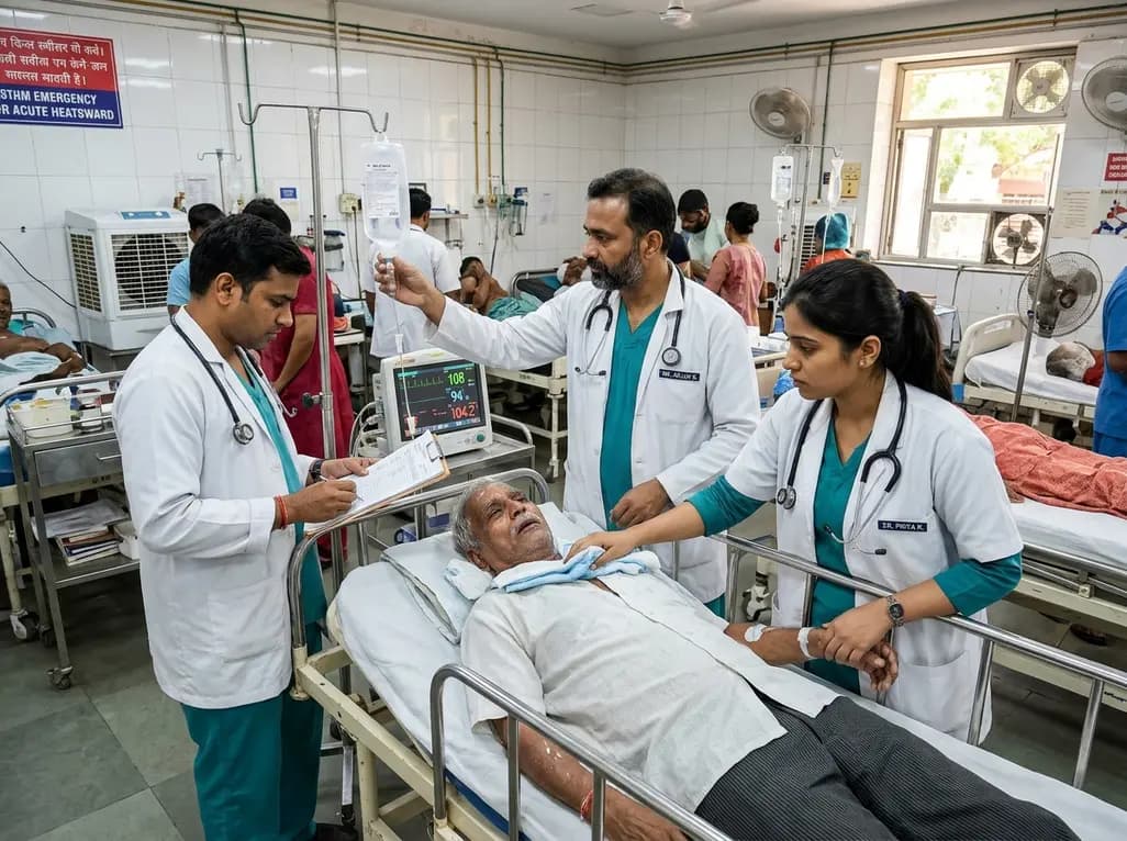
Recent epidemiological data reveals a dramatic surge in summer heat mortality driven by human-induced climate warming. This analysis explores the pathophysiological impacts on cardiac and renal systems, diagnostic dilemmas in death certification, and essential emergency cooling protocols for acute clinical care.
Today

Odisha has established India's first state-level clinical protocol specifically tailored for pregnant women with sickle cell disease. Developed with NHM and UNICEF, the framework provides structured antenatal screening, crisis mitigation, standardized referral pathways, and specialized interdisciplinary care.
Today

India is advancing preventive healthcare by integrating genomics, artificial intelligence, and quantum technologies. With the SANGAM programme sequencing one million Indian genomes, clinicians can unlock personalized therapies, detect cardiometabolic risks early, and curb the burden of diabetes and heart disease.
Today
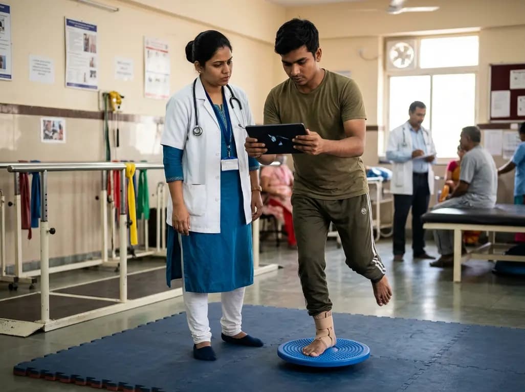
A randomized controlled trial demonstrates that an eight-week dual-task training intervention significantly improves static and dynamic postural control over standard balance exercises in individuals with chronic ankle instability, addressing critical neurocognitive deficits during athletic and daily activities.
Today
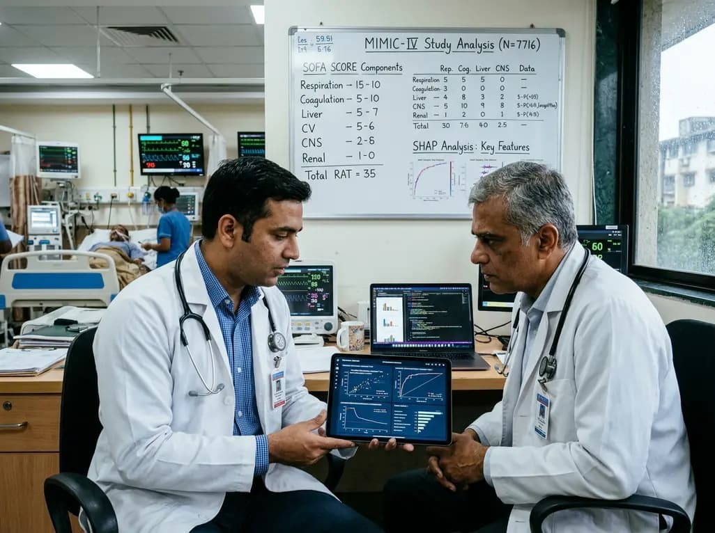
A retrospective cohort study of 7,716 ICU patients from the MIMIC-IV database demonstrates that Gradient Boosting Decision Trees significantly outperform traditional SOFA scores in predicting in-hospital mortality among patients with sepsis and diabetes, with SHAP analysis providing clinical interpretability.
Today
