.webp&w=384&q=75)

Loading, please wait...
Loading, please wait...
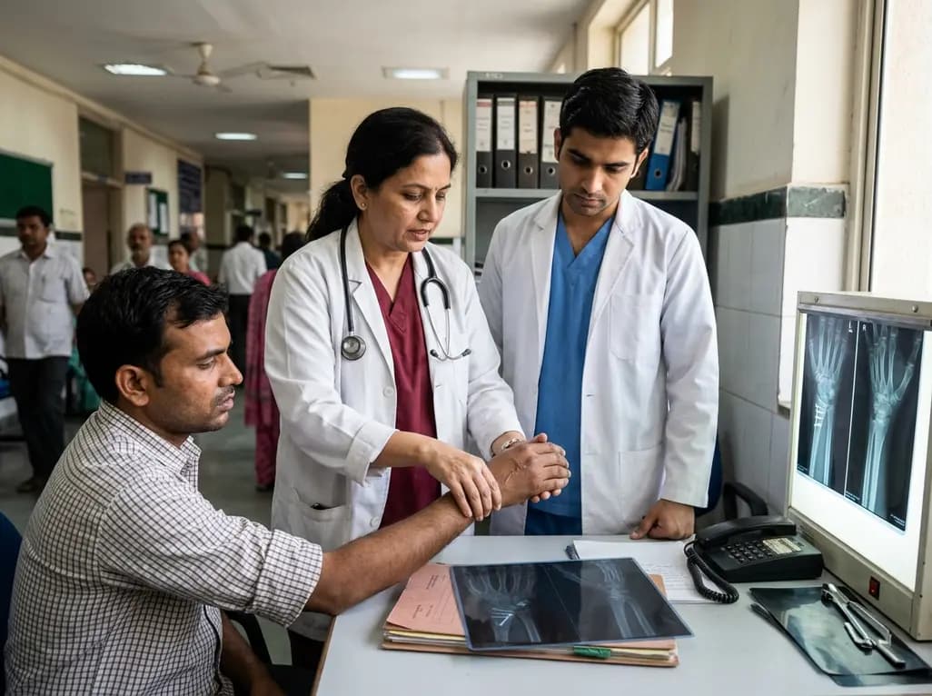
Chronic wrist pain and mechanical dysfunction often stem from complex pathology involving the triangular fibrocartilage complex. For orthopedic surgeons, achieving durable distal radioulnar joint stability remains a significant operative challenge. Recent surgical advances demonstrate that performing an arthroscopically assisted TFCC foveal repair alongside dorsal capsuloligamentous reinforcement offers a powerful solution for patients presenting with severe instability. This combined reconstructive approach directly addresses both the deep foveal anchoring mechanism and the dorsal restraint system. Consequently, patients achieve reliable pain relief, improved grip strength, and restored wrist mechanics.
The triangular fibrocartilage complex acts as the premier soft-tissue stabilizer of the distal radioulnar joint. When traumatic avulsions or chronic degenerative tears disrupt the foveal attachment of the radioulnar ligaments, severe rotational and translational instability often ensues. Patients typically report debilitating ulnar-sided wrist pain, weakness during gripping tasks, and painful joint subluxation during forearm pronation and supination.
Historically, isolated foveal repairs have yielded satisfactory results in acute settings. However, chronic tears present unique structural challenges. Over time, persistent joint subluxation stretches the secondary dorsal capsular constraints and attenuates surrounding tissue quality. As a result, standard suture fixation to the ulnar fovea may fail to restore sufficient stability against rotational torque.
Furthermore, long-standing joint laxity alters wrist kinematics, accelerating degenerative changes across the distal radioulnar articulation. Orthopedic specialists must identify patients with high-grade joint laxity who will benefit from structural augmentation. Recognizing the combined loss of deep foveal anchoring and dorsal capsular competence allows surgeons to plan comprehensive reconstruction strategies that optimize long-term joint function.
The foveal footprint of the triangular fibrocartilage complex represents the primary isometric anchor for both the dorsal and volar radioulnar ligaments. Consequently, restoring this deep anatomical attachment is the fundamental cornerstone of any reconstructive intervention. Transosseous foveal repair achieves this objective by securing torn ligamentous tissue directly to a prepared osseous bed in the distal ulnar fovea using transosseous suture tunnels.
Arthroscopic assistance significantly refines this procedure. It enables direct visualization of intra-articular tear patterns, dynamic assessment of joint laxity, and precise preparation of the bony footprint without extensive open dissection. Through targeted bone tunnels, high-strength sutures re-approximate the deep ligament limbs back to their native insertion.
Nevertheless, biomechanical investigations reveal that severe distal radioulnar joint instability involves multidirectional tissue laxity. While isolated transosseous foveal fixation successfully restores primary axial resistance, it may leave residual dorsal joint translation unaddressed under physiologic loads. Therefore, combining transosseous foveal anchorage with secondary dorsal reinforcement creates a synergistic construct. This comprehensive construct replicates the natural force-coupling mechanism of the wrist, protecting the primary healing repair from early mechanical overload.
The combined reconstructive technique integrates arthroscopic precision with targeted soft-tissue augmentation. First, the surgeon performs diagnostic wrist arthroscopy to debride non-viable tissue and prepare the ulnar fovea. Subsequently, the surgical team drills transosseous bone tunnels through the distal ulna to facilitate anatomically aligned suture passage and foveal reconstruction.
Following transosseous fixation, the surgical team addresses dorsal instability through a focused dorsal approach. Surgeons introduce an allogeneic tendon graft to reinforce the attenuated dorsal radioulnar joint capsule and dorsal radioulnar ligament fibers. The allograft provides an immediate, robust structural scaffold that bridges the dorsal joint interval.
Surgeons securely anchor the tendon graft across the dorsal margins of the sigmoid notch and the distal ulna using suture anchors or interference fixation. This allograft reinforcement augments the repaired dorsal capsule without causing excessive joint tightness. Importantly, allogeneic graft tissue eliminates donor site morbidity associated with autologous tendon harvest. By establishing immediate mechanical restraint against dorsal subluxation, this reinforcement protects the healing transosseous foveal junction throughout the critical early remodeling phase.
Recent long-term clinical data highlight the effectiveness of combining transosseous foveal repair with dorsal allograft reinforcement in patients with severe chronic distal radioulnar joint instability. In an extensive clinical series followed over a mean period of 44.5 months, patients experienced profound and sustained symptomatic relief. Notably, visual analog scale pain scores dropped dramatically from a preoperative average of 5.5 down to 0.7 at final follow-up.
Moreover, objective functional parameters demonstrated substantial improvements. Mean grip strength increased from 75.1% to 86.6% compared to the unaffected contralateral limb. Functional assessment scores also reflected remarkable progress; the modified Mayo wrist score surged from 68.9 points preoperatively to an impressive 90.3 points postoperatively.
Additionally, patient-reported outcome measures underscored substantial clinical benefits. The Patient-Rated Wrist Evaluation score decreased significantly from 50.4 to 8.9 points. Because every single patient achieved a reduction greater than the established 24-point minimal clinically important difference, the procedure demonstrated exceptional reliability. These sustained long-term metrics confirm that augmenting foveal repair with dorsal grafting effectively resolves persistent joint subluxation while restoring pain-free upper limb function.
These compelling clinical findings carry critical implications for practicing orthopedic surgeons, sports medicine physicians, and hand reconstructive specialists. Chronic triangular fibrocartilage complex tears accompanied by severe distal radioulnar joint instability represent a distinct, high-risk subset of wrist injuries. Treating these challenging presentations with isolated suture fixation or debridement carries a heightened risk of persistent joint subluxation and premature procedural failure.
Therefore, clinicians should incorporate rigorous dynamic stability assessments into their diagnostic workup. Clinicians should evaluate forearm ballottement, stress radiography, and specialized magnetic resonance imaging to gauge the true extent of capsuloligamentous disruption. When clinical examination confirms severe multidirectional laxity, surgeons should proactively consider dual-component reconstruction.
Furthermore, utilizing allogeneic tendon graft material streamlines operative efficiency while sparing patients the morbidity and pain associated with harvesting autografts like the palmaris longus. Although prospective randomized controlled trials are warranted to further compare combined reconstruction against isolated foveal repair, current therapeutic evidence strongly supports dual stabilization for chronic, unstable patterns. Hand surgeons can confidently apply this technique to restore joint congruency and salvage wrist function in active individuals.
Postoperative rehabilitation plays a crucial role in safeguarding the reconstructed distal radioulnar joint while facilitating functional recovery. Following combined transosseous foveal fixation and dorsal reinforcement, the care team initially immobilizes the operative extremity in an above-elbow splint or cast with the forearm positioned in neutral rotation. This early immobilization period lasts approximately four to six weeks, shielding the healing foveal bone-tendon interface and dorsal allograft from disruptive rotational shear forces.
Subsequently, patients transition into a protected, removable wrist splint to initiate gentle, active-assisted range of motion exercises. Physical therapists guide progressive forearm supination, pronation, and wrist flexion-extension while avoiding aggressive passive manipulation. Strengthening protocols typically commence at eight to twelve weeks postoperatively, focusing on dynamic wrist stabilizers including the extensor carpi ulnaris and pronator quadratus muscles.
Ultimately, this gradual, phased rehabilitation trajectory allows the allograft tissue to incorporate securely and mature structurally. Patients generally return to demanding manual labor and athletic activities between four and six months post-surgery. Long-term surveillance confirms that this structured rehabilitation framework preserves joint stability, prevents recurrent subluxation, and fosters durable functional recovery.
Isolated foveal repair directly reattaches the primary radioulnar ligaments to the ulnar fovea. However, chronic and severe distal radioulnar joint instability typically involves extensive attenuation or tearing of secondary stabilizers, including the dorsal joint capsule. Without augmenting these lax dorsal structures, persistent rotational shear forces can overpower the isolated foveal suture line. Consequently, adding dorsal reinforcement provides essential mechanical constraint, preventing recurrent subluxation and protecting the healing transosseous repair.
Combining transosseous foveal repair with dorsal allograft reinforcement delivers comprehensive multiplanar joint stability. Clinically, this combined approach dramatically relieves chronic ulnar-sided wrist pain, significantly increases grip strength, and restores forearm rotational mechanics. Objective outcome tools show substantial improvements in modified Mayo wrist scores and Patient-Rated Wrist Evaluation metrics. Furthermore, utilizing an allograft eliminates donor site morbidity, enabling patients to achieve predictable functional recovery and return to athletic activities safely.
Rehabilitation begins with four to six weeks of rigid immobilization in neutral forearm rotation to protect the tendon graft and bone tunnels. Afterward, patients transition into a removable splint and begin guided active-assisted wrist and forearm motion. Progressive isometric and isotonic strengthening exercises start around eight to twelve weeks post-surgery, emphasizing dynamic stabilizing muscles. Patients usually resume unrestricted manual work and sports between four and six months once joint stability is established.
Disclaimer: This content is for informational and educational purposes only and does not constitute medical advice or establish a doctor-patient relationship. Healthcare professionals must exercise their independent clinical judgment. Refer to the latest local and national guidelines for clinical practice.
References

Read summarized clinical updates, watch expert medical content, and earn CME certifications right from your smartphone.
Open your smartphone camera to scan and install.





Transosseous TFCC foveal repair combined with dorsal reinforcement offers significant pain relief, improved grip strength, and restored stability in chronic DRUJ injury.
last month
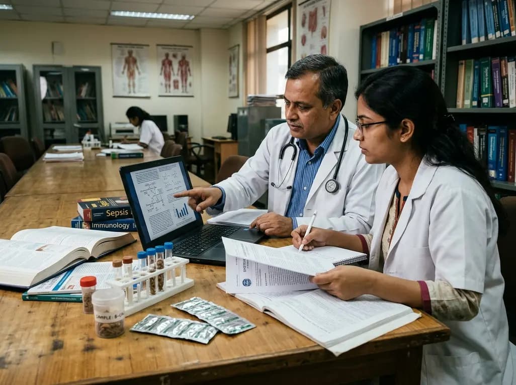
A systematic review of 41 preclinical studies reveals that baicalin dampens systemic inflammation, inhibits NF-κB and NLRP3 pathways, and improves organ preservation in sepsis models. However, substantial study heterogeneity and a complete lack of human clinical trials highlight the need for further translational research.
Today

Discover the crucial distinctions between mesonephric and mesonephric-like proliferations of the female genital tract. Learn the key histomorphologic, immunohistochemical, and molecular differences essential for accurate diagnosis and clinical management.
Today

A large UK Biobank study reveals that plasma proteomic signatures capture preclinical organ damage and improve multiorgan risk prediction across early CKM syndrome stages 0 to 2 beyond traditional PREVENT clinical models.
Today

A narrative review investigates whether the articularis genus muscle functions as an independent anatomical entity or blends with the vastus intermedius. We evaluate its morphology, role in retracting the suprapatellar bursa, and direct clinical significance in anterior knee pain and arthroplasty.
Today

A Bayesian multilevel meta-analysis reveals that aerobic training combined with moderate carbohydrate restriction modestly lowers HbA1c in type 2 diabetes. However, sparse data and very low certainty leave incremental benefits over exercise or diet alone unproven, highlighting the need for individualized care.
Today
