

Loading, please wait...
Loading, please wait...

Syringomyelia represents an uncommon spinal pathology characterized by fluid-filled cavitation within the spinal cord parenchyma. Clinicians typically encounter this condition alongside hindbrain anomalies, trauma, tumors, or chronic arachnoiditis. However, syringomyelia arising secondary to degenerative spinal canal stenosis represents a distinct and underdiagnosed clinical occurrence. Degenerative changes in the cervical and thoracic spine compress the neural elements and alter cerebrospinal fluid circulation. Consequently, patients develop progressive spinal cord cavitation without congenital hindbrain herniation. Understanding this rare manifestation enables neurosurgeons and spine specialists to formulate timely, targeted interventions.
Degenerative spinal canal stenosis stems primarily from advancing spondylotic degeneration across the cervical and thoracic vertebrae. As intervertebral discs dehydrate and collapse, biomechanical stresses prompt reactive osteophyte formation, uncovertebral arthrosis, and ligamentum flavum hypertrophy. These progressive anatomical distortions gradually compromise the functional dimensions of the spinal canal. Consequently, circumferential or focal narrowing impinges directly upon the spinal cord and its delicate vascular supply. Although cervical spondylotic myelopathy occurs frequently among aging adults, concurrent syringomyelia remains remarkably infrequent in clinical practice. Systematic reviews demonstrate that this secondary cavitation occurs across a wide adult age spectrum, presenting predominantly in men in their fifth and sixth decades of life. Because general clinical awareness remains low, physicians frequently mistake the resulting symptoms for isolated spondylotic myelopathy or idiopathic syringomyelia. Additionally, delayed recognition may expose vulnerable neural pathways to prolonged mechanical compression and ischemic damage. Therefore, identifying degenerative canal stenosis as the primary driver of intramedullary cyst formation changes the overall diagnostic and surgical approach.
The pathogenesis linking spinal canal stenosis to intramedullary syrinx formation involves interconnected mechanical and hydrodynamic events. Severe extradural stenosis obstructs the subarachnoid space and disrupts laminar cerebrospinal fluid oscillations. Consequently, cardiac systole creates an abnormal pressure gradient across the constricted spinal segment. Elevated pulse pressures drive subarachnoid fluid through widened Virchow-Robin spaces into the spinal cord parenchyma. Additionally, repetitive flexion and extension against spondylotic osteophytes induce localized mechanical microtrauma. This chronic microvascular strain impairs cord perfusion and triggers focal ischemic necrosis. Over time, ischemic myelomalacia promotes tissue cavitation and coalesces into a continuous syrinx cavity. Furthermore, localized epidural venous compression elevates intramedullary venous pressure, reducing interstitial fluid clearance and impairing glymphatic function. Together, these persistent hydrodynamic disturbances and cellular injuries perpetuate intramedullary fluid accumulation within the spinal cord. Clinicians must recognize that relieving the mechanical obstruction reverses these destructive physical forces, allowing spontaneous fluid reabsorption.
Patients exhibiting syringomyelia secondary to degenerative canal stenosis present along two distinct clinical pathways. First, symptomatic individuals comprise approximately fifty-seven percent of reported cases. These patients manifest classic signs of spinal cord compromise driven by both mechanical compression and cystic cavitation. Motor weakness represents the predominant complaint, occurring in seventy-five percent of symptomatic patients. Individuals frequently report progressive hand clumsiness, reduced grip strength, and spastic gait disturbances. Sensory deficits appear in half of symptomatic cases, often manifesting as thermoanalgesic dissociation or widespread paresthesias across the limbs. Furthermore, axial neck stiffness and radicular pain afflict nearly thirty-eight percent of individuals. In contrast, asymptomatic patients account for roughly forty-three percent of recognized cases. Radiologists typically identify these incidental syrinxes during routine imaging investigations for benign cervical strain or neck pain. Crucially, long-term retrospective observation indicates that asymptomatic incidental syrinxes often remain completely quiescent without surgical intervention. Differentiating these phenotypes ensures tailored therapeutic decision-making and prevents unnecessary surgical operations.
Accurate clinical diagnosis demands comprehensive neuroimaging evaluation of the complete craniospinal axis. Magnetic resonance imaging serves as the definitive gold standard modality for assessing both stenosis and intramedullary cavitation. High-resolution sagittal and axial T2-weighted scans delineate the extent of canal narrowing, ligamentous thickening, and syrinx dimensions. Furthermore, clinicians must administer intravenous gadolinium contrast to definitively exclude underlying intramedullary neoplasms, including ependymomas and hemangioblastomas. In addition, imaging of the posterior fossa remains mandatory to rule out cerebellar tonsillar herniation characteristic of Chiari I malformations. Phase-contrast cine magnetic resonance imaging provides exceptional diagnostic assistance by capturing real-time cerebrospinal fluid flow dynamics. This functional sequence confirms focal subarachnoid obstruction at the spondylotic level while verifying patent basilar cisterns. Similarly, neuroimaging eliminates post-traumatic encephalomalacia and inflammatory arachnoiditis from the differential diagnosis. Establishing this clear etiology prevents hazardous misdiagnoses, unnecessary diagnostic procedures, and inappropriate interventions.
Managing symptomatic patients requires careful surgical planning focused on eliminating the inciting hydrodynamic barrier. Historically, surgeons frequently treated primary syringomyelia using direct intramedullary drainage procedures. However, modern neurosurgical consensus strongly advocates extradural decompression alone for stenosis-induced cavitation. Direct syrinx shunting requires invasive myelotomy, carrying elevated risks of cord tethering, direct neural injury, and recurrent catheter obstruction. Conversely, extradural decompression restores physiological subarachnoid cerebrospinal fluid circulation without breaching the spinal cord parenchyma. Surgeons choose between anterior cervical discectomy and fusion, cervical corpectomy, or posterior decompressive laminectomy and laminoplasty based on compressive architecture. Systematic reviews reveal that extradural decompression alone achieves syrinx regression in fifty-eight percent of operated individuals. Posterior decompressive procedures account for the substantial majority of documented radiological resolutions. Therefore, spine specialists reserve direct syrinx drainage exclusively for exceptional cases that fail to improve after definitive mechanical decompression and canal widening.
Postoperative recovery following extradural decompression demonstrates favorable clinical and functional trajectories across published series. Decompressing the pinched cord arrests myelopathic progression and facilitates marked functional recuperation. Most treated individuals regain motor strength, restore coordinated hand function, and experience significant gait improvement. Sensory disturbances and radicular pain also diminish substantially, although chronic spinothalamic injury occasionally produces lingering paresthesias. Radiographically, postoperative imaging demonstrates partial cavity collapse or complete syrinx resolution in the majority of patients. Nevertheless, radiographic reduction does not always mirror functional recovery in a linear fashion. Some individuals experience remarkable neurological restoration despite small persistent fluid cavities. Conversely, irreversible myelomalacia may leave residual deficits despite complete morphological collapse. For asymptomatic individuals, conservative observation remains highly safe, as serial scans reveal stable or spontaneously regressing fluid collections. Ongoing clinical vigilance and structured long-term surveillance ensure sustained patient stability over time.
Severe degenerative canal stenosis compresses the subarachnoid space and blocks pulsatile cerebrospinal fluid flow. This mechanical obstruction creates a focal pressure dissociation across the cardiac cycle, forcing fluid into the spinal cord parenchyma through perivascular channels. Additionally, localized cord ischemia and microtrauma induce intramedullary tissue necrosis, promoting cavitation.
Asymptomatic syrinxes identified incidentally alongside degenerative canal stenosis generally warrant conservative management rather than immediate surgery. Available evidence indicates that these cavities typically remain stable or regress spontaneously over time. Clinicians recommend regular clinical surveillance and serial magnetic resonance imaging, reserving surgical decompression exclusively for progressive neurological deterioration or expanding cysts.
Extradural decompression directly corrects the underlying etiology by restoring physiological cerebrospinal fluid circulation across the stenotic spinal segment. In contrast, primary intradural shunting requires direct spinal cord myelotomy, introducing significant risks of cord injury, arachnoid scarring, and shunt malfunction. Consequently, decompression alone achieves syrinx regression without exposing the cord to invasive drainage.
Disclaimer: This content is for informational and educational purposes only... Refer to the latest local and national guidelines for clinical practice.
References

Read summarized clinical updates, watch expert medical content, and earn CME certifications right from your smartphone.
Open your smartphone camera to scan and install.





A systematic review explores syringomyelia secondary to degenerative spinal canal stenosis. Extradural decompression alone effectively resolves intramedullary cavitations, whereas incidental asymptomatic syrinxes often remain stable under conservative observation.
Today

A secondary analysis of the prospective international Hyponatremia Intervention Trial (HIT) evaluates how volume status and underlying etiologies in hypotonic hyponatremia shift across adult age groups, highlighting crucial diagnostic and therapeutic challenges for geriatric and internal medicine practice.
Today

A new study evaluates a multilingual digital health platform combining rule-based cervical cancer risk assessment, retrieval-augmented generation AI, and geospatial navigation. The tool dramatically reduces clinical hallucinations while providing actionable health guidance for underserved populations.
Today

A case-control study demonstrates significant lipid profile alterations in myocardial infarction patients, highlighting elevated total cholesterol, triglycerides, and LDL-C alongside decreased HDL-C, emphasizing early lipid screening and aggressive secondary cardiovascular prevention.
Today
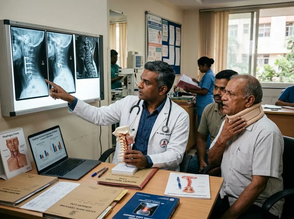
A large registry study reveals that postoperative dysphagia occurs more frequently and persists longer after multilevel anterior cervical spine surgery than single-level procedures. Key risk factors include baseline dysphagia, diabetes, upper cervical segment involvement, and medical comorbidities.
Today

A landmark UK Biobank study by Karolinska Institutet researchers reveals that optimal rest mitigates neurodegeneration in cardiometabolic illness. Patients maintaining high sleep quality demonstrated superior cognitive scores and larger brain volumes, highlighting sleep optimization as an actionable clinical strategy.
Today
