

Loading, please wait...
Loading, please wait...

The coexistence of degenerative lumbar disease and end-stage hip osteoarthritis represents a pervasive clinical dilemma known as hip-spine syndrome. While orthopedic surgeons frequently analyze how spinal stiffness affects hip prostheses, the reciprocal impact of total hip arthroplasty on lumbar spinal fusion has historically received far less scrutiny. A comprehensive systematic review examining 271,008 patients now illuminates this critical reciprocal relationship. The evidence demonstrates that surgical sequencing directly governs patient-reported recovery, implant longevity, and adverse event profiles. Consequently, understanding how joint replacement interacts with subsequent or prior spinal interventions is essential for reconstructive surgeons. Clinicians must carefully evaluate biomechanical adaptations before scheduling elective interventions.
Spino-pelvic alignment relies on dynamic communication between the mobile lumbosacral spine, pelvis, and acetabulofemoral joints. When degenerative osteoarthritis compromises the hip, the pelvis frequently compensates by tilting anteriorly or posteriorly to preserve upright horizontal gaze. Similarly, degenerative disc pathology limits sagittal flexibility and alters normal pelvic tilt during standing and sitting transitions. When surgeons fuse lumbar segments, they eliminate normal spinal compensations and force the hip joint to accommodate exaggerated motion arcs. Conversely, restoring hip range of motion through total hip replacement shifts mechanical loads back onto adjacent spinal segments. Therefore, an intervention in one anatomic region immediately transforms the stress environment of the other. If clinicians fail to recognize this kinetic chain, secondary mechanical failure often follows. Furthermore, anatomical stiffness in an arthritic hip may mask subtle spinal stenosis symptoms, leading to diagnostic confusion. Comprehensive clinical examination and postural radiographs in both standing and sitting positions remain indispensable. By mapping dynamic pelvic parameters before operating, surgical teams can anticipate biomechanical strain and mitigate progressive spinal instability.
Clinical data reveal that patient outcomes vary markedly based on procedural order. Specifically, patients who undergo total hip arthroplasty prior to lumbar spinal fusion exhibit unique functional profiles. In large multicenter cohorts, individuals receiving hip arthroplasty first demonstrated higher baseline Oswestry Disability Index scores compared to patients treated in reverse order. In addition, their Harris Hip Scores were comparatively lower during initial evaluations. These metrics suggest that severe functional impairment often prompts surgeons to address the hip joint initially. Nevertheless, completing hip reconstruction first offers undeniable biomechanical advantages. By correcting hip flexion contractures and restoring pelvic mobility, hip arthroplasty can re-establish physiologic sagittal balance. As a result, some patients experience notable relief of low back symptoms, which occasionally eliminates or postpones the need for extensive spinal arthrodesis. However, if spinal surgery remains unavoidable, surgeons must monitor persistent axial disability closely. Coordinated rehabilitation protocols should focus on core muscle activation and pelvic stability. Consequently, multidisciplinary planning ensures that initial arthroplasty creates a favorable mechanical baseline for subsequent spinal stabilization.
Performing hip arthroplasty after previous spinal stabilization introduces substantial mechanical and structural hazards for the lumbar column. In the systematic review, total hip arthroplasty performed after spinal fusion correlated with higher rates of adjacent segment disease. Moreover, these patients experienced increased incidences of pseudarthrosis, mechanical hardware failure, and revision lumbar procedures. When spinal fusion creates a rigid, non-compliant column, subsequent altered gait mechanics and pelvic tilt changes generate focal stress concentrations at the fusion margins. Consequently, the junctional vertebrae absorb repetitive torsional loads during normal ambulatory motion. Over time, these elevated mechanical forces accelerate intervertebral disc degeneration directly above or below the fused segments. Furthermore, poor pelvic compliance can compromise implant seated stability, triggering revision surgeries. Surgeons must therefore scrutinize prior instrumentation integrity before undertaking subsequent total joint replacement. Modifying prosthetic acetabular cup orientation based on functional sitting radiographs can diminish catastrophic impingement. Ultimately, preserving spine-pelvis harmony protects both the joint replacement and the existing lumbar hardware from premature failure.
Beyond structural mechanical issues, surgical timing substantially influences systemic perioperative morbidity. The systematic review highlighted that performing spinal fusion within two years after hip arthroplasty significantly elevates several serious clinical risks. Specifically, these patients demonstrated higher rates of deep vein thrombosis, acute neurological complications, and prolonged postoperative opioid dependency. When two major orthopedic operations occur in close succession, cumulative surgical trauma intensifies the systemic inflammatory response. Moreover, impaired postoperative mobility between sequential procedures magnifies venous stasis and thromboembolic hazards. Chronic pain pathways also become hyper-sensitized when structural pain persists across multiple anatomic regions. As a result, patients frequently develop opioid tolerance, complicating postoperative analgesic management. Clinicians must therefore implement aggressive multimodal thromboprophylaxis regimens when managing short-interval procedures. Additionally, preoperative opioid tapering programs and multimodal pain protocols can mitigate dependency risks. Close neurological surveillance remains imperative, as altered spinal biomechanics and scar tissue can heighten nerve root vulnerability during instrumentation. Careful staging allows physiological recovery before initiating another major surgical insult.
Navigating concurrent hip and spine degeneration requires seamless multidisciplinary collaboration between adult reconstruction specialists and spine surgeons. First, clinicians must accurately identify the primary symptom generator. High-resolution diagnostic intra-articular hip injections can isolate joint pathology from radicular spine pain with exceptional accuracy. If hip osteoarthritis represents the predominant functional barrier, prioritizing total hip arthroplasty is generally prudent. Indeed, eliminating hip flexion contractures often restores pelvic lordosis and resolves secondary postural back discomfort. Conversely, if severe neurological deficits or progressive motor weakness exist, urgent spinal decompression and stabilization must take precedence. When both procedures are inevitable, surgical teams should avoid rushing into the second procedure within two years unless clinically mandated. Furthermore, advanced preoperative planning must incorporate standing and relaxed-seated lateral spinopelvic radiographs. These functional views identify whether a patient exhibits a flexible or rigid spine. For instance, rigid lumbar columns require customized acetabular anteversion angles to prevent prosthetic impingement. By aligning surgical sequencing with individual spinopelvic kinematics, surgeons can dramatically reduce complications and optimize long-term patient recovery.
Postoperative rehabilitation plays a crucial role in safeguarding both spinal constructs and hip arthroplasty components. Physical therapists must design structured exercise programs that emphasize hip abductor strength, lumbo-pelvic coordination, and hamstring flexibility. In addition, therapists should avoid aggressive spinal flexion or hyperextension during early rehabilitation phases, particularly in patients with combined procedures. Restoring balanced kinematics decreases abnormal shear stresses across transitional spinal zones and protects prosthetic cup interfaces. Meanwhile, orthopedic research must continue investigating customized surgical workflows. Advanced technological solutions, including robotic-assisted arthroplasty and patient-specific navigation, offer unprecedented component placement accuracy. These emerging technologies allow surgeons to simulate dynamic postural changes before making a single bone resection. Furthermore, prospective clinical registries should track patient-reported functional outcomes alongside revision rates over extended multi-decade periods. Such longitudinal datasets will clarify the subtle trade-offs associated with alternative surgical intervals. Ultimately, bridging the gap between spine and arthroplasty disciplines will elevate standards of care for aging populations worldwide.
Surgeons generally prioritize total hip arthroplasty when hip arthritis predominantly drives symptoms or limits extension. Restoring hip range of motion can normalize pelvic tilt and alleviate compensatory back discomfort. Conversely, progressive neurologic compression or severe spinal instability dictates performing spinal decompression and stabilization first to prevent irreversible nerve damage.
Lumbar arthrodesis immobilizes lumbosacral segments, preventing normal pelvic retroversion during sitting and standing postural transitions. Consequently, the prosthetic hip experiences abnormal mechanical stress, increased impingement, and higher dislocation rates. Furthermore, altered gait mechanics and reduced spinal shock absorption transmit destructive forces directly across the new joint replacement components.
Undergoing spinal fusion within two years following hip arthroplasty significantly increases risks of deep vein thrombosis, neurological deficits, and long-term opioid use. Cumulative surgical trauma, limited early mobility, and sustained nociceptive signaling compound systemic vulnerability. Staging procedures beyond this recovery window allows physiologic stabilization and optimal tissue healing.
Disclaimer: This content is for informational and educational purposes only and does not constitute medical advice, diagnosis, or treatment. Healthcare professionals should exercise their independent clinical judgment when making treatment decisions. Guidelines and evidence-based practices should always be considered in the context of individual patient care. Refer to the latest local and national guidelines for clinical practice.
References:

Read summarized clinical updates, watch expert medical content, and earn CME certifications right from your smartphone.
Open your smartphone camera to scan and install.





A systematic review of 271,008 patients reveals how the surgical sequencing of total hip arthroplasty and lumbar spinal fusion impacts lumbar complications, revision rates, and functional outcomes.
Today

A qualitative study evaluated the INCLUDE trial, a culturally tailored mobile diabetes prevention intervention. Findings highlight digital accessibility and community coaching as key facilitators, while time constraints and rigid dietary elements posed barriers, offering vital lessons for clinical practice.
Today

A large cohort study demonstrates that fetal growth restriction in a first pregnancy raises maternal cardiovascular risk within five years postpartum, including elevated rates of heart failure, stroke, and myocardial infarction. Clinicians must initiate proactive postpartum screening and cardiovascular prevention.
Today
An NHANES analysis reveals that meeting 24-hour movement guidelines significantly lowers the odds of cardiovascular disease, obesity, hypertension, and diabetes among adults with disabilities, emphasizing the power of combined physical activity, sleep, and limited sedentary time.
Today
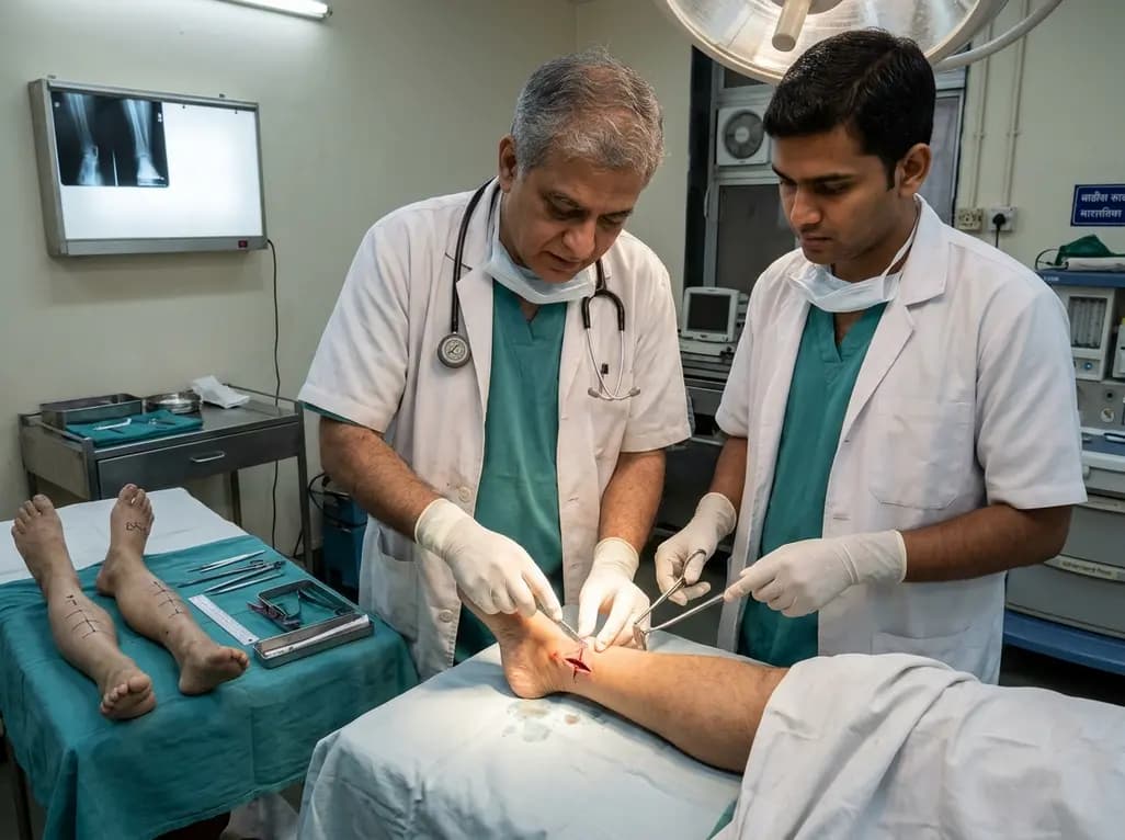
A paired cadaveric study compared triple and quintuple percutaneous Achilles tendon lengthening. While quintuple hemisection showed numerically greater elongation, differences were not statistically significant.
Today
A randomized trial in California evaluated a grocery support program for low-income, chronically ill adults. The intervention improved nutrition security by 2.8%, increased vegetable intake, boosted mental health scores, and reduced cost-related healthcare delays by 3.8%, showing the power of food-as-medicine.
Today
