.webp&w=384&q=75)

Loading, please wait...
Loading, please wait...
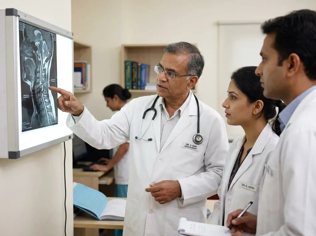
Anterior cervical meningomyelocele is an exceptionally rare congenital neural tube anomaly characterized by herniation of spinal meninges and cord elements through an anterior vertebral defect. When present alongside Klippel-Feil syndrome, a condition defined by abnormal congenital fusion of cervical vertebrae, surgical management becomes immensely challenging. Patients with this complex malformation often present in adulthood with progressive neurological deficits due to spinal cord tethering or mechanical compression. Surgical interventions must address neural decompression, dural repair, and structural spinal stabilization simultaneously. A landmark case report details the successful surgical treatment of a 71-year-old patient suffering from this rare combined lesion.
Klippel-Feil syndrome arises from failure of normal segmentation of cervical somites during early embryonic development. Consequently, affected individuals display fused vertebral segments that alter normal spinal biomechanics and place excessive mechanical strain on adjacent unfused levels. Although posterior neural tube defects like posterior spina bifida occur frequently, an anterior neural sac represents a distinct anatomical anomaly. In these cases, herniation occurs anteriorly into the vertebral bodies or retropharyngeal space.
The co-occurrence of congenital vertebral fusion and anterior neural protrusion creates complex biomechanical forces. Spinal cord tethering within the herniated sac combines with abnormal cervical motion, progressively stretching neural elements during routine neck movements. Consequently, clinical presentation may be delayed until middle or late adulthood when age-related degenerative changes worsen existing anatomical compromise. Surgeons must thoroughly understand both the osseous fusion patterns and the dural defect configuration before attempting invasive repair. Detailed multiplanar imaging helps clinicians appreciate how fusion blocks alter regional stress distribution and exacerbate cord distortion.
Accurate identification of anterior neural defects requires advanced imaging modalities. Magnetic resonance imaging remains the gold standard for evaluating spinal cord pathology, dural sac morphology, and neural tissue herniation. Additionally, high-resolution computed tomography with three-dimensional reconstruction essentializes the visualization of complex bony arches, fused vertebral segments, and defect boundaries.
In the described clinical scenario, a 71-year-old patient presented with chronic neck pain, upper limb paresthesias, and progressive distal motor weakness. Diagnostic spine imaging identified an anterior cervical meningomyelocele digging deeply into the C7 vertebral body, accompanied by adjacent congenital vertebral fusion blocks. MRI confirmed that neural tissue protruded into the bony defect, causing significant cord compression and local cord edema. Identifying these precise spatial relationships before surgery is essential to avoid intraoperative injury to vulnerable anterior vascular structures, including the vertebral arteries and carotid sheath. Clinicians must meticulously review sagittal and axial images to plan safer surgical trajectories.
Operative management of cervical anterior meningomyelocele combined with Klippel-Feil deformity demands advanced microsurgical expertise. The primary surgical objectives include releasing tethered spinal cord tissue, obliterating the dural defect, and stabilizing the altered spine segment. Because the lesion resides anteriorly, an anterior cervical approach provides direct anatomical visualization.
The surgical strategy in this index case utilized a partial cervical corpectomy at the C7 level to access the lesion directly. Using high-power microsurgical visualization, surgeons carefully dissected the herniated meninges and neural elements away from the bony edges of the C7 defect. Next, the tethered spinal cord was gently reinserted back into the primary spinal canal. Surgeons then performed micro-suturing and applied dural substitutes to achieve a watertight dural closure, preventing cerebrospinal fluid leakage. Reinserting neural elements into their proper anatomical compartment relieves mechanical tension and prevents further neurological decline. However, achieving complete neural decompression without inflicting iatrogenic cord injury requires extreme patience, precise instrument manipulation, and continuous intraoperative neurophysiological monitoring.
Once neural decompression and dural closure are complete, reconstructive surgical techniques must restore structural integrity. Removing vertebral body bone during partial corpectomy creates an osseous void that requires structural support. Furthermore, because adjacent congenital fusion blocks already restrict motion and alter biomechanics, the reconstructed segment must withstand significant biomechanical stress.
In this landmark case, anterior vertebral reconstruction was achieved through the placement of a structural interbody graft reinforced with anterior plate instrumentation. Structural bone grafting or expandable interbody cages provide immediate mechanical stability, while anterior cervical plating prevents graft displacement and promotes solid bony fusion. Fusion across the decompressed segment distributes axial loads more evenly across the cervical spine, preventing progressive post-corpectomy kyphosis. Achieving solid spinal fusion is vital because spinal instability can compromise dural repair and precipitate recurrent neural compression. Subsequent clinical follow-up confirmed that rigid stabilization combined with neural decompression yielded excellent neurological recovery and complete resolution of upper limb paresthesias.
Surgical intervention for anterior neural tube defects inherently carries substantial risks that surgeons must anticipate and mitigate. The anterior neck contains critical vital structures, such as the esophagus, trachea, recurrent laryngeal nerve, and major carotid vessels. Dissecting anteriorly near C7 requires careful soft tissue retraction to avoid pharyngeal injury or postoperative dysphagia.
Furthermore, cerebrospinal fluid leak represents one of the most serious surgical complications following dural repair in the anterior cervical spine. Inadequate dural closure can lead to persistent CSF fistulas, pseudomeningocele formation, or life-threatening meningitis. Therefore, utilizing biological glues, muscle grafts, or synthetic dural patches alongside primary microsurgical closure is strongly advocated. Additionally, surgeons must manage spinal cord reperfusion injury after decompressing chronically ischemic cord tissue. Careful blood pressure management and judicious perioperative steroid administration may mitigate neurological swelling. Preoperative counseling must thoroughly outline these potential hazards so that patients understand both the risks and the substantial potential benefits of reconstructive surgery.
A review of existing medical literature highlights how extraordinarily uncommon anterior cervical meningomyelocele remains, accounting for less than one percent of all spinal neural tube defects. Most meningomyeloceles present posteriorly during infancy, whereas anterior cervical variants often escape early detection due to their hidden location within the deep neck structures.
Historically, surgical management options for anterior meningomyeloceles varied widely, ranging from conservative observation to complex posterior-anterior staged operations. However, recent evidence confirms that single-stage anterior corpectomy, microsurgical cord reinsertion, and anterior vertebral reconstruction offer a safe and definitive solution. Combining decompression with rigid instrumentation directly resolves neural tethering while re-establishing cervical spine alignment. This effective paradigm provides valuable guidance for neurosurgeons and orthopedic spine surgeons dealing with concurrent embryological neural abnormalities and complex spinal deformities. Continued publication of detailed case reports and multicenter clinical registries will further refine surgical algorithms and optimize long-term outcomes for complex spinal malformations.
Klippel-Feil syndrome is a rare congenital skeletal disorder characterized by the abnormal fusion of two or more cervical vertebrae. This condition restricts neck mobility, shortens neck length, and alters spinal biomechanics, often causing premature degenerative changes, chronic neck pain, and increased susceptibility to spinal cord injury or neurological deficits.
Anterior cervical meningomyelocele involves neural tissue herniating anteriorly into the vertebral column or deep neck space. Operating in this anterior anatomical region requires navigating past vital structures, such as the esophagus, carotid vessels, and trachea. Reinserting neural elements safely and achieving watertight dural closure without causing CSF leaks or cord injury is technically demanding.
Partial corpectomy allows direct anterior access to release tethered neural tissue and repair the dural defect. Subsequent anterior vertebral reconstruction using interbody grafts and metal instrumentation restores structural stability, prevents progressive spinal deformity or kyphosis, and ensures optimal spinal alignment, facilitating smooth neurological recovery and pain relief.
Disclaimer: This content is for informational and educational purposes only and should not be used as a substitute for professional medical advice, diagnosis, or treatment. Refer to the latest local and national guidelines for clinical practice.
References

Read summarized clinical updates, watch expert medical content, and earn CME certifications right from your smartphone.
Open your smartphone camera to scan and install.





A 71-year-old patient with Klippel-Feil syndrome and anterior cervical meningomyelocele underwent successful partial corpectomy, microsurgical cord reinsertion, and anterior vertebral reconstruction, demonstrating excellent neurological recovery in complex spinal malformations.
last month

Researchers in Hebei, China identified two novel HIV-1 unique recombinant forms, BDNY017 and BDK025, among patients failing antiretroviral therapy. Genomic analysis revealed complex mosaic structures involving CRF07_BC, CRF08_BC, and subtype B, alongside key drug resistance mutations including M184V, A98G, and E138K.
Today

A nationwide study covering 3.7 million Swedish births reveals that prenatal exposure to TORCH pathogens significantly elevates long-term risks of intellectual disability and autism. Healthcare providers must recognize clinical indicators, promote prenatal prevention, and track early pediatric neurodevelopment.
Today
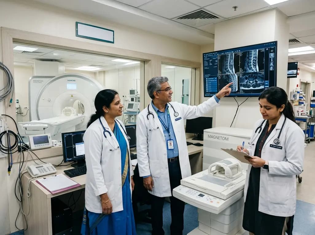
Under India's PLI initiative, domestic production of 57 critical medical devices has commenced, including MRI and CT scanners, cath labs, and heart valves. Global partnerships and local investments are expanding high-end technology, curbing import reliance, and strengthening clinical accessibility nationwide.
Today

A longitudinal study reveals that patients undergoing staged bilateral hip arthroscopy experience progressive contralateral labral deterioration during surgical intervals. However, structural degradation does not compromise 2-year postoperative clinical outcomes or patient-reported satisfaction scores.
Today

SWEDEPAD-1 trial insights show paclitaxel-coated devices do not improve long-term limb salvage in patients with chronic limb-threatening ischemia and tissue loss. While one-year reinterventions decreased, the devices were linked to a higher risk of major amputation at three months.
Today
