.webp&w=384&q=75)

Loading, please wait...
Loading, please wait...
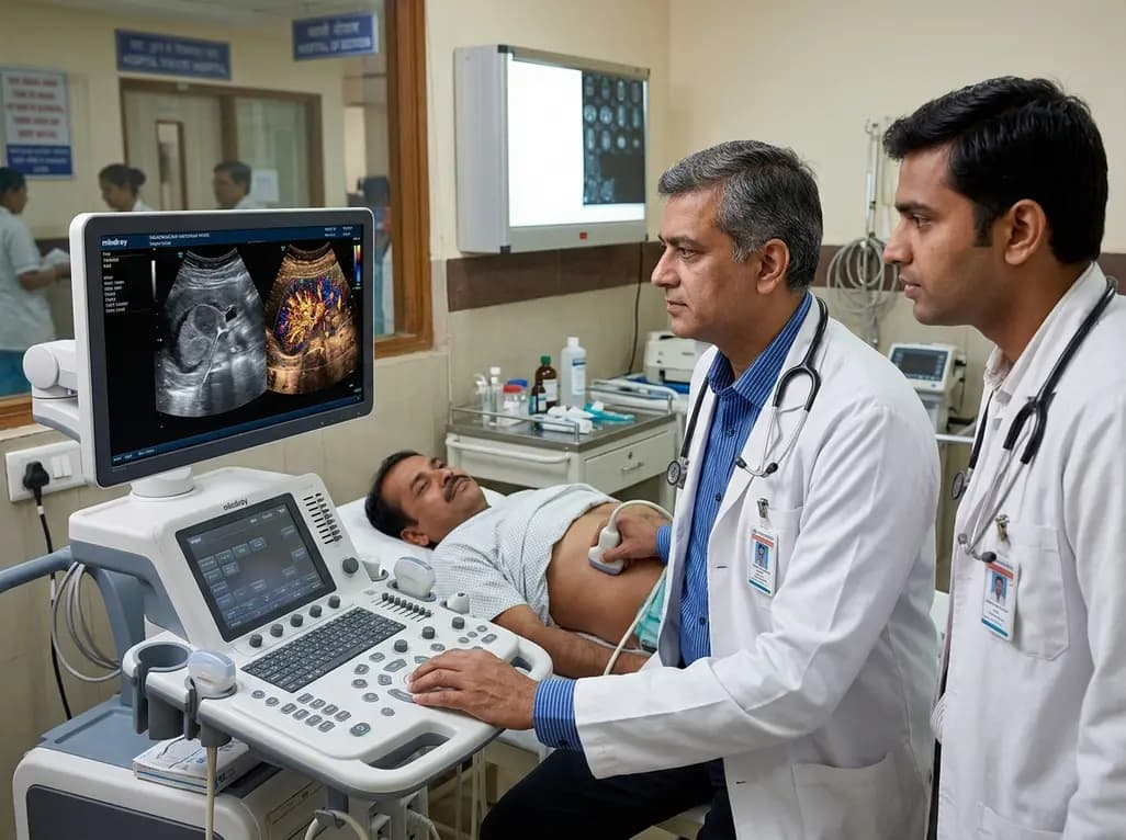
Accurate differentiation between benign and malignant renal solid lesions remains a substantial diagnostic challenge in routine urological and radiological practice. Clinicians frequently encounter incidental renal masses on routine abdominal imaging, requiring dependable non-invasive characterization to avoid unnecessary nephrectomies or invasive biopsy procedures. Consequently, modern diagnostic strategies increasingly focus on high-resolution microvascular architecture. Emerging imaging modalities, particularly super-resolution ultrasound, offer unprecedented visualization of capillary networks beyond the acoustic diffraction limit. By combining acoustic localization techniques with microbubble dynamics, clinicians can now evaluate complex vascular signatures in real time.
Incidental detection of small renal masses has expanded dramatically because of widespread cross-sectional imaging utilization. However, conventional B-mode ultrasound and Doppler techniques often struggle to distinguish benign entities, such as oncocytoma and lipid-poor angiomyolipoma, from malignant renal cell carcinoma. Gray-scale ultrasound lacks specific morphological criteria, while conventional color Doppler fails to resolve tiny, slow-flowing capillary beds. Consequently, many patients undergo surgical resection or radical nephrectomy for benign disease, which introduces unnecessary perioperative morbidity and renal function impairment. Although contrast-enhanced computed tomography and magnetic resonance imaging provide valuable anatomical and enhancement data, they carry risks related to ionizing radiation and nephrotoxic contrast agents. Therefore, nephrologists and urologists need safe, highly accurate, and non-invasive functional imaging modalities. High-resolution vascular profiling has emerged as a promising approach because tumor angiogenesis produces distinct architectural patterns. Malignant neoplasms generate chaotic, disorganized microvessel networks characterized by abnormal branching, blind ends, and arteriovenous shunts. In contrast, benign renal masses generally retain structured and orderly vascular configurations. Accurately characterizing these microvascular differences non-invasively represents a critical step forward in renal oncology.
Contrast-enhanced ultrasound utilizes gas-filled microbubble contrast agents that remain entirely within the intravascular space. These microbubbles act as ideal blood-pool tracers, enabling real-time assessment of macrovascular and macro-perfusion kinetics without renal toxicity. However, conventional contrast-enhanced imaging remains constrained by wave diffraction limits, which prevent the visualization of microvessels smaller than several hundred micrometers. To overcome this fundamental physical boundary, super-resolution ultrasound leverages ultrasound localization microscopy principles. By administering dilute microbubble concentrations, the system detects, localizes, and tracks individual microbubble centroids across thousands of ultrafast ultrasound frames. Subsequently, sophisticated motion-correction and tracking algorithms reconstruct microvascular tracks with micrometer-level spatial resolution. Furthermore, this advanced processing computes precise microvascular flow velocities and directional trajectories within deep tissue beds. When researchers combine super-resolution ultrasound with conventional contrast-enhanced ultrasound, they obtain both macroscopic perfusion dynamics and microscopic vascular morphometry simultaneously. This dual-layer hemodynamic and structural mapping provides comprehensive insights into renal tumor angiogenesis, enabling clinicians to visualize vessel tortuosity, branching density, and spatial distribution with exceptional fidelity.
A recent prospective clinical trial evaluated 114 pathologically confirmed solid renal lesions, comprising 88 malignant tumors and 26 benign masses. All participating patients underwent comprehensive multi-modal sonographic evaluations, including conventional B-mode ultrasound, contrast-enhanced ultrasound, and super-resolution ultrasound. Investigators analyzed both qualitative vascular patterns and quantitative microvascular metrics across all modalities. Notably, malignant renal lesions exhibited significantly more tortuous vessels and markedly higher microvascular image complexity compared to benign neoplasms. Because malignant angiogenesis involves rapid, disorganized endothelial proliferation, cancerous lesions demonstrated abnormal vascular caliber variations and chaotic branching networks. In contrast, benign renal masses typically displayed orderly, parallel, or peripherally organized vascular arrangements with regular vessel spacing. Univariate analysis revealed that quantitative parameters derived from microvessel tracking, such as vascular density, vessel diameter distribution, and directional entropy, differed significantly between benign and malignant cohorts. Furthermore, qualitative assessment of microvascular irregularity reinforced these objective findings. These data demonstrate that sub-diffraction microvascular imaging successfully captures distinct pathological hallmarks of malignancy in clinical renal lesions.
To maximize diagnostic accuracy, investigators developed comprehensive diagnostic scoring panels combining significant parameters from conventional ultrasound, contrast-enhanced ultrasound, and super-resolution imaging. They subsequently performed exploratory receiver operating characteristic analyses to compare single-modality evaluations against integrated multiparametric panels. While conventional ultrasound achieved modest diagnostic performance, the addition of contrast-enhanced parameters significantly improved sensitivity and specificity. However, the multiparametric panel incorporating super-resolution ultrasound parameters achieved the highest overall area under the curve value. Specifically, the integration of quantitative microvascular complexity metrics with contrast enhancement kinetics drastically reduced false-positive classifications among benign lesions. In addition, the combined panel demonstrated superior diagnostic accuracy for small renal masses under four centimeters, which typically present the greatest diagnostic ambiguity. By synthesizing morphological, macro-perfusion, and microvascular characteristics into a unified diagnostic score, clinicians achieved robust discrimination of renal cell carcinomas from benign mimics. Therefore, multiparametric sonographic evaluation establishes a new benchmark for non-invasive renal tumor characterization.
Integrating super-resolution microvascular assessment into urological pathways holds transformative potential for patient management and surgical decision-making. In clinical practice, distinguishing indolent or benign lesions from aggressive renal cell carcinoma dictates whether a patient requires active surveillance, thermal ablation, partial nephrectomy, or radical surgery. Because super-resolution ultrasound provides microscopic resolution without ionizing radiation or nephrotoxicity, it offers an ideal surveillance tool for elderly patients or individuals with pre-existing chronic kidney disease. Furthermore, accurate pre-operative microvascular characterization helps surgeons evaluate tumor margins and vascular pedicle architecture before executing complex nephron-sparing surgeries. In addition, this advanced modality could assist oncologists in monitoring early vascular responses to anti-angiogenic targeted therapies or immune checkpoint inhibitors. Reductions in microvascular complexity and vessel tortuosity may indicate therapeutic response well before gross dimensional shrinkage occurs on standard cross-sectional scans. Consequently, super-resolution ultrasound bridges the gap between macroscopic radiology and histopathological microvascular analysis.
Despite its remarkable diagnostic potential, super-resolution ultrasound faces several practical limitations that require refinement before widespread clinical adoption. First, microbubble localization microscopy algorithms demand substantial computational processing power, which often prevents immediate real-time bedside display. Second, physiological motion from patient respiration, cardiac pulsation, and probe instability can introduce tracking artifacts, necessitating robust motion-compensation algorithms. Furthermore, acoustic attenuation in deep tissues or high body mass index patients can degrade signal-to-noise ratios, limiting microbubble detection in deeply seated renal lesions. Standardizing acquisition protocols, microbubble dosing, and quantitative analysis software will be essential for reproducible multicenter application. In the future, integrating artificial intelligence and deep learning architectures into ultrasound reconstruction pipelines will accelerate processing speeds and automate microvascular feature extraction. Moreover, volumetric three-dimensional super-resolution ultrasound promises complete volumetric characterization of renal vascular networks. As technology matures, super-resolution imaging will become a vital pillar of personalized urological oncology.
Conventional Doppler ultrasound relies on phase shifts to detect blood flow but remains strictly limited by acoustic diffraction, visualizing only macrovessels larger than several hundred micrometers. In contrast, super-resolution ultrasound tracks individual microbubble contrast agents over consecutive ultrafast frames. This innovative method surpasses diffraction boundaries, accurately reconstructing microvessels under one hundred micrometers. Consequently, clinicians can evaluate fine capillary networks and vessel tortuosity that Doppler cannot resolve.
Malignant renal tumors undergo rapid, dysregulated angiogenesis driven by pro-angiogenic factors such as vascular endothelial growth factor. This uncoordinated vascular proliferation produces disorganized, fragile, and tortuous capillary networks with abnormal branching and irregular calibers. Conversely, benign tumors maintain structured, organized vascular architecture. Detecting elevated microvascular tortuosity and geometric complexity therefore provides a powerful diagnostic biomarker to distinguish malignant lesions from benign renal neoplasms non-invasively.
Super-resolution ultrasound provides unprecedented non-invasive microvascular details, significantly improving diagnostic confidence and reducing unnecessary biopsies or surgical interventions. However, it does not completely replace histopathological or immunohistochemical analysis for definitive staging, grading, and genetic profiling. Instead, clinicians utilize this advanced imaging modality as a complementary, risk-free diagnostic tool that enhances active surveillance decisions, guides targeted biopsy needles, and informs personalized surgical planning.
Disclaimer: This content is for informational and educational purposes only and does not substitute professional medical judgment. Refer to the latest local and national guidelines for clinical practice.
References

Read summarized clinical updates, watch expert medical content, and earn CME certifications right from your smartphone.
Open your smartphone camera to scan and install.





A prospective clinical study evaluates the diagnostic utility of super-resolution ultrasound (SRUS) combined with contrast-enhanced ultrasound (CEUS) to differentiate benign from malignant renal tumors based on quantitative and qualitative microvascular characteristics.
last month

Modern renal nutrition is shifting toward additive-aware dietary guidance. This paradigm differentiates between intrinsic nutrients in whole foods and hyper-bioavailable inorganic additives, offering practical strategies to mitigate additive stacking and toxicity without restricting nutritious whole foods.
Today

A multiparametric MRI approach combining quantitative T2 shading, apparent diffusion coefficient (ADC) measurements, and morphological signs provides superior diagnostic accuracy over subjective evaluation to differentiate ovarian endometriomas from benign hemorrhagic cysts.
Today

A landmark case report reveals a donor heart beating for a century in a 63-year-old recipient, challenging conventional age limits in cardiac transplantation and offering critical lessons on organ longevity, donor selection, and long-term graft survival.
Today

A new randomized crossover trial reveals that a hinged ulnar gutter orthosis significantly improves hand function and weight-bearing tolerance compared to standard volar splints in patients with degenerative triangular fibrocartilage complex lesions, supporting functionally permissive orthotic designs.
Today

New research reveals that M1 macrophage-derived exosomes aggravate diabetic nephropathy by transferring WTAP to stabilize S1PR2 mRNA. Silencing WTAP in these vesicles attenuates endothelial injury and renal fibrosis, pointing toward innovative nanomedicine therapies.
Yesterday
