.webp&w=384&q=75)

Loading, please wait...
Loading, please wait...
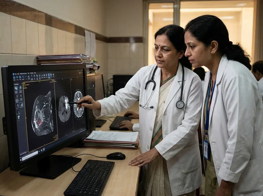
Neoadjuvant therapy (NAT) is now a central pillar in multidisciplinary breast cancer management. Clinicians routinely strive to achieve pathological complete response (pCR), because it heralds substantially improved long-term survival. However, accurate noninvasive response monitoring remains challenging. Standardized breast cancer MRI evaluation plays an indispensable role in guiding surgical decisions and tracking tumor regression. Radiologists frequently debate the respective clinical utility of dynamic contrast-enhanced magnetic resonance imaging (DCE-MRI) versus diffusion-weighted imaging (DWI). Recent clinical evidence introduces an asymmetric complementarity framework. This strategy establishes subtype-specific interpretations that significantly enhance visual assessment precision across heterogeneous breast tumors.
Neoadjuvant therapy produces diverse biological changes depending on receptor status. Consequently, clinicians cannot rely on a generic imaging protocol for every patient. In a recent retrospective cohort of 248 patients, investigators evaluated visual interpretations of DCE-MRI and DWI across three core biological subtypes. These groups included hormone receptor-positive/HER2-negative (HR+/HER2-), HER2-positive (HER2+), and triple-negative breast cancer (TNBC). Two independent radiologists visually assessed radiological complete response (rCR) and measured residual tumor size while blinded to pathological results.
Furthermore, interobserver agreement proved remarkably robust across both imaging modalities. The radiologists achieved a kappa value of 0.77 for DCE-MRI and 0.85 for DWI. In addition, the intraclass correlation coefficient for interobserver tumor size measurement reached 0.96 for both sequences. Therefore, visual assessment is highly reproducible in clinical practice. Nevertheless, the study highlighted that biological heterogeneity dictates sequence performance. Radiologists must interpret functional diffusion changes and vascular perfusion through specific molecular subtypes.
When evaluating therapy response, DCE-MRI and DWI exhibit distinctly divergent diagnostic strengths. Specifically, DCE-MRI visualizes tumor microvasculature through contrast kinetics. In contrast, DWI captures cellular density and membrane integrity on apparent diffusion coefficient (ADC) maps. In this cohort, DCE-MRI demonstrated significantly higher overall specificity, positive predictive value (PPV), and diagnostic accuracy. Therefore, when DCE-MRI indicates complete vascular absence, clinicians can reliably anticipate true histological clearance.
Conversely, DWI exhibited markedly higher sensitivity across the overall patient cohort. Because diffusion restriction resolves rapidly as cytotoxic agents lyse neoplastic cells, DWI detects microscopic changes before macrovascular shutdown occurs. However, persistent post-treatment inflammation and necrosis can cause residual diffusion restriction. As a result, DWI in isolation can produce false-positive readings. Meanwhile, cytotoxic regimens may normalize vascular permeability without eliminating all tumor cells. Consequently, combining functional diffusion data with vascular enhancement establishes a balanced diagnostic perspective. This paired approach significantly reduces interpretive errors during clinical evaluations.
The true clinical value of this framework emerges from its subtype-specific strategy. In HER2-positive breast cancer, DCE-MRI and DWI demonstrate true mutual complementarity. Specifically, DWI delivers an exceptional negative predictive value (NPV), reliably excluding residual malignancy when diffusion normalizes. Concurrently, DCE-MRI provides high positive predictive value, accurately confirming persistent tumor when focal enhancement persists. Therefore, clinicians should combine both sequences to guide surgical de-escalation in HER2-positive disease.
However, a distinctly different pattern appears in HR-positive/HER2-negative malignancies. In this subtype, DCE-MRI serves as the primary anchor, whereas DWI offers limited additive value. Hormone receptor-positive tumors frequently exhibit scattered regression patterns and dense fibrous stroma. Consequently, baseline cellular density remains relatively low, which restricts the diagnostic discriminatory capability of ADC maps. Moreover, triple-negative breast cancer findings showed promising trends requiring larger validation cohorts. Thus, tailoring sequence weighting directly to receptor profiles maximizes diagnostic accuracy.
Accurate residual tumor sizing directly determines whether a patient qualifies for breast-conserving surgery or requires mastectomy. In this critical domain, DCE-MRI demonstrated clear superiority over DWI. Specifically, DCE-MRI achieved high concordance with pathological measurement, recording an intraclass correlation coefficient (ICC) of 0.91. In sharp contrast, DWI demonstrated poor concordance, with an ICC of only 0.51. This statistically significant difference underscores a vital limitation of diffusion imaging for surgical mapping.
Furthermore, post-therapeutic biological alterations explain why diffusion-based sizing falters. Cytotoxic chemotherapy causes macrophage infiltration, peritumoral edema, and extensive granulation tissue. These reactive processes restrict water diffusion, falsely exaggerating tumor dimensions on high b-value images. Consequently, surgeons relying solely on DWI measurements risk performing unnecessarily extensive resections. In contrast, DCE-MRI relies on active angiogenesis to outline residual malignant foci. Therefore, breast surgeons must use contrast-enhanced sequences as the definitive blueprint for determining resection margins.
In Indian oncology practice, optimizing imaging strategies carries profound clinical and socioeconomic importance. Many patients present with locally advanced breast cancer at baseline, necessitating neoadjuvant chemotherapy. Furthermore, specialized breast imaging resources vary across public and private cancer centers. Implementing standardized multiparametric protocols that unite DCE-MRI and DWI ensures equitable, high-precision care. Radiologists should incorporate subtype-informed reporting templates that explicitly align imaging findings with initial immunohistochemistry status.
Moreover, multidisciplinary tumor boards in Indian institutions should actively utilize this asymmetric framework. When evaluating a HER2-positive patient undergoing targeted blockade, clinicians can leverage DWI's high NPV to confirm complete response. If breast conservation is planned, surgical oncologists should reference post-treatment DCE-MRI for definitive residual tumor margins. Additionally, avoiding over-reliance on standalone DWI prevents inappropriate conversions from lumpectomy to mastectomy. Integrating molecular subtype data with multiparametric MRI optimizes personalized oncological care across tertiary centers.
In HER2-positive breast cancer, diffusion-weighted imaging exhibits a remarkably high negative predictive value, reliably confirming complete biological response when restriction resolves. Meanwhile, dynamic contrast-enhanced imaging delivers superior positive predictive value by verifying true residual tumor vascularization. Consequently, pairing both modalities creates an asymmetric complementarity where DWI reliably excludes remaining cancer, while DCE-MRI confirms true residual disease. This dual assessment provides surgeons with maximum diagnostic certainty prior to breast conservation.
DCE-MRI demonstrates superior concordance with surgical pathology because it highlights active tumor angiogenesis. In contrast, diffusion-weighted imaging often miscalculates lesion boundaries. Post-chemotherapy tissue changes, such as treatment-related macrophage infiltration, peritumoral edema, and wound granulation, restrict water molecule diffusion similarly to viable tumor cells. Therefore, DWI frequently overestimates residual lesion size. Consequently, breast surgeons should exclusively rely on contrast-enhanced sequences to define resection margins and avoid unnecessarily extensive surgeries.
Hormone receptor-positive and HER2-negative tumors frequently regress in a scattered, concentric, or non-mass fashion rather than shrinking as a solid sphere. Furthermore, these lesions typically feature prominent fibrous stroma and lower baseline cellularity compared to HER2-positive or triple-negative tumors. These unique histological characteristics limit the discriminatory accuracy of apparent diffusion coefficient maps on DWI. Therefore, clinicians must utilize DCE-MRI as the primary anchor modality to evaluate post-treatment vascular regression in hormone receptor-positive disease.
Disclaimer: This content is for informational and educational purposes only and does not constitute medical advice. Consult qualified healthcare professionals for diagnosis and treatment decisions. Refer to the latest local and national guidelines for clinical practice.
References

Read summarized clinical updates, watch expert medical content, and earn CME certifications right from your smartphone.
Open your smartphone camera to scan and install.





A recent study reveals asymmetric complementarity between DCE-MRI and DWI visual assessment for evaluating neoadjuvant therapy in breast cancer. DCE-MRI offers superior specificity and residual tumor sizing, while DWI provides high sensitivity in HER2+ disease, enabling subtype-tailored surgical planning.
Today

A randomized trial demonstrates that breaking up sedentary behavior with frequent 5-minute brisk walks matches a 45-minute continuous bout for daily energy expenditure while uniquely reducing prolonged sitting, offering a flexible lifestyle prescription for adults with overweight or obesity.
Today

A landmark single-cell study reveals that MEHP, a major phthalate metabolite, damages human ovarian tissue beyond follicles. By disrupting glial-stromal communication, cellular adhesion, and mitochondrial respiration, environmental plasticizers impair ovarian integrity at realistic exposure levels.
Today

The Delhi High Court granted interim relief to beverage manufacturers against the FSSAI ban on the energy drink descriptor. Discover the regulatory dispute, caffeine safety thresholds, metabolic concerns, and the vital clinical recommendations for healthcare practitioners advising young consumers in India.
Today

A scoping review reveals severe gaps in long-term assistive technology provision following humanitarian emergencies. Addressing post-disaster rehabilitation requires systematic follow-up, workforce training, and integration into national health systems to sustain mobility and recovery.
Today
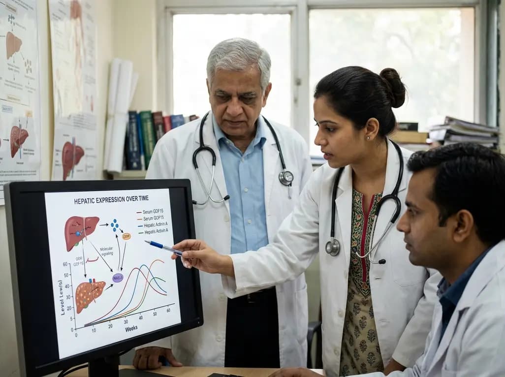
A landmark preclinical evaluation highlights temporal discordance across circulating and hepatic activin A, follistatin, and GDF15 during MASLD development. The findings offer clinicians crucial insights into hepatokine kinetics, SMAD2/3 signaling shifts, and stage-specific biomarker patterns.
Today
