.webp&w=384&q=75)

Loading, please wait...
Loading, please wait...
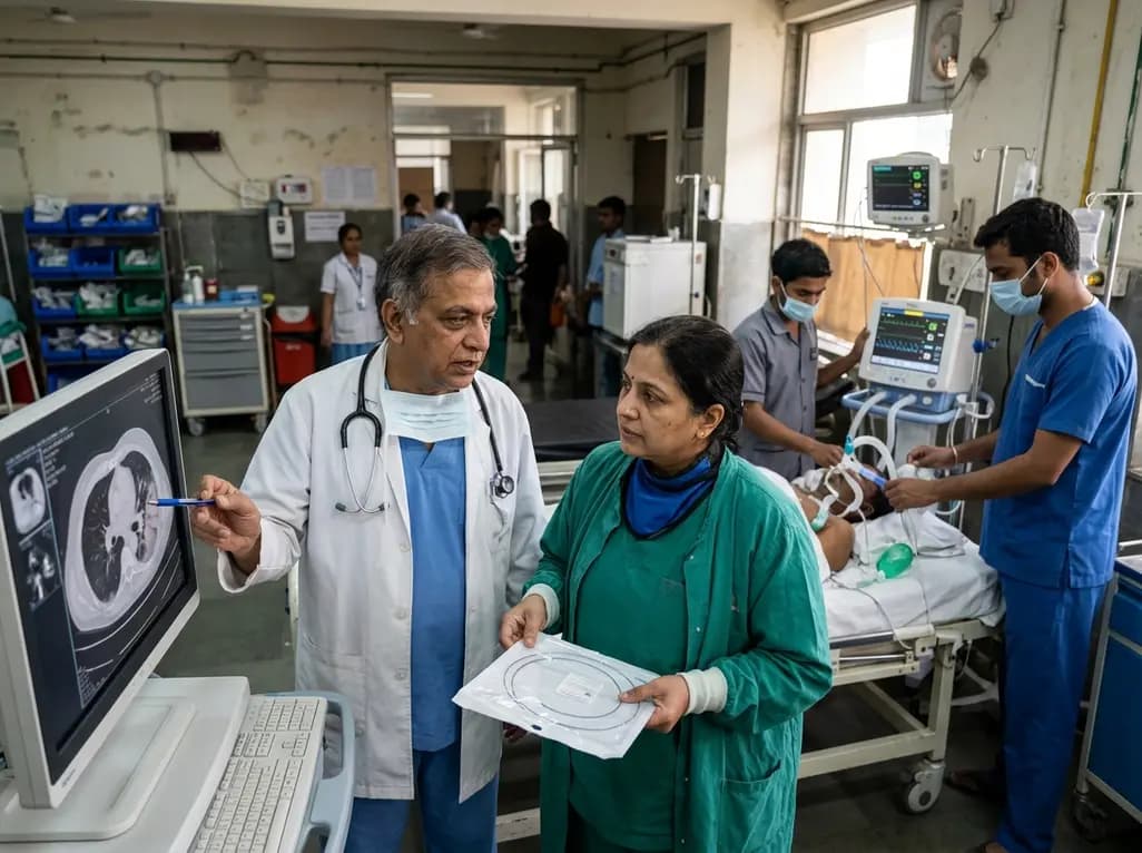
Acute hemoptysis represents one of the most frightening and perilous emergencies in respiratory medicine. When catastrophic bleeding occurs, asphyxiation or severe hemorrhagic shock can cause death within minutes. Historically, fragmented care pathways, delayed specialist consultations, and substantial regional disparities have compromised survival rates during acute episodes. Consequently, respiratory and thoracic societies have established expert recommendations for standardized hemoptysis centers. Modeled after successful stroke and acute chest pain networks, these dedicated units integrate emergency resuscitation, advanced vascular imaging, and interventional pulmonology. This structured framework elevates institutional preparedness, standardizes clinical workflows, and accelerates lifesaving therapeutic decisions across hospital networks.
Pulmonary hemorrhage frequently arises secondary to endemic or chronic parenchymal lung diseases. Common underlying etiologies include active or prior pulmonary tuberculosis, extensive bronchiectasis, invasive fungal infections, and cavitary bronchogenic malignancies. Furthermore, the rapid expansion of invasive diagnostic and therapeutic bronchoscopy has created an additional risk of procedural bleeding. In life-threatening hemoptysis, asphyxiation secondary to blood flooding the tracheobronchial tree represents a much more immediate danger than hemorrhagic exsanguination. Therefore, medical teams must intervene within an exceptionally narrow therapeutic window.
Unfortunately, conventional hospital pathways often evaluate severe hemoptysis through fragmented, sequential consultations. Emergency physicians typically evaluate the patient first, followed by delayed referrals to pulmonology, interventional radiology, or cardiothoracic surgery. Consequently, institutional latency and diagnostic bottlenecks increase patient morbidity and mortality significantly. To address this widespread clinical vulnerability, dedicated hemoptysis centers consolidate clinical expertise and diagnostic resources into a cohesive emergency infrastructure. Furthermore, this standardized framework ensures uninterrupted hemodynamic monitoring, rapid bedside triage, and prompt access to specialized hemostatic interventions across diverse hospital systems.
The clinical foundation of a specialized center relies on a highly coordinated multidisciplinary team structure. Specifically, core institutional teams must unite pulmonologists, intensivists, interventional radiologists, thoracic surgeons, and emergency physicians into a unified response system. In addition, experienced critical care nurses and trained respiratory therapists constitute an indispensable component of this rapid response architecture. Accredited institutions must designate a centralized clinical coordinator who oversees emergency code activation and directs clinical workflow across departments.
Beyond essential personnel, establishing an effective center demands dedicated physical infrastructure and advanced therapeutic technologies. For instance, the primary resuscitation bay requires immediate, bedside availability of large-bore endotracheal tubes, flexible video bronchoscopes, and rigid bronchoscopy equipment. Moreover, round-the-clock access to digital subtraction angiography suites and hybrid operating rooms remains vital for prompt vascular embolization. Institutional leadership must also guarantee priority blood bank allocation, standardized massive transfusion protocols, and bedside point-of-care coagulation testing. By establishing these rigorous operational benchmarks, centers eliminate bureaucratic delays and ensure seamless emergency delivery.
During acute hemorrhagic episodes, medical teams must prioritize definitive airway management above exhaustive diagnostic testing. Asphyxia represents the primary cause of immediate mortality in life-threatening pulmonary bleeding. Therefore, attending clinicians must immediately place the patient in the lateral decubitus position, keeping the suspected bleeding lung in the dependent orientation. This simple positioning maneuver protects the contralateral lung from blood aspiration and preserves vital gas exchange.
Simultaneously, resuscitation teams must establish dependable vascular access using multiple large-bore peripheral cannulae or central venous lines. Fluid resuscitation, judicious administration of systemic antifibrinolytic agents, and aggressive correction of underlying coagulopathies proceed concurrently. If respiratory failure ensues, emergency physicians perform endotracheal intubation using a large endotracheal tube measuring at least eight millimeters in internal diameter. This generous lumen accommodates therapeutic bronchoscopes and facilitates efficient clot evacuation. Furthermore, clinicians can deploy bronchial blockers to isolate the hemorrhagic lung segment when unilateral bleeding occurs. Accordingly, standardized emergency resuscitation algorithms prevent catastrophic airway obstruction while preparing patients for definitive endovascular intervention.
Once resuscitation teams stabilize respiratory function and vital signs, accurate localization of the bleeding vessel becomes paramount. Multidetector computed tomography angiography represents the initial diagnostic investigation of choice in stable patients. Specifically, bronchial and systemic artery computed tomography angiography rapidly identifies hypertrophied bronchial vessels, aberrant systemic collaterals, and underlying parenchymal abnormalities. Furthermore, cross-sectional imaging reveals subtle vascular lesions, such as Rasmussen aneurysms or pseudoaneurysms, which guide subsequent catheterization.
Concurrently, flexible or rigid bronchoscopy provides indispensable real-time visual assessment of the tracheobronchial tree. Interventional pulmonologists utilize endoscopic tools to clear obstructive blood clots, identify the involved lobar segment, and administer topical hemostatic measures. For example, direct endobronchial cold saline lavages, topical vasoconstrictor instillation, and balloon catheter tamponade can achieve temporary hemostasis. However, clinicians must never allow prolonged endoscopic procedures to delay definitive vascular embolization when imaging identifies the culprit vessel. Therefore, standardized center algorithms integrate computed tomography angiography and targeted bronchoscopy into an efficient, complementary diagnostic workflow.
Transcatheter bronchial artery embolization serves as the premier definitive therapy for moderate to severe pulmonary hemorrhage. Because more than ninety percent of life-threatening bleeding originates from the high-pressure bronchial circulation, transcatheter vascular occlusion provides immediate hemostasis. Interventional radiologists systematically catheterize culprit bronchial arteries, anomalous branches, and non-bronchial systemic collaterals. Consequently, the selective delivery of microparticles, gelatin sponge slurries, or metallic coils successfully arrests active hemorrhage while preserving viable pulmonary parenchyma.
Nevertheless, endovascular embolization may occasionally prove unsuccessful, or bleeding may arise from high-pressure pulmonary arterial lesions. Under these critical circumstances, emergent cardiothoracic surgery remains the ultimate rescue modality. Experienced thoracic surgeons perform targeted anatomical wedge resections, segmentectomies, or formal lobectomies when baseline cardiopulmonary reserve permits. Moreover, surgical resection offers definitive eradication for localized structural pathologies, including complex mycetomas and chronic cavitary abscesses. Thus, the integrated network of a hemoptysis center guarantees rapid, friction-free transfer from interventional angiography suites directly to the operating room without dangerous delays.
Clinicians activate a hemoptysis emergency response whenever patients exhibit life-threatening bleeding. Specific criteria include expectoration exceeding one hundred milliliters of blood over a brief duration, visible hemodynamic instability, or impending airway compromise. Furthermore, acute oxygen desaturation or persistent coughing that impairs ventilation warrants immediate multidisciplinary mobilization. Standardized protocols bypass routine consultation delays, ensuring rapid bedside arrival of airway specialists, intensivists, and interventional teams.
Hemodynamic and respiratory stability dictates the diagnostic sequence. In stable or stabilized patients, multidetector computed tomography angiography remains the primary investigation. It maps culprit bronchial arteries and underlying parenchymal pathology before intervention. Conversely, in patients with persistent severe bleeding or acute asphyxiation risk, teams prioritize immediate bronchoscopy. Endoscopic evaluation clears blood clots, stabilizes the airway, and localizes the bleeding source prior to definitive embolization.
Transcatheter bronchial artery embolization serves as the premier minimally invasive intervention for controlling massive hemorrhage. Because the majority of pulmonary bleeding originates from high-pressure systemic bronchial arteries, selective catheterization delivers immediate hemostatic control. Interventional radiologists deploy calibrated particles or microcoils into culprit vessels, successfully stopping active bleeding in over eighty percent of cases. Consequently, this procedure stabilizes high-risk patients and minimizes the necessity for emergency open surgery.
Disclaimer: This content is for informational and educational purposes only, and does not substitute professional medical advice, diagnosis, or treatment. Healthcare professionals should utilize individual clinical judgment and correlate recommendations with patient-specific factors. Refer to the latest local and national guidelines for clinical practice.
References

Read summarized clinical updates, watch expert medical content, and earn CME certifications right from your smartphone.
Open your smartphone camera to scan and install.





Expert recommendations outline the establishment of standardized hemoptysis centers to streamline emergency triage, rapid airway protection, interventional radiology, and multidisciplinary respiratory care, significantly reducing mortality from life-threatening pulmonary hemorrhage.
Today
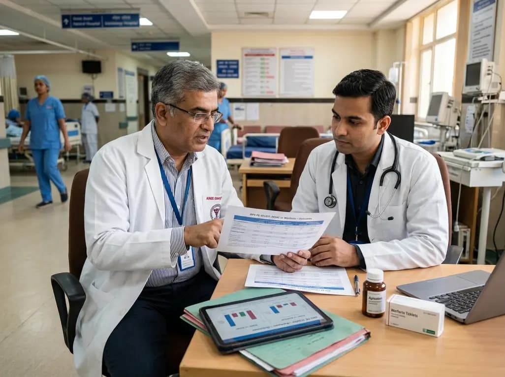
A retrospective study compares DOACs and warfarin for pulmonary thromboembolism in non-triple-positive antiphospholipid syndrome, demonstrating comparable recurrence rates and bleeding risks across both therapeutic cohorts.
Today
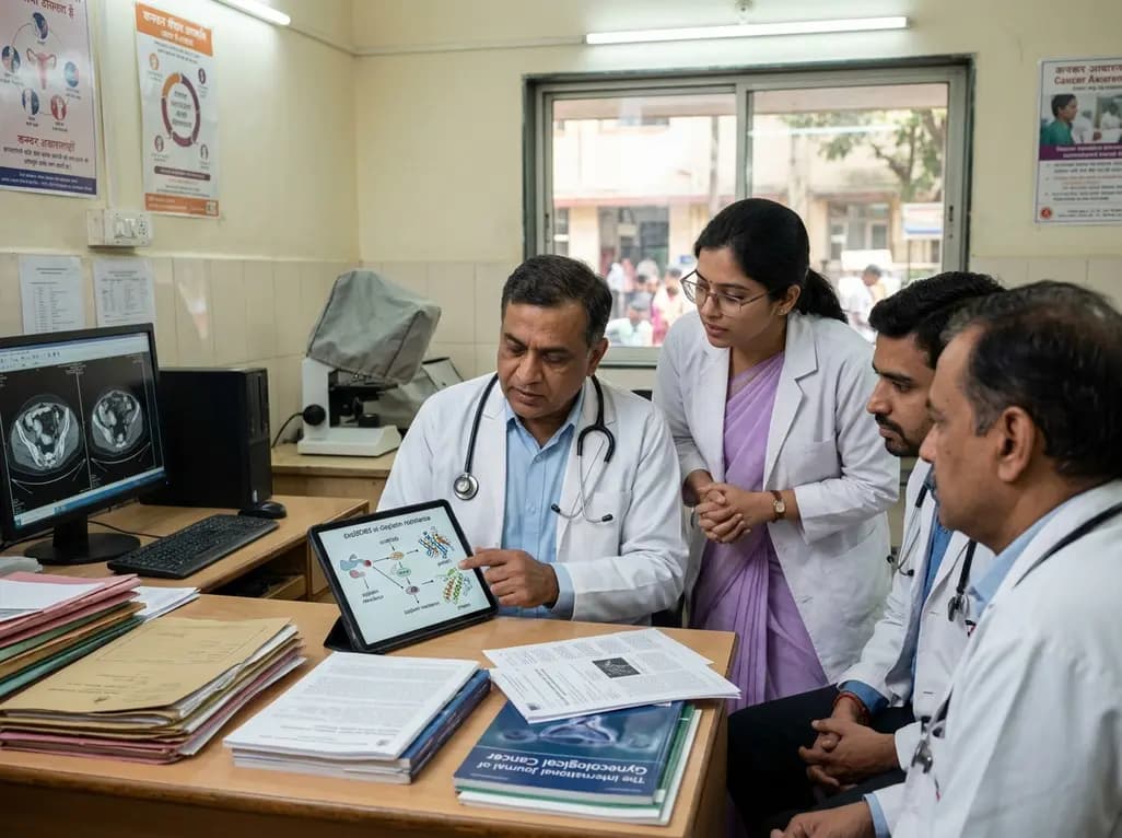
A landmark study reveals that circMCM3 promotes cisplatin resistance in cervical squamous cell carcinoma by recruiting USP49 to stabilize PTBP1 and CCND1. Targeting this newly identified molecular axis presents a promising therapeutic avenue to restore chemotherapy sensitivity and improve gynaecological outcomes.
Today

A 24-week trial shows that patients starting amlodipine/benazepril maintain stable blood pressure across all seasons, whereas those on benazepril/hydrochlorothiazide require significantly more up-titration when initiated in winter (18.9% vs. 5.0%), highlighting amlodipine's superior seasonal hemodynamic stability.
Today

A rare case of a pediatric pathological femoral fracture caused by occult telangiectatic osteosarcoma demonstrates the risks of misdiagnosis after trauma, where intramedullary nailing prompted iatrogenic tumor dissemination and required modular total endoprosthetic reconstruction.
Today
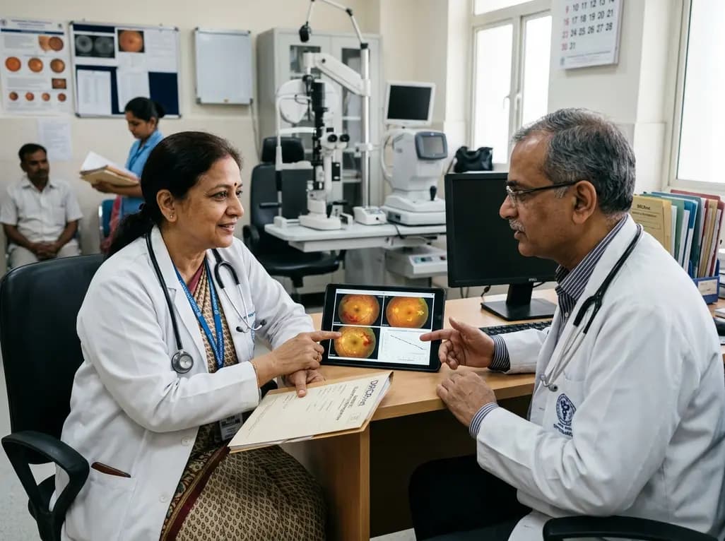
A comprehensive analysis of DRCRnet studies reveals that 15.1% of participants discontinue diabetic retinopathy clinical trials. Identifying baseline demographic and clinical predictors helps ophthalmologists and endocrinologists mitigate real-world treatment dropouts and protect long-term visual outcomes.
Today
