.webp&w=384&q=75)

Loading, please wait...
Loading, please wait...
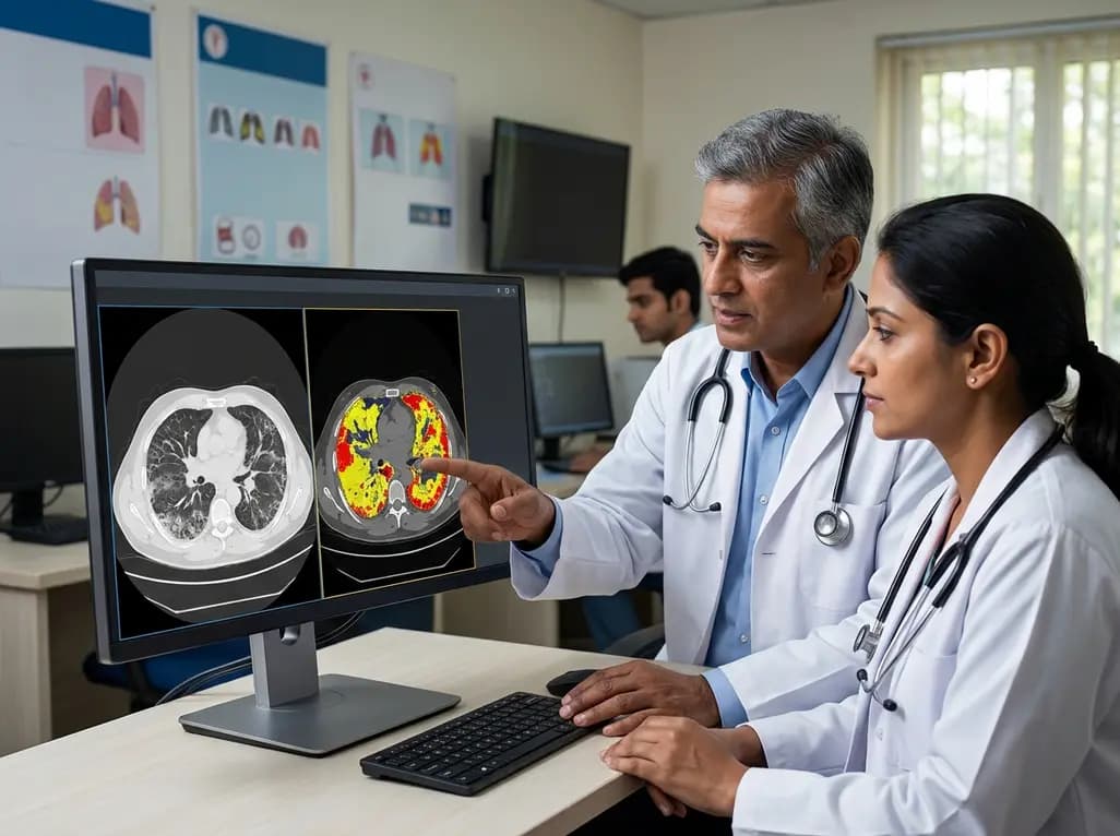
Systemic sclerosis (SSc) is a complex multi-system autoimmune disorder characterized by vasculopathy and extensive tissue fibrosis. Among its many complications, interstitial lung disease (ILD) has emerged as the primary cause of morbidity and mortality for patients worldwide, including within the growing clinical cohorts in India. Identifying **SSc-ILD progression HRCT** markers at an early stage is a critical clinical objective, as early intervention can significantly alter the patient's long-term trajectory. However, the traditional reliance on human visual assessment often falls short due to the subtle nature of early parenchymal changes. Consequently, clinicians are increasingly looking toward technological advancements to improve diagnostic precision and prognostic accuracy.
Furthermore, the current standard of care involves frequent monitoring through pulmonary function tests (PFTs) and periodic high-resolution computed tomography (HRCT). While PFTs provide a functional snapshot, they often reflect damage that has already occurred rather than predicting imminent decline. The integration of automated software into the diagnostic workflow represents a major shift in how we approach this challenge. By providing objective, reproducible data, automated systems aim to remove the subjective variability inherent in human interpretation. This transition is especially relevant for pulmonologists and rheumatologists who require granular data to justify the escalation of immunosuppressive or antifibrotic therapies in a timely manner.
For decades, the visual interpretation of chest imaging by experienced radiologists has been the gold standard for diagnosing lung involvement in systemic sclerosis. However, this method is inherently semi-quantitative and suffers from significant inter-observer variability. Even highly trained radiologists may struggle to quantify minute changes in ground-glass opacities (GGO) or early reticulation across multiple scans. In contrast, automated quantitative analysis utilizes sophisticated algorithms to evaluate every pixel of the HRCT scan. This study highlights how software like Thoracic VCAR can provide a more nuanced view of the lung parenchyma, identifying shifts in tissue density that are invisible to the naked eye.
Moreover, automated systems can segment the lung into specific zones and calculate the exact percentage of various patterns, such as fibrosis, honeycombing, and ground-glass opacities. While a radiologist might score an increase in fibrosis as a slight categorical jump, automated software can detect a fractional percentage increase that correlates more closely with physiological changes. In the context of **SSc-ILD progression HRCT** monitoring, this precision is vital. The ability to distinguish between stable disease and slow progression allows for a more personalized approach to patient management. As we move toward precision medicine, these objective tools are becoming indispensable in the high-stakes environment of rheumatological care.
To evaluate the efficacy of these different assessment methods, researchers conducted a retrospective longitudinal study involving patients diagnosed with SSc-ILD. These patients underwent baseline HRCT scans and pulmonary function tests, followed by subsequent evaluations at one year and two years. This longitudinal design was essential for capturing the dynamic nature of the disease. Two expert radiologists performed blinded visual scoring, while the automated analysis was conducted independently using standardized software. By comparing these results against functional outcomes, specifically the decline in Forced Vital Capacity (FVC), the study sought to determine which imaging modality served as a better predictor of clinical worsening.
In addition to standard imaging metrics, the study utilized the Erice and INBUILD criteria to define functional progression. These criteria are widely recognized in the global medical community for identifying clinically significant lung function decline. By mapping the imaging changes between the first two time points to the functional outcomes at the third time point, the researchers could assess the predictive value of early imaging markers. This methodology provides a robust framework for understanding how early morphological changes in the lung contribute to long-term disability. For Indian practitioners, understanding these correlations is essential for adopting evidence-based protocols in busy clinical settings where early detection saves lives.
The results of the study provide compelling evidence for the superiority of automated analysis. While both visual and automated assessments were capable of detecting the progression of established fibrosis, only the automated software could identify early increases in ground-glass opacities and the corresponding reduction in normal lung tissue. This is a crucial distinction because ground-glass opacities often represent the active, potentially reversible phase of the disease. If a clinician relies solely on visual assessment, they might miss these subtle warning signs until permanent fibrotic damage has already set in, leading to poorer patient outcomes.
Furthermore, the software-derived parameters showed a significantly stronger correlation with changes in PFTs compared to visual scores. Specifically, the automated detection of reduced normal lung volume was a highly sensitive indicator of future functional decline. This suggests that the loss of healthy lung units is a more reliable prognostic marker than the mere presence of fibrotic tissue. In the realm of **SSc-ILD progression HRCT**, these findings underscore the limitations of human perception. By quantifying what the eye cannot see, automated analysis provides a more comprehensive picture of the disease burden. This allows for a proactive rather than reactive treatment strategy, which is the cornerstone of modern rheumatology and pulmonology.
One of the most significant takeaways from this research is the predictive power of early ground-glass opacity changes. The study found that an automatically detected increase in GGO during the first year of monitoring was a strong predictor of subsequent functional decline in the second year. Specifically, a software-detected change yielded a sensitivity of 81% and a specificity of 77% for identifying patients who would go on to suffer significant FVC loss. This level of predictive accuracy is rarely achieved through visual assessment alone, making it a game-changer for risk stratification in systemic sclerosis patients.
Consequently, integrating automated HRCT analysis into routine clinical practice could revolutionize how we monitor SSc-ILD. For Indian doctors, who often manage high volumes of patients, having an objective score can streamline decision-making. If the software indicates an early increase in GGO, the clinician can consider escalating therapy or increasing the frequency of monitoring before the patient becomes symptomatic. This approach minimizes the risk of irreversible lung damage and improves the overall quality of life. As technology becomes more accessible, the adoption of these automated tools should be prioritized in tertiary care centers to ensure that SSc-ILD patients receive the most accurate and timely care possible.
The management of systemic sclerosis-associated interstitial lung disease is entering a new era. The evidence clearly indicates that automated quantitative HRCT analysis offers a level of precision and predictive power that visual assessment simply cannot match. By focusing on early markers like GGO changes and the loss of normal lung tissue, clinicians can identify at-risk patients much earlier in the disease course. This allows for the timely initiation of therapies that can preserve lung function and reduce mortality. The transition from subjective to objective monitoring is not just a technological upgrade; it is a necessary evolution in patient care.
Ultimately, the goal of monitoring **SSc-ILD progression HRCT** is to improve long-term functional outcomes. While visual assessment will always have a place in radiology, its limitations in the context of progressive ILD must be acknowledged. Software-based analysis provides the objective data required to navigate the complexities of SSc management. As we look to the future, the routine use of such tools will likely become the standard of care, ensuring that every patient with systemic sclerosis has the best possible chance of maintaining their respiratory health. Practitioners are encouraged to stay updated on these advancements and consider how automated tools can be integrated into their own clinical workflows for better patient management.
Automated analysis uses specialized software to provide objective, pixel-by-pixel measurements of lung tissue, whereas visual assessment relies on a radiologist’s subjective interpretation. Automated tools can quantify subtle changes in ground-glass opacities and normal lung volume that are often missed by the human eye. This objectivity reduces inter-observer variability and provides more precise data for monitoring disease progression over time, which is critical for long-term management.
Research indicates that an increase in ground-glass opacities (GGO) and a decrease in the volume of normal lung tissue are the strongest predictors of subsequent functional decline. Specifically, early changes detected by automated software in the first year of monitoring have shown high sensitivity and specificity in predicting a significant drop in Forced Vital Capacity (FVC) in the following year, allowing for much earlier clinical intervention.
Quantifying normal lung tissue is vital because its reduction often precedes the appearance of extensive fibrosis. Automated software can detect the subtle conversion of healthy lung parenchyma into ground-glass opacities or reticulation. This loss of "normal" tissue is a highly sensitive indicator of disease activity and progression. Monitoring this metric helps clinicians identify patients who are losing functional reserve even when their visual fibrosis scores appear relatively stable.
Disclaimer: This content is for informational and educational purposes only and does not constitute medical advice, diagnosis, or treatment. Always seek the advice of a qualified healthcare provider with any questions regarding a medical condition. The use of automated tools should complement, not replace, clinical judgment and professional radiological interpretation. Refer to the latest local and national guidelines for clinical practice.
References
Motta F et al. Automated high-resolution computed tomography analysis outperforms visual assessment in predicting interstitial lung disease progression in systemic sclerosis. Rheumatology (Oxford). 2026 Jul 09. doi: undefined. PMID: 42426553.
Goh NS et al. Interstitial Lung Disease in Systemic Sclerosis: An Update on Management. Lancet Rheumatology. 2023;5(10):e580-e592.
Hoffmann-Vold AM et al. Evidence-based algorithm for the early detection of systemic sclerosis-associated interstitial lung disease. Lancet Rheumatology. 2020;2(2):e71-e83.

Read summarized clinical updates, watch expert medical content, and earn CME certifications right from your smartphone.
Open your smartphone camera to scan and install.





A new study reveals that automated quantitative HRCT analysis significantly outperforms visual assessment in predicting the progression of interstitial lung disease (ILD) in systemic sclerosis (SSc) patients, offering better correlation with functional decline and early detection of ground-glass opacities.
3 months ago

Discover the crucial role of glymphatic dysfunction in stroke, where perivascular clearance failure and neuroinflammation exacerbate cerebral edema and post-stroke brain injury.
Today

A prenatal case highlights severe glutaric acidemia type II presenting as bilateral hyperechoic cystic kidneys at 18 weeks. Whole-exome sequencing revealed a novel homozygous ETFDH frameshift variant, demonstrating the power of rapid genetic testing in fetal renal anomalies.
Today

Review how tailored exercise programs benefit frail older patients with heart failure. Explore pathophysiological mechanisms, individualized training prescriptions, and strategies to overcome adherence barriers in clinical practice.
Yesterday

This comparative study evaluates posterior labral tears with and without glenohumeral instability, highlighting distinct demographic patterns, MRI underestimation in stable shoulders, and successful arthroscopic repair outcomes.
Yesterday

A clinical study demonstrates that early intervention with tocilizumab, either as monotherapy or bridged with corticosteroids, achieves significant proptosis reduction and resolves active inflammation in treatment-naive thyroid eye disease.
Yesterday
