

Loading, please wait...
Loading, please wait...

Social anxiety disorder represents a pervasive psychiatric condition marked by intense fear of negative scrutiny in social environments. Recent neuroimaging advances indicate that clinical symptoms do not stem from isolated regional impairments. Instead, abnormal interactions across distributed brain networks drive these persistent cognitive distortions. Research into social anxiety functional connectivity explores how neural pathways reorganize when individuals transition between acute distress and peaceful states. Clinicians frequently encounter patients whose inner apprehension persists stubbornly even within secure surroundings. Therefore, delineating the neural signatures underlying these emotional shifts is essential for clinical practice. Functional neuroimaging reveals that frontolimbic circuits, default mode networks, and sensory regions interact continuously. In healthy individuals, these networks adapt flexibly to shifting cognitive demands. However, patients with social anxiety display rigid synchronization patterns that sustain hypervigilance. Understanding these connectivity alterations clarifies why standard therapeutic cues sometimes fail. Moreover, investigating functional couplings during stress and quiet recovery elucidates the persistent carryover effects that maintain social distress.
To explore these continuous neural transitions, investigators conducted a controlled neuroimaging study evaluating thirty-six patients with social anxiety and thirty-two healthy controls. The experimental design included four discrete five-minute fMRI runs. Specifically, participants engaged in anxiety-provoking mental imagery, followed by quiet rest, relaxing imagery, and a final resting period. By counterbalancing the script order, the researchers eliminated procedural bias. Furthermore, the protocol utilized seed-based functional connectivity analysis to probe six critical anatomical hubs. These targeted regions included the bilateral amygdala, left hypothalamus, bilateral dorsomedial prefrontal cortex, left ventromedial prefrontal cortex, and left posterior cingulate cortex. These hubs regulate autonomic arousal, top-down cognitive appraisal, and self-referential thoughts. In addition, the authors applied connectome-based predictive modeling. This machine-learning technique tested whether distributed connectivity patterns could predict scores on the Brief Fear of Negative Evaluation scale. By recording signals across active imagery and passive rest, this methodology provides clear insights into neural flexibility.
The neuroimaging analyses revealed distinct connectivity differences across task engagement and resting states. Most notably, significant group-by-task interaction effects emerged in functional coupling between the left amygdala and right cerebellum. Additionally, the left posterior cingulate cortex showed altered connectivity with the bilateral superior frontal gyrus and right posterior middle temporal gyrus. These pathways bridge salience detection with self-referential mentalizing networks. In healthy controls, these circuits decouple efficiently during externally focused activities. Conversely, patients with social anxiety exhibited persistent disruptions, reflecting an inability to quieten introspective distress. Group-by-engagement interactions further revealed altered connectivity between the left hypothalamus and bilateral dorsomedial prefrontal cortex, alongside disrupted ventromedial-to-dorsomedial prefrontal coupling. Furthermore, three-way interaction models highlighted aberrant links between the right amygdala and left ventromedial prefrontal cortex, the dorsomedial and left dorsolateral prefrontal cortex, and the ventromedial cortex with the supplementary motor area. Consequently, these compromised connections explain why socially anxious individuals fail to suppress autonomic alarm responses.
A particularly striking finding involves the neural responses recorded during relaxation-inducing conditions. Conventional clinical logic assumes that relaxing imagery attenuates autonomic arousal and calms emotional circuits. However, the study demonstrated that patients with social anxiety displayed maladaptive neural responses during relaxation blocks. Instead of experiencing neural soothing, their brains showed inappropriate functional coupling between default mode and limbic regions. Clinically, this pattern reflects relaxation-induced anxiety, wherein intentional attempts to unwind provoke intrusive worry and heightened self-monitoring. When external structure diminishes during relaxation, default mode activity naturally increases. For socially anxious individuals, this introspective shift reactivates self-critical thoughts and fears of social failure. The observed decoupling between the ventromedial prefrontal cortex and regulatory hubs illustrates an impaired ability to achieve tranquil baseline states. As a result, therapeutic periods designed for recuperation often generate elevated subjective discomfort. Recognizing this neurobiological vulnerability enables clinicians to anticipate distress during unguided mindfulness and tailor psychological interventions accordingly.
Beyond identifying categorical group differences, the investigators used connectome-based predictive modeling to assess clinical relevance. Remarkably, intrinsic functional connectivity patterns accurately predicted individual scores on the Brief Fear of Negative Evaluation scale across imagery segments. This predictive capacity confirms that observed network dysregulations represent core pathophysiological features of social anxiety disorder. When prefrontal regulatory centers fail to balance limbic and default mode activity, fear of negative evaluation escalates. The machine-learning algorithm successfully identified distributed connection networks that differentiated symptom severity among participants. Interestingly, predictive networks incorporated functional connections active during both guided tasks and unguided resting states. This observation indicates that baseline neural tone carries substantial prognostic value. By validating these brain connections against validated clinical metrics, the study establishes an objective neurobiological framework for social anxiety assessment. In future psychiatric practice, functional connectome profiling could assist clinicians in stratifying patient risk and tracking treatment response objectively.
These neuroimaging findings provide valuable practical insights for psychiatrists and mental health professionals treating social anxiety disorder. First, the presence of aberrant prefrontal-amygdala connectivity emphasizes the importance of cognitive techniques that actively counter self-referential rumination. While behavioral exposure helps patients confront social environments, targeting inner cognitive appraisal remains equally essential. Second, the discovery of paradoxical neural hyperactivity during relaxation scripts indicates that clinicians should apply unguided relaxation cautiously. In patients experiencing severe fear of evaluation, unstructured relaxation exercises can aggravate introspective apprehension. Therefore, clinicians might prioritize structured cognitive behavioral strategies, task-focused attention training, or biofeedback protocols. Third, these objective neuroimaging findings encourage further exploration of targeted neuromodulation therapies. Modulating the dorsolateral or dorsomedial prefrontal cortex could potentially strengthen inhibitory control over autonomic and limbic structures. Finally, recognizing prolonged post-stress neural carryover explains why patients experience lingering exhaustion after social encounters, supporting more compassionate and targeted patient psychoeducation.
In social anxiety disorder, functional connectivity between prefrontal regulatory regions, the amygdala, and default mode structures is significantly altered. While healthy individuals transition flexibly between active tasks and resting states, patients with social anxiety exhibit rigid, aberrant couplings that sustain excessive self-referential processing and autonomic distress across both conditions.
Quiet relaxation reduces external cognitive demands, causing default mode network activity to rise. In patients with social anxiety, compromised prefrontal inhibitory control allows this introspective state to trigger involuntary rumination, fear of evaluation, and paradoxical autonomic arousal, a phenomenon documented on functional neuroimaging as maladaptive neural synchronization.
Connectome-based predictive modeling utilizes whole-brain functional connectivity signatures to forecast individual clinical scores, such as fear of negative evaluation. This advanced approach demonstrates that specific frontolimbic and default mode connectivity patterns directly mirror symptom severity, offering potential biomarkers for objective psychiatric diagnosis and treatment monitoring.
Disclaimer: This content is for informational and educational purposes only and should not be considered medical advice. Always consult a qualified healthcare professional regarding any medical condition or treatment. Refer to the latest local and national guidelines for clinical practice.
References

Read summarized clinical updates, watch expert medical content, and earn CME certifications right from your smartphone.
Open your smartphone camera to scan and install.





A task and resting-state fMRI study reveals disrupted social anxiety functional connectivity across prefrontal-limbic and default mode networks, demonstrating paradoxical hyperarousal during relaxation states and validating connectome predictive modeling for fear of negative evaluation.
Today

A large retrospective cohort study demonstrates that explainable machine learning models, notably XGBoost, accurately predict hospital admission at emergency triage. By analyzing routine triage parameters, hospitals can streamline bed allocation, decrease boarding times, and alleviate acute care overcrowding.
Today

A new quality-improvement initiative reveals that universal maternal mental health screening in the NICU increased screening rates from 29.5% to 95.0%, with nearly 60% of screened mothers requiring psychosocial referrals, highlighting the urgent need for embedded mental health resources.
Today
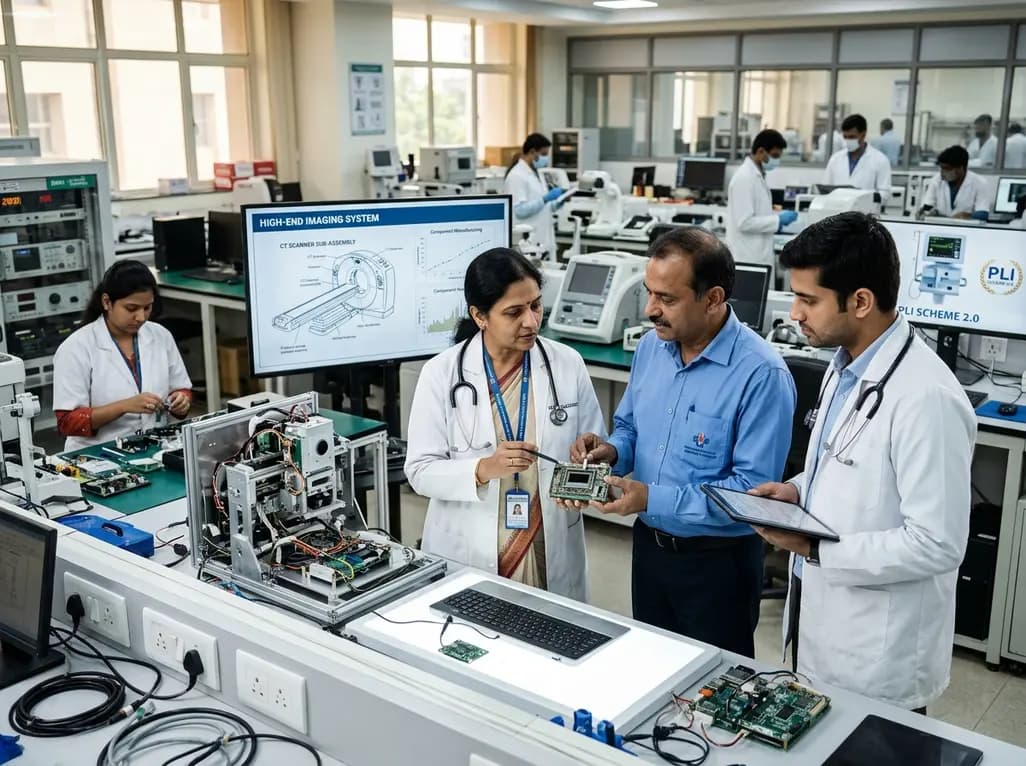
The Department of Pharmaceuticals has extended the medical devices PLI scheme to 2035-36 with a Rs 2,700 crore outlay. Featuring relaxed investment barriers and eliminated sales thresholds, the revised framework targets deep manufacturing, high-end diagnostics, critical MedTech components, and supply resilience.
Today
A metabolomic study reveals that aging delays glutathione synthesis and gamma-glutamylcysteine accumulation following muscle injury. Although precursor supplementation targets this enzymatic bottleneck, in vitro tests show no immediate effect on myogenesis, highlighting the complexity of muscle repair in older adults.
Today

A systematic review evaluates psychological interventions for smoking cessation in diabetes, showing short-term success but highlighting challenges in long-term abstinence.
Today
