

Loading, please wait...
Loading, please wait...

Acute vertigo accounts for millions of emergency visits worldwide each year, presenting an urgent diagnostic puzzle for clinicians. However, distinguishing between benign peripheral vestibulopathies and life-threatening posterior circulation strokes remains notoriously difficult at the bedside. In this high-stakes environment, smartphone nystagmus detection has emerged as a promising frontier for objective point-of-care assessment. Frontline emergency physicians often lack specialized neuro-otology training to interpret subtle, fleeting ocular motor signs accurately. Consequently, misdiagnosis rates for acute vestibular syndrome remain uncomfortably high during early presentations.
Furthermore, conventional magnetic resonance imaging exhibits a notable sensitivity deficit during the initial forty-eight hours of posterior fossa ischemia. Bedside examination frameworks, particularly the Head Impulse, Nystagmus, and Test of Skew protocol, offer superior diagnostic accuracy when clinicians perform them proficiently. Nevertheless, standard laboratory video-oculography equipment remains prohibitively costly, cumbersome, and largely unavailable outside tertiary neurology centers. Emergency departments urgently require scalable, rapid, and objective tools to record patient eye movements reliably. Therefore, leveraging omnipresent mobile devices to capture ocular oscillations represents a vital and pragmatic clinical innovation.
Capturing diagnostic-quality ocular kinematics using mobile devices has historically faced severe technical obstacles outside laboratory settings. Specifically, handheld recordings suffer from erratic camera jitter, variable ambient lighting, patient restlessness, and sudden eyelid closures. In an innovative feasibility study, investigators evaluated an advanced engineering pipeline applied to sixty-seven real-world emergency department video recordings. Rather than relying on static mechanical mounts, the research team implemented a salient feature-based tracking algorithm that monitors ocular and facial landmarks simultaneously.
Because patient head movements and camera shakes compromise raw coordinate traces, the algorithm subtracts stationary facial landmarks from the moving ocular centroid in real time. Consequently, the pipeline cancels out gross image instability, isolating true horizontal eye velocity and displacement relative to the cranium. Moreover, natural blinking frequently triggers tracking failures and coordinate drift in automated systems. To resolve this hurdle, the developers incorporated an automated blink-detection safeguard that momentarily halts tracking during eyelid closure. Subsequently, a third-degree polynomial Savitzky-Golay filter smooths the displacement waveform. This localized filtering suppresses sensor noise while preserving steep, high-velocity fast phases that define true nystagmus.
The study evaluated real-world recordings derived from the open-access SMART-VERTIGO database gathered during busy emergency department shifts. Despite challenging lighting variations and unconstrained operator movements, initial feature tracking proved technically feasible in ninety-four percent of recordings. Specifically, the system successfully tracked landmarks across every video captured at thirty frames per second and in over ninety-two percent of sixty-frame-per-second files. Thus, standard commercial smartphones generate sufficiently robust video data for complex computational kinematic extraction without requiring external attachments.
Furthermore, the computational signals demonstrated remarkable fidelity when researchers compared extracted displacement traces against expert clinical video verification. Most impressively, the algorithm successfully captured very low-amplitude nystagmus beats that were virtually invisible to the naked human eye during routine bedside inspection. In acute clinical practice, low slow-phase velocities frequently characterize subtle central vestibular strokes. Because human observers often miss these faint ocular drifts under standard ambient lighting, automated signal processing provides critical diagnostic sensitivity. Therefore, this digital methodology elevates subjective visual inspection into an objective, reproducible physiological measurement.
Accurate characterization of nystagmus morphology plays a pivotal role in distinguishing peripheral vestibular conditions from acute central cerebrovascular events. For instance, acute vestibular neuritis characteristically induces unidirectional, horizontal-torsional nystagmus that increases in intensity when the patient gazes toward the fast phase. In contrast, central lesions within the cerebellum or brainstem frequently produce direction-changing gaze-evoked nystagmus or purely vertical beats. In addition, central pathologies often display markedly lower slow-phase velocities than vigorous peripheral vestibular disorders.
Unfortunately, emergency clinicians operating under severe time constraints struggle to classify complex waveforms through simple physical inspection alone. Furthermore, visual fixation often suppresses peripheral nystagmus, inadvertently masking vital diagnostic clues unless clinicians employ specialized equipment like Frenzel goggles. Handheld smartphone recordings paired with algorithmic tracking offer an accessible pathway to overcome these practical diagnostic barriers. By converting raw video footage into calibrated displacement waveforms, clinicians can objectively identify beat direction, amplitude, and waveform constancy. Consequently, this technology helps clinicians detect dangerous stroke mimics and true central deficits with substantially greater diagnostic confidence.
The validation of handheld video acquisition carries profound ramifications for emergency triage and contemporary acute vestibular care pathways. First, smartphones represent an ubiquitous global technology that requires zero additional institutional capital investment from resource-strained hospitals. Frontline doctors, triage nurses, and paramedics can rapidly record brief ocular video clips during active vertigo bouts. As a result, critical dynamic physical findings receive permanent digital preservation before transient vestibular signs spontaneously resolve or attenuate.
Moreover, this technological leap creates groundbreaking opportunities for decentralized telemedicine and telestroke networks worldwide. Regional and community healthcare centers frequently operate without on-site neurologists or neuro-otologists to assess acute dizziness presentations. However, local practitioners can securely transmit standardized smartphone recordings to remote tertiary experts for immediate review. When coupled with automated cloud algorithms, remote specialists can analyze objective displacement tracings within minutes of patient arrival. Thus, rapid tele-vestibular triage ensures equitable care delivery, reduces unwarranted cranial computed tomography scans, and accelerates life-saving thrombolytic therapies.
Furthermore, integrating real-time computer vision models into mobile applications will soon provide instant bedside feedback directly to emergency staff. Additionally, multicenter prospective trials will confirm algorithmic generalizability across diverse patient demographics, varying iris pigmentation, and complex oculomotor pathologies. In summary, handheld video oculography stands at the precipice of revolutionizing emergency vertigo care, bridging the divide between complex neuro-otology and routine emergency practice.
The tracking algorithm simultaneously monitors moving ocular points and stationary facial landmarks around the eyes. By continuously calculating and subtracting facial centroid movement from eye coordinate displacements across frames, the software cancels out camera jitter and head translations. Consequently, this differential analysis isolates true horizontal eye movement relative to the skull.
Yes, computer vision algorithms detect subtle, low-amplitude ocular oscillations that escape the unaided eye. By measuring microscopic pixel displacements and filtering high-frequency noise, the software uncovers low-velocity slow phases. This heightened sensitivity proves crucial in emergency settings, where faint nystagmus often signifies an underlying central lesion or early posterior circulation stroke.
Automated tracking converts short smartphone video clips into objective ocular motor waveforms that clinicians can transmit instantly across digital networks. Remote neuro-otologists can review precise graphical tracings alongside video footage to distinguish stroke from peripheral vestibulopathies. Consequently, this remote capability expedites time-sensitive interventions and reduces unnecessary neuroimaging in resource-limited emergency departments.
Disclaimer: This content is for informational and educational purposes only... Refer to the latest local and national guidelines for clinical practice.
References

Read summarized clinical updates, watch expert medical content, and earn CME certifications right from your smartphone.
Open your smartphone camera to scan and install.





A recent feasibility study demonstrates that handheld smartphone videos and facial feature-tracking algorithms can accurately detect nystagmus in acute vertigo presentations within busy emergency departments, offering a low-cost, scalable diagnostic tool for acute vestibular care.
Today

Gastrointestinal smooth muscle cells rely on nanoscale membrane domains to integrate mechanosensitive currents. Recent insights show that nanodomain architecture, rather than channel properties alone, orchestrates motility, opening new therapeutic avenues for clinical dysmotility and gastrointestinal aging.
Today
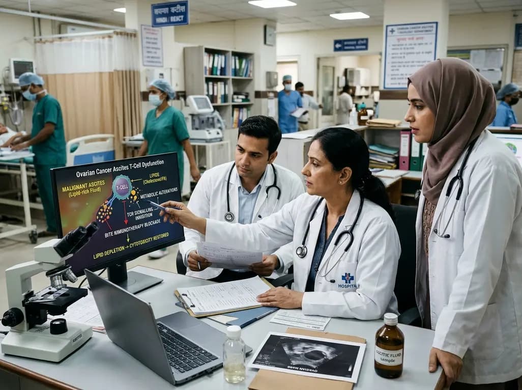
A landmark study reveals that lipid-rich malignant ascites alters the T-cell metabolic transcriptome, causing cholesterol efflux and impaired fatty acid oxidation. This suppresses TCR signalling and blocks BiTE immunotherapy, while lipid depletion restores antitumour cytotoxicity in ovarian cancer.
Today
Novel clinical evidence demonstrates the feasibility of combining VA-ECMO with the NyokAssist self-expanding transvalvular pump for active left ventricular unloading in infarct-related cardiogenic shock, achieving successful stabilization and decannulation.
Today
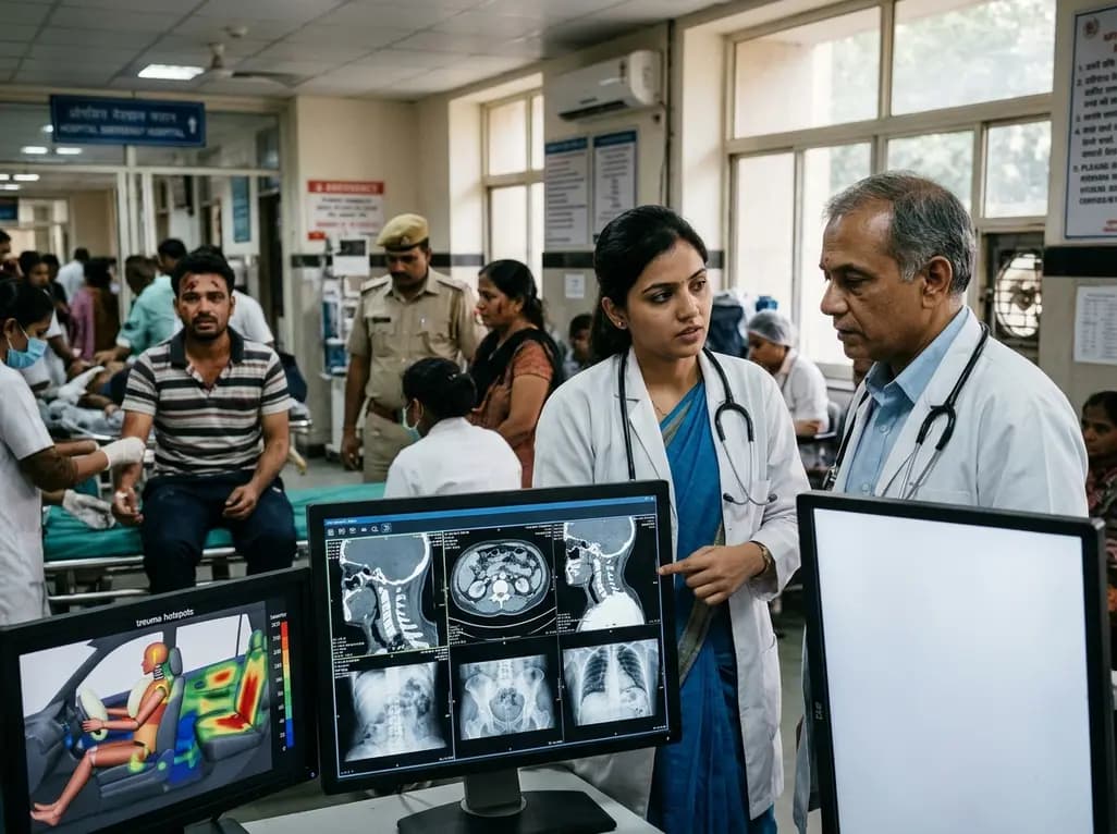
Recent crash analyses demonstrate significant variations in traffic injury severity across vehicle seating positions. Electric vehicle passengers face disproportionately higher trauma risks than drivers, particularly during airbag deployments, demanding adapted emergency assessment and clinical vigilance.
Today

Understand the causal role of Lipoprotein(a) in cardiovascular disease, why international guidelines recommend once-in-a-lifetime molar testing, and the clinical implications of recent phase 3 RNA outcome trials.
Today
