.webp&w=384&q=75)

Loading, please wait...
Loading, please wait...
Distinguishing disorders of arousal from sleep-related hypermotor epilepsy presents a major clinical challenge in contemporary neurology. Both conditions manifest during non-rapid eye movement sleep and frequently display overlapping motor features. Although video polysomnography serves as the standard diagnostic tool, electroencephalographic markers distinguishing these nocturnal phenomena remain insufficiently clear. Recent clinical investigation highlights slow wave sleep fragmentation as a pivotal biomarker that offers fresh diagnostic clarity. Slow-wave sleep represents a crucial physiological phase supporting cognitive recovery and neural restoration. Consequently, disruptions during deep sleep stages significantly impair sleep quality and alter physiological brain activity. While clinicians recognized arousal disruptions in disorders of arousal, researchers previously lacked comprehensive data regarding how hypermotor epilepsy influences slow-wave sleep architecture. Therefore, evaluating deep sleep instability across both pediatric and adult patient groups provides vital insight for differential diagnosis. By examining specific microstructural shifts, clinicians can better differentiate epileptic events from non-epileptic parasomnias. Consequently, accurate characterization reduces diagnostic delays and prevents inappropriate anti-seizure medication administration.
To investigate these distinct sleep patterns, researchers conducted a multicenter study analyzing eighty-seven patients. The study cohort comprised forty-seven individuals diagnosed with disorders of arousal and forty patients with sleep-related hypermotor epilepsy. The researchers enrolled both adult and pediatric cohorts, reflecting the broad demographic presentation of these nocturnal sleep conditions. Furthermore, all participants underwent comprehensive overnight video polysomnography with blinded scoring to eliminate diagnostic bias. The clinical investigators quantified deep sleep disruption by measuring slow, mixed, and fast arousals throughout the night. Additionally, the research team tracked the overnight trajectory of these arousal events to identify temporal variations. The primary physiological parameters evaluated included total sleep time, overall sleep efficiency, stage N2 percentage, stage N3 percentage, and rapid eye movement sleep proportions. Interestingly, total sleep time and sleep efficiency showed no statistically significant differences between the two patient cohorts. Both groups spent similar proportions of time in deep slow-wave sleep during the recorded night. However, stage N2 sleep percentage appeared significantly lower in patients with arousal disorders compared to epilepsy patients.
Although macro-architectural sleep metrics revealed minimal differences in total deep sleep duration, microstructural analysis uncovered striking distinctions. Patients suffering from disorders of arousal exhibited markedly higher slow wave sleep fragmentation compared to those with hypermotor epilepsy. Specifically, arousal disorder patients demonstrated a predominant elevation in slow and mixed arousal indices during deep sleep. These slow arousals represent synchronized high-voltage electroencephalographic oscillations that reflect an intrinsic sleep-preserving mechanism. In contrast, patients with sleep-related hypermotor epilepsy demonstrated a significantly higher frequency of fast arousals during deep sleep. Fast arousals typically feature desynchronized high-frequency activity indicative of abrupt cortical arousal. Therefore, the physiological composition of sleep disruption differs fundamentally between these two patient groups. In arousal disorders, the brain attempts to maintain sleep state stability despite disruptive stimuli, resulting in fragmented slow waves. Conversely, epileptic discharges trigger sharp, abrupt desynchronization that manifests as fast micro-arousals. Consequently, analyzing the specific subtype of arousal provides critical diagnostic information that standard sleep staging cannot reveal.
Understanding how sleep instability evolves across the night provides additional diagnostic leverage for treating clinicians. Using linear mixed models, researchers identified significant time-dependent interactions for arousal indices between the two patient groups. Specifically, patients with disorders of arousal demonstrated a pronounced reduction in sleep fragmentation toward the final hours of the night. This decline aligns directly with homeostatic sleep pressure dissipation, as slow-wave sleep propensity naturally diminishes during later sleep cycles. Consequently, slow and mixed arousal indices in arousal disorders peak early in the night when homeostatic sleep drive is highest. In contrast, patients with sleep-related hypermotor epilepsy maintained a relatively stable frequency of fast sleep interruptions throughout the entire nocturnal recording. Epileptic discharges occur independently of homeostatic sleep pressure, leading to persistent sleep fragmentation across all sleep cycles. Therefore, tracking overnight arousal trajectories helps clinicians differentiate homeostatic parasomnias from persistent epileptic activity. Furthermore, this dynamic pattern explains why parasomnia events occur predominantly during the first third of the night.
To translate these physiological findings into actionable diagnostic tools, researchers performed receiver-operating characteristic curve analyses. The analysis defined highly specific cutoff values for slow-wave sleep disruption parameters, particularly in adult populations. Specifically, the overall fragmentation index and the slow/mixed arousal index achieved remarkable diagnostic accuracy, yielding area under the curve values of 0.81 and 0.84, respectively. These statistical values demonstrate robust diagnostic performance, allowing clinicians to confidently separate non-epileptic arousal disorders from nocturnal epilepsy. Furthermore, establishing quantitative threshold values eliminates subjective interpretation during polysomnography scoring. Clinicians can now utilize standardized microstructural metrics to complement clinical history and video recordings. Consequently, incorporating these quantitative indices reduces diagnostic uncertainty when clinical manifestations remain ambiguous. Additionally, identifying slow arousal predominance supports a diagnosis of non-epileptic parasomnia, reassuring patients and avoiding unnecessary anti-seizure pharmacotherapy. Conversely, detecting low slow-wave fragmentation alongside high fast arousal indices strongly points toward nocturnal epilepsy, prompting appropriate anti-seizure treatment initiation. Therefore, objective microstructural cutoff values serve as powerful diagnostic adjuncts in specialized centers.
The identification of distinctive sleep microstructure markers marks a significant advancement in clinical sleep medicine and neurology. Misdiagnosing sleep-related hypermotor epilepsy as a disorder of arousal can lead to unmanaged nocturnal seizures and potential physical injury. Conversely, misdiagnosing non-epileptic parasomnias as epilepsy results in long-term, unnecessary anti-seizure therapy with potential adverse side effects. Therefore, implementing precise polysomnographic markers substantially enhances patient safety and therapeutic efficacy. Clinicians treating patients with nocturnal motor events should actively evaluate slow-wave sleep micro-architecture during overnight studies. Moreover, sleep laboratories should integrate specialized scoring protocols that distinguish slow, mixed, and fast arousal subtypes. Consequently, routine polysomnography will yield significantly richer diagnostic data without requiring additional invasive testing. In addition, future research may refine these diagnostic algorithms for pediatric cohorts, further broadening their clinical utility. As clinical practice incorporates advanced sleep analysis, patient care will become increasingly personalized and accurate. Ultimately, understanding sleep fragmentation dynamics bridges the gap between complex neurophysiology and practical patient management.
Slow-wave sleep fragmentation refers to repeated micro-interruptions during deep non-rapid eye movement sleep. These disruptions involve slow, mixed, or fast arousal responses that disturb sleep continuity. Quantifying these fragmented events helps clinicians distinguish non-epileptic arousal disorders from sleep-related hypermotor epilepsy during overnight polysomnography evaluations.
Disorders of arousal feature higher total slow-wave sleep fragmentation with predominant slow and mixed arousal subtypes that decline overnight. Conversely, sleep-related hypermotor epilepsy demonstrates lower overall deep sleep fragmentation but features a higher proportion of fast desynchronized arousals that remain consistent throughout the entire sleep period.
Accurate differential diagnosis prevents misdiagnosis and inappropriate treatment. Misdiagnosing epilepsy as a parasomnia leaves dangerous nocturnal seizures untreated. Conversely, mistaking arousal disorders for epilepsy leads to unnecessary, lifelong anti-seizure medication exposure. Quantitative sleep fragmentation markers offer objective clarity, ensuring patients receive targeted and effective clinical management.
Disclaimer: This content is for informational and educational purposes only and does not constitute medical advice, diagnosis, or treatment. Always seek the advice of a qualified healthcare provider with any questions regarding a medical condition. Refer to the latest local and national guidelines for clinical practice.
References
Cordani R et al. Slow-Wave Sleep Fragmentation in Disorders of Arousal and Sleep-Related Hypermotor Epilepsy: A Multicenter Polysomnographic Study. Neurology. 2026 Jul 14. doi: 10.1212/WNL.0000000000218158. PMID: 42308435.
Halász P, Szűcs A. Disorders of arousal and sleep-related hypermotor epilepsy are interrelated. Some new viewpoints. Seizure. 2025 Sep;131:185-189.

Read summarized clinical updates, watch expert medical content, and earn CME certifications right from your smartphone.
Open your smartphone camera to scan and install.





A multicenter study reveals that slow-wave sleep fragmentation is significantly higher in disorders of arousal compared to sleep-related hypermotor epilepsy, providing crucial objective microstructural biomarkers for accurate differential diagnosis.
Today

A growing number of individuals on GLP-1 receptor agonists experience hair shedding due to rapid weight loss and nutrient gaps. Known as telogen effluvium, this temporary condition can be prevented through dietary adjustments, adequate protein intake, and medical oversight without stopping therapy.
Today

The National Human Rights Commission has directed the Rajasthan government to act on maternal deaths and severe injuries in Kota linked to substandard oxytocin injections. The intervention follows complaints regarding batch quality failures, acute renal injury requiring dialysis, and regulatory oversights.
2 days back
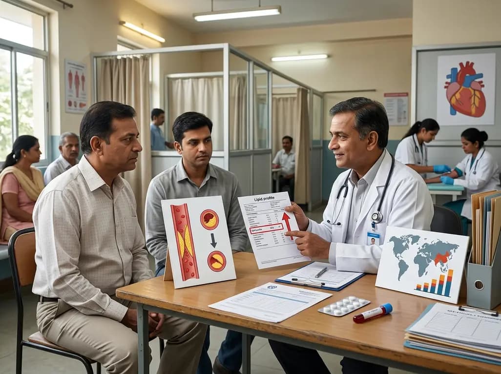
A JAMA study using Global Burden of Disease 2023 data reveals high LDL cholesterol caused 3.6 million deaths globally, with India and China bearing one-third of the disease burden. Addressing systemic barriers to statin therapy and strengthening primary care lipid screening remain critical clinical priorities.
2 days back

Anterior cruciate ligament reconstruction failure lacks uniform definition. A narrative review proposes an integrative framework incorporating objective and subjective instability, persistent pain, restricted motion, graft rupture, and secondary meniscal injury to standardize clinical reporting.
Last week

A comprehensive review highlights major modifiable dementia risk factors in India, emphasizing early-onset diabetes, sensory deficits, undernutrition, and lifestyle interventions to protect cognitive health across diverse populations.
3 days back
