.webp&w=384&q=75)

Loading, please wait...
Loading, please wait...
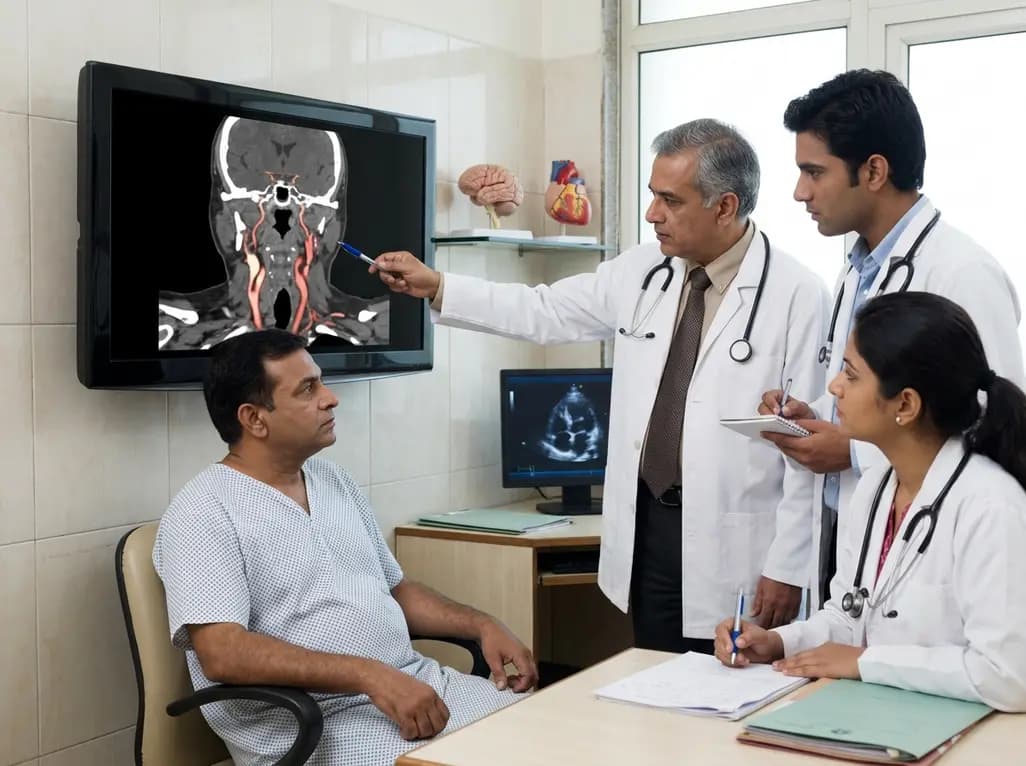
The rare occurrence of bilateral common carotid occlusion represents one of the most challenging presentations in vascular neurology. While unilateral common carotid artery occlusion (CCAO) is documented in medical literature, the sequential involvement of both carotid arteries is exceptionally scarce. In the Indian clinical context, where the burden of stroke is increasingly high, understanding the etiology of such atypical vascular patterns is essential. Typically, CCAO results from atherosclerotic progression, aortic dissection, or Takayasu arteritis. However, when the presentation is acute and sequential, clinicians must investigate alternative mechanisms. Consequently, the discovery of underlying hypercoagulable states becomes a priority. This specific case involving a 56-year-old man highlights how unexpected systemic factors can lead to catastrophic neurological events. Furthermore, the rapid progression from a left-sided occlusion to a right-sided brachiocephalic involvement within a week underscores the volatility of these conditions. Therefore, early identification of non-atherosclerotic drivers is vital to prevent recurrent large-vessel strokes. By analyzing these rare events, medical professionals can better refine their diagnostic algorithms for cryptogenic stroke patients who do not fit the typical profile of peripheral vascular disease.
Protein C deficiency is a well-known inherited thrombophilia that significantly increases the risk of thromboembolic events. Traditionally, this condition is associated with venous thromboembolism, such as deep vein thrombosis or pulmonary embolism. However, its role in arterial thrombosis, including bilateral common carotid occlusion, remains a subject of intense investigation. In the coagulation cascade, protein C acts as a natural anticoagulant by inactivating factors Va and VIIIa. When levels are insufficient, the body enters a prothrombotic state, favoring the formation of clots. In addition to venous risks, several studies suggest that protein C deficiency can contribute to arterial ischemic events, especially when other structural cardiac abnormalities are present. Specifically, in this case, the patient’s laboratory evaluation confirmed a significant deficiency, which likely served as the primary catalyst for recurrent clot formation. Moreover, the sequential nature of the carotid occlusions suggests a persistent systemic drive toward thrombosis. Clinicians should therefore consider protein C levels in patients presenting with recurrent arterial occlusions of unknown origin. This highlights the importance of a multidisciplinary approach involving hematologists to manage long-term anticoagulation strategies effectively.
The presence of a patent foramen ovale (PFO) creates a potential pathway for paradoxical embolism, where a venous thrombus enters the arterial circulation through a right-to-left shunt. This mechanism is frequently implicated in cryptogenic strokes among younger patients. However, its role in causing large-vessel events like bilateral common carotid occlusion is less common but equally critical. When a patient has both protein C deficiency and a PFO, they possess a dangerous combination of a clot-forming environment and a bypass for those clots to reach the brain. Transesophageal echocardiography remains the gold standard for identifying such shunts, as demonstrated in this illustrative case. Furthermore, the hemodynamic changes during the postoperative period can exacerbate the risk of right-to-left shunting. Consequently, a thrombus formed in the venous system, fueled by protein C deficiency, can cross the PFO and embolize into the carotid or brachiocephalic arteries. This pathway explains why the patient suffered sequential occlusions on opposite sides of the vascular tree. Recognizing this synergy between structural heart defects and blood chemistry is fundamental for secondary stroke prevention and deciding on PFO closure.
Diagnosing bilateral common carotid occlusion requires a high index of suspicion, especially when symptoms recur despite initial successful treatment. Initially, magnetic resonance angiography and digital subtraction angiography (DSA) are the preferred modalities to visualize the extent of the loss of flow. In this case, the first event involved the left internal carotid and middle cerebral arteries, which was managed with aspiration thrombectomy. However, the subsequent development of new neurological deficits on the seventh postoperative day necessitated repeat imaging. This imaging revealed a new occlusion in the brachiocephalic artery and the right common carotid artery. Such atypical vascular patterns, where occlusions occur in separate vascular territories sequentially, strongly suggest an embolic source rather than localized plaque rupture. Furthermore, the transition from a localized carotid issue to a more proximal brachiocephalic involvement is a red flag for systemic embolization. Therefore, a comprehensive workup including transesophageal echocardiography and a full thrombophilia profile is mandatory. Ultimately, the diagnosis of sequential bilateral CCAO relies on the synthesis of high-resolution imaging and meticulous laboratory testing to uncover the underlying systemic vulnerabilities.
Management of acute bilateral common carotid occlusion involves rapid recanalization to save salvageable brain tissue. Emergency aspiration thrombectomy is often the first line of defense for large-vessel occlusions. In the described case, this technique successfully achieved recanalization of the left carotid artery during the first event. Subsequently, carotid artery stenting was utilized to address residual stenosis at the origin of the internal carotid artery. This combined approach aims to both remove the acute obstruction and stabilize the vessel wall. However, the recurrence of an occlusion on the contralateral side within a week poses significant therapeutic dilemmas. Should the clinician prioritize mechanical intervention again, or is systemic stabilization more urgent? In this instance, the involvement of the brachiocephalic artery added further complexity to the surgical landscape. Furthermore, the timing of initiating aggressive anticoagulation is critical, especially when balancing the risk of hemorrhagic transformation in a new infarct. Consequently, the treatment plan must be individualized, considering the patient’s overall stability and the potential for further paradoxical embolic events originating from the venous side.
This illustrative case provides several vital lessons for the management of bilateral common carotid occlusion and cryptogenic stroke. First, clinicians must look beyond traditional atherosclerotic risk factors when encountering atypical vascular distributions. Specifically, the presence of sequential occlusions should immediately trigger an evaluation for right-to-left shunts and inherited thrombophilias. Protein C deficiency, while rare, can have devastating arterial consequences when combined with a PFO. Second, the success of initial thrombectomy does not guarantee long-term stability if the underlying embolic source is not addressed. Therefore, long-term management must include robust anticoagulation and potentially structural heart interventions, such as PFO closure, to mitigate the risk of recurrence. Furthermore, early collaboration between neurologists, cardiologists, and hematologists ensures a comprehensive care plan. In the Indian healthcare setting, where access to specialized testing may vary, prioritizing these evaluations in high-risk patients can significantly improve outcomes. Ultimately, a thorough understanding of the interplay between blood coagulability and cardiac anatomy is the key to preventing the life-altering consequences of recurrent large-vessel strokes and sequential carotid artery failures.
Protein C deficiency primarily creates a hypercoagulable state by failing to inactivate specific clotting factors in the blood. While it most commonly results in venous clots, these thrombi can enter the arterial system through a patent foramen ovale. Additionally, some evidence suggests protein C deficiency may directly contribute to arterial wall thrombosis under specific inflammatory conditions. This leads to large-vessel occlusions and subsequent ischemic strokes in affected patients.
A patent foramen ovale (PFO) acts as a gateway that allows blood to bypass the lungs and move from the right atrium to the left. In the presence of a hypercoagulable state like protein C deficiency, venous clots can travel through this opening into the systemic circulation. This process, known as paradoxical embolism, can result in sudden blockages of major arteries, including the common carotid, leading to severe neurological deficits.
Patients typically present with sudden-onset neurological symptoms, such as motor aphasia, hemiparesis, or vision loss. If the occlusion is sequential, a patient might recover from an initial stroke on one side only to develop new, distinct symptoms on the opposite side days later. Imaging usually reveals a loss of flow in the carotid arteries. These recurrent, alternating deficits are a hallmark of an embolic source requiring urgent systemic and vascular investigation.
Disclaimer: This content is for informational and educational purposes only. It is not intended as medical advice or a substitute for professional healthcare. Always seek the advice of a physician or other qualified health provider with any questions regarding a medical condition. Refer to the latest local and national guidelines for clinical practice.
References
Kitamura Y et al. Sequential bilateral common carotid artery occlusion in protein C deficiency and patent foramen ovale: illustrative case. J Neurosurg Case Lessons. 2026 Jul 20. doi: undefined. PMID: 42475756.
Mattuizzi C, Lippi G. Protein C deficiency and cardiovascular disease. Ann Transl Med. 2020;8(12):798.
Schuchlenz HW et al. The role of a patent foramen ovale in patients with cryptogenic stroke. Karadeniz Medical Journal. 2021;14(2):88-94.

Read summarized clinical updates, watch expert medical content, and earn CME certifications right from your smartphone.
Open your smartphone camera to scan and install.





This case study examines a 56-year-old man who experienced sequential bilateral common carotid artery occlusion. Findings revealed protein C deficiency and a patent foramen ovale (PFO), suggesting paradoxical embolism as the primary cause. This highlights the need for thorough thrombophilia screening.
3 months ago

As monsoon seasons elevate arboviral disease threats, Tamil Nadu has initiated an ICMR-monitored evaluation of the tetravalent dengue vaccine in Tenkasi district. Clinicians emphasize clinical monitoring, public prevention, and high-quality early pediatric care to reduce disease morbidity across endemic regions.
Today

Leucine Rich Bio has introduced South Asia's first comprehensive vaginal microbiome test, BugSpeaks. By profiling microbial dysbiosis, pathogens, and antimicrobial resistance, this next-generation diagnostic platform provides clinicians with deeper insights into recurrent infections, fertility, and reproductive health.
Today

Review how tailored exercise programs benefit frail older patients with heart failure. Explore pathophysiological mechanisms, individualized training prescriptions, and strategies to overcome adherence barriers in clinical practice.
Today

This comparative study evaluates posterior labral tears with and without glenohumeral instability, highlighting distinct demographic patterns, MRI underestimation in stable shoulders, and successful arthroscopic repair outcomes.
Today

A clinical study demonstrates that early intervention with tocilizumab, either as monotherapy or bridged with corticosteroids, achieves significant proptosis reduction and resolves active inflammation in treatment-naive thyroid eye disease.
Today
