.webp&w=384&q=75)

Loading, please wait...
Loading, please wait...

Evaluating maxillofacial injuries remains a demanding challenge in acute emergency care. Computed tomography serves as the primary imaging modality for facial trauma, yet subtle cortical disruptions frequently elude initial interpretation. Clinicians frequently miss occult craniofacial fractures when clear primary cortical step-offs are absent. Consequently, identifying indirect radiologic clues becomes essential to expedite therapeutic interventions, avoid permanent cosmetic deformity, and prevent severe functional complications.
Emergency clinicians often encounter patients with significant facial blunt impact who show no overt cortical disruptions on preliminary axial scans. However, indirect imaging features provide vital diagnostic alerts that warrant meticulous re-evaluation. For instance, opacification or fluid-debris levels within the paranasal sinuses strongly signal adjacent bony wall damage. Similarly, the presence of unexplained orbital emphysema or intracranial pneumocephalus points toward a breached bony barrier. Soft tissue alterations also serve as invaluable secondary guides during acute trauma triage. Specifically, asymmetric fat stranding within the masticator space, pterygopalatine fossa, or buccal space highlights local mechanical disruption. Furthermore, focal periodontal air collections or small soft-tissue hematomas frequently point directly to an underlying bone failure. When interpreting these subtle signs, radiologists should not dismiss isolated sinus haziness as pre-existing inflammatory sinonasal disease. Instead, practitioners must maintain a high suspicion for subtle bone trauma whenever soft tissue or gas abnormalities appear. Therefore, recognizing these indirect clues prevents early discharge of unstable patients and drives targeted diagnostic follow-up.
Complex three-dimensional architecture makes specific craniofacial regions particularly susceptible to hidden structural disruption. Recent clinical data demonstrate that occult injuries cluster predominantly in areas with thin cortical bone and multiplanar complexity. Specifically, the mandibular condyle represents one of the most frequent sites of hidden fractures. These condylar injuries often present without blatant displacement, manifesting merely as subtle joint-space asymmetry or mild surrounding capsular swelling. Similarly, the thin nasal bones frequently harbor non-displaced fracture lines that easily blend with normal suture anatomy. Furthermore, trauma teams often overlook non-displaced fractures of the delicate orbital floor and lamina papyracea. Consequently, patients with these missed injuries may subsequently develop progressive enophthalmos, diplopia, or ocular muscle entrapment. In addition, the zygomaticomaxillary buttresses and pterygoid plates require focused inspection because unnoticed impactions can destabilize the midfacial framework. Thus, physicians should consistently scrutinize these fragile anatomical zones, especially when initial axial slices seem deceptively unremarkable.
Technical limitations significantly undermine the sensitivity of initial emergency trauma scans. For example, relying exclusively on thick axial reconstructions dramatically increases the risk of overlooking subtle fractures oriented parallel to the scan plane. Moreover, moderate patient motion artifact, common in agitated or intoxicated trauma victims, readily obscures hairline cortical disruptions. Dental restorations and metallic foreign bodies generate severe streak artifacts that obscure adjacent alveolar structures and condylar anatomy. As a result, the median time to final fracture confirmation often extends to twenty-four hours or longer. This diagnostic delay carries substantial clinical implications for acute patient care. Specifically, patients subjected to diagnostic delays experience higher odds of unplanned management changes, including delayed surgical stabilization. Furthermore, prolonged delays elevate the risk of inappropriate airway management, progressive soft tissue edema, and chronic malocclusion. Therefore, trauma centers must optimize image acquisition protocols to minimize artifacts and eliminate preventable interpretation blind spots.
Overcoming subtle diagnostic pitfalls requires modern post-processing techniques and structured image evaluation. Routine implementation of thin-section multiplanar reformations in coronal and sagittal planes dramatically enhances cortical boundary tracking. In fact, subspecialty re-review using sub-millimeter isotropic reconstructions routinely uncovers previously occult fractures along the orbital walls and mandibular condyles. Furthermore, cone-beam computed tomography provides superior spatial resolution for dentoalveolar and temporomandibular joint trauma while delivering a lower radiation dose. When primary imaging remains inconclusive despite persisting clinical symptoms, clinicians should actively obtain targeted follow-up imaging. Dedicated maxillofacial computed tomography protocols, rather than generic whole-head scans, ensure optimal beam collimation and high-resolution bone algorithms. Additionally, artificial intelligence algorithms and automated fracture detection tools are emerging as valuable adjuncts to reduce human oversight. By embedding systematic multiplanar reconstructions into routine trauma workflows, emergency departments ensure prompt identification and avoid unnecessary diagnostic delays.
Detecting an occult injury is not merely an academic exercise; it directly alters clinical management. Nearly forty percent of patients with belatedly recognized facial fractures require significant treatment modifications. For instance, identifying an undisplaced mandibular condyle fracture mandates immediate dietary modifications, bite registration, and intermaxillary fixation to preserve dental occlusion. Similarly, confirming an orbital blowout fracture necessitates prompt anti-inflammatory therapy, nose-blowing restrictions, and surgical evaluation to prevent globe herniation. Moreover, discovering occult frontal sinus wall fractures guides antibiotic prophylaxis and neurosurgical consultation to prevent ascending intracranial infections. In contrast, missing these injuries frequently leads to secondary malunion, persistent facial asymmetry, and chronic temporomandibular joint dysfunction. Consequently, healthcare providers must understand that timely diagnosis preserves long-term form and function. Through vigilant multiplanar review and swift cross-specialty collaboration between emergency physicians, radiologists, and maxillofacial surgeons, trauma teams achieve superior clinical outcomes.
The most frequent secondary CT signs include fluid or blood accumulation within the paranasal sinuses, localized soft-tissue fat stranding, and orbital emphysema. Clinicians also commonly observe periodontal gas collections, intracranial pneumocephalus, and subtle asymmetry within the temporomandibular joint space. Because these secondary indicators highlight underlying mechanical disruption, their presence should always prompt a rigorous search for subtle cortical fractures across multiple imaging planes.
These anatomical structures feature complex multiplanar curvature and exceptionally delicate cortical margins. On standard axial computed tomography scans, fracture lines often run parallel to the imaging slices, making them virtually invisible. Furthermore, prominent dental metal artifacts and patient motion artifacts obscure the temporomandibular joint. Consequently, clinicians frequently miss non-displaced condylar and nasal fractures unless they examine thin-section coronal and sagittal reconstructions or use dedicated maxillofacial imaging protocols.
Diagnostic delays exceeding twenty-four hours significantly increase the likelihood of unplanned treatment alterations and adverse functional outcomes. For example, missed fractures can lead to malocclusion, progressive enophthalmos, chronic temporomandibular pain, and cosmetic deformities. Additionally, late identification may necessitate delayed surgical reductions, which are technically challenging due to early fibrosis. Prompt recognition through secondary imaging signs ensures immediate conservative or operative stabilization, optimizing healing and functional recovery.
Disclaimer: This content is for informational and educational purposes only. It should not be used as a substitute for professional medical advice, diagnosis, or treatment. Refer to the latest local and national guidelines for clinical practice.
References

Read summarized clinical updates, watch expert medical content, and earn CME certifications right from your smartphone.
Open your smartphone camera to scan and install.





A retrospective trauma analysis highlights the pivotal role of secondary CT signs—such as sinus fluid and orbital emphysema—in identifying occult craniofacial fractures. Recognizing these subtle markers prevents diagnostic delays and directly alters clinical management in over a third of cases.
Today

A recent Journal of Clinical Oncology study demonstrates that industry payments to oncologists directly sway prescribing behavior. Oncologists who received marketing transfers favored promoted therapies over non-promoted alternatives, highlighting critical ethical concerns for evidence-based cancer practice.
Today

A recent qualitative study examines how college students seek digital sexual health information under persistent uncertainty. Discover the concept of functional trust, the clinical hazards of search abandonment, and evidence-based approaches clinicians can adopt to guide youth toward verified reproductive healthcare.
Today
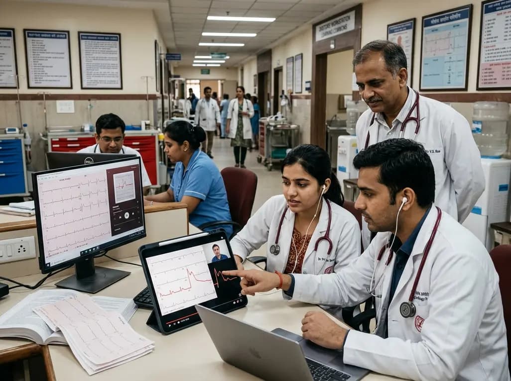
A randomized controlled trial demonstrates that curated FOAMed videos and podcasts produce superior gains in high-risk ECG interpretation and acute coronary syndrome clinical decision-making compared with print-based materials among interprofessional emergency care teams.
Today
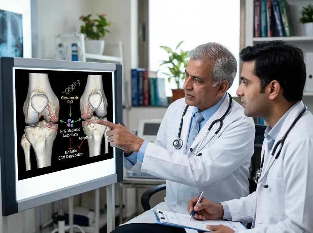
A study reveals that Ginsenoside Rg3 attenuates osteoarthritis progression by modulating the Nrf2-mediated autophagy pathway, reducing chondrocyte apoptosis, and curbing extracellular matrix degradation, offering promising chondroprotective potential.
Today
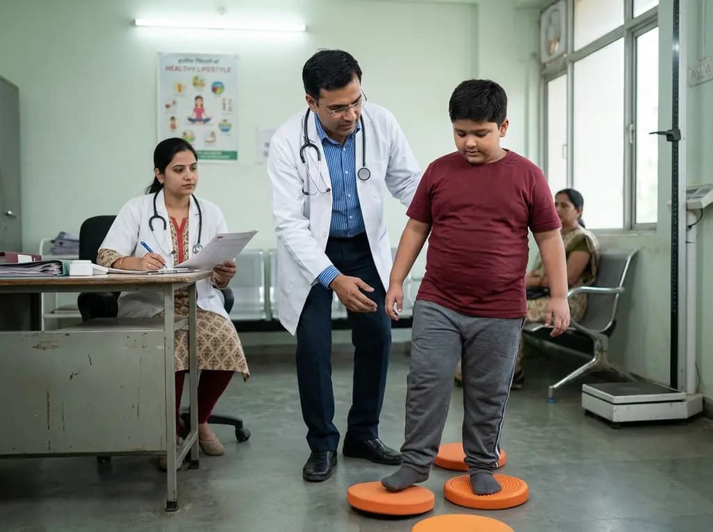
A cross-sectional study demonstrates that lower pediatric motor competence strongly correlates with adverse metabolic syndrome markers, including central adiposity, elevated triglycerides, and low HDL cholesterol, emphasizing early neuromuscular interventions in youth with overweight and obesity.
Today
