.webp&w=384&q=75)

Loading, please wait...
Loading, please wait...
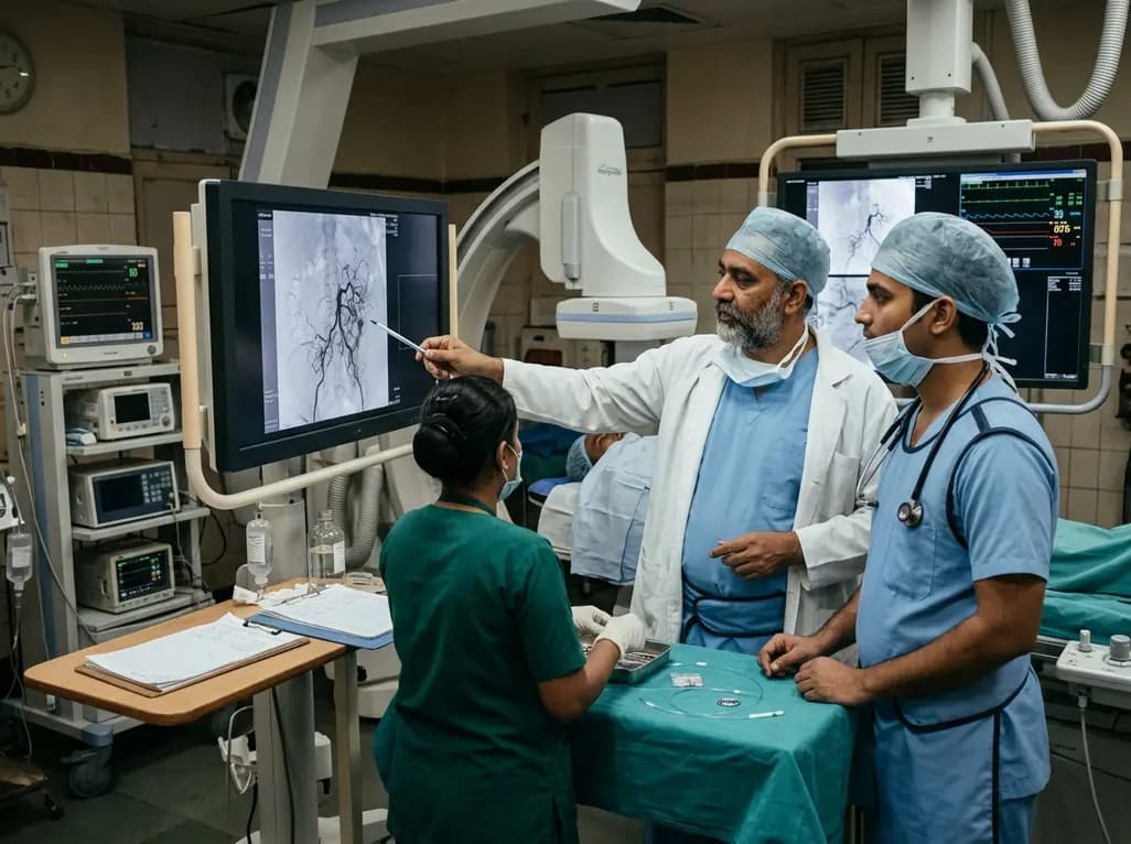
A sacral dural arteriovenous fistula represents an uncommon yet debilitating spinal vascular pathology that frequently challenges neurovascular clinicians. Because symptoms often mimic common degenerative lumbar disorders, delayed identification remains a widespread clinical concern. Recent multicentre clinical evidence provides crucial insights into the vascular architecture, diagnostic pitfalls, and therapeutic results of these lesions, highlighting how timely intervention substantially improves neurological outcomes.
Diagnosing this condition requires heightened clinical vigilance because its early symptoms often overlap with common spinal disorders. Patients typically present with progressive lower limb weakness, sensory disturbances, gait impairment, and sphincter dysfunction. Unfortunately, clinicians initially misdiagnose one-third of these patients with lumbar canal stenosis, peripheral neuropathy, or transverse myelitis. Consequently, patients frequently endure months or years of progressive disability before receiving an accurate diagnosis. Magnetic resonance imaging serves as an essential preliminary evaluation tool. It characteristically reveals extensive spinal cord oedema and dilated perimedullary venous plexuses. However, clinicians often fail to identify the fistula site on conventional lumbar scans because the lesion resides in the sacrum. Therefore, when unexplained conus medullaris hyperintensity appears on imaging, neurovascular teams must extend spinal investigations into the sacral region. Digital subtraction angiography remains the definitive diagnostic gold standard. Careful catheterisation of the internal iliac arteries allows clinicians to pinpoint the arteriovenous shunt precisely. Early angiographic confirmation prevents irreversible ischaemic myelopathy and fosters prompt therapeutic planning.
Understanding the intricate vascular anatomy of sacral lesions is crucial for selecting an optimal interventional strategy. In comprehensive clinical cohorts, the majority of sacral fistulae originate at the first sacral segment, followed by the second and third segments. The arterial supply exhibits distinct patterns that differentiate these malformations from thoracolumbar variants. Specifically, the lateral sacral artery arising from the internal iliac artery serves as the primary feeding vessel in over eighty percent of cases. Additionally, branches from the external iliac artery and the median sacral artery occasionally supply the lesion. These feeding vessels generally follow a straight, calibre-favourable course, which significantly facilitates superselective endovascular catheterisation. Venously, the shunt drains exclusively through the vena medullaris terminalis. This drainage vein subsequently carries retrograde arterialised blood into the coronal and perimedullary venous networks of the lower spinal cord. As a result, substantial venous hypertension develops throughout the conus medullaris. This elevated hydrostatic pressure ultimately reduces spinal perfusion pressure, producing progressive congestive myelopathy.
Endovascular embolisation has emerged as a premier first-line treatment for most patients with sacral lesions. Because the lateral sacral artery provides a relatively straight and accessible arterial conduit, interventional radiologists can navigate microcatheters directly to the fistula nidus with high precision. Liquid embolic agents, such as Onyx or n-butyl cyanoacrylate, effectively penetrate the distal feeding branch and the proximal draining vein. Consequently, endovascular intervention achieves immediate angiographic occlusion in the vast majority of treated individuals. Postoperative magnetic resonance imaging typically confirms the resolution of perimedullary venous engorgement and significant alleviation of spinal cord oedema. Nevertheless, endovascular therapy carries a modest risk of recurrence or residual shunting. In clinical cohorts, approximately one-fifth of embolised patients experience recurrent vascular flow during long-term monitoring. These recurrences frequently arise from incomplete occlusion of the early draining vein or collateral revascularisation. Therefore, interventional teams must schedule rigorous radiological follow-up to detect delayed recanalisation early.
Microsurgical fistulectomy offers a robust, highly reliable definitive therapeutic solution for sacral lesions. Although endovascular embolisation serves as the primary modality for most straightforward cases, microsurgery plays an indispensable role when embolisation fails or proves technically unfeasible. Surgical intervention entails a targeted sacral laminectomy or laminotomy to directly expose the dura mater and the arteriovenous junction. Surgeons meticulously identify the arterial feeders and isolate the arterialised draining vein before applying permanent vascular clips or bipolar coagulation. Notably, clinical trials demonstrate exceptional durability following microsurgical fistulectomy, with complete shunt obliteration and zero recurrence across longitudinal follow-up. Furthermore, patients presenting with residual flow after initial endovascular embolisation achieve definitive cure following secondary surgical disconnection. The direct visual confirmation of venous interruption ensures permanent elimination of the pathological arteriovenous shunt. Thus, multidisciplinary teams should seamlessly integrate microsurgical options into their institutional management algorithms for complex sacral vascular malformations.
Successful interruption of the arteriovenous fistula halts venous hypertension and fosters substantial clinical improvement. Clinicians evaluate neurological recovery using validated tools, such as the modified Aminoff-Logue scale for gait and micturition. Following successful surgical or endovascular occlusion, patients experience statistically significant reductions in disability scores. Motor strength typically improves first, allowing many ambulatory patients to regain independent mobility. In addition, sensory deficits and neuropathic pain gradually subside as spinal venous congestion resolves. However, longstanding bladder and bowel dysfunction may show slower or incomplete recovery due to permanent ischaemic tissue injury. Postoperative magnetic resonance imaging plays a pivotal role in confirming the regression of intramedullary T2 hyperintensity and disappearance of abnormal perimedullary flow voids. Moreover, follow-up digital subtraction angiography at six to twelve months remains essential to verify durable occlusion. Multidisciplinary neuro-rehabilitation further enhances physical recovery, ensuring patients achieve optimal functional independence.
A sacral dural arteriovenous fistula is an abnormal vascular connection between arteries and veins in the sacral dura mater. It diverts high-pressure arterial blood into perimedullary veins. This shunting causes severe spinal venous hypertension, progressive conus medullaris oedema, lower extremity weakness, and sphincter dysfunction.
Endovascular embolisation is preferred because the primary feeding vessel, the lateral sacral artery, typically has a straight, wide course. This favourable anatomy allows interventional radiologists to navigate microcatheters easily to the fistula site. The procedure provides effective shunt occlusion with minimal invasiveness and low complication rates.
Microsurgical fistulectomy is recommended when endovascular embolisation is unfeasible, incomplete, or complicated by recurrence. Surgery provides direct visualization of the dural shunt and allows complete interruption of the arterialised draining vein. Consequently, microsurgical disconnection delivers exceptionally durable results with negligible rates of fistula recurrence.
Disclaimer: This content is for informational and educational purposes only. It is not intended as a substitute for professional medical advice, diagnosis, or treatment. Always seek the advice of your physician or other qualified healthcare provider with any questions you may have regarding a medical condition or treatment options. Never disregard professional medical advice or delay in seeking it because of something you have read in this article. The findings and conclusions in this report are those of the authors and do not necessarily represent the official position of any affiliated institutions. Refer to the latest local and national guidelines for clinical practice.
References
Duan Y et al. Long-term outcome in a cohort of 36 patients with sacral dural arteriovenous fistulae after endovascular embolisation or microsurgery. Stroke Vasc Neurol. 2025 Oct 27. doi: 10.1136/svn-2024-003463. PMID: 39947741.
Barić H, Vukić M, Ozretić D, et al. Spinal dural arteriovenous fistulas: a report on outcomes in a single-center retrospective cohort treated surgically and/or endovascularly. Croat Med J. 2021;62(3):240-249.
Szmygin M, Sojka M, Pyra K, et al. Endovascular embolisation as minimally-invasive treatment for spinal dural arteriovenous fistulas — evaluation of long-term results. Neurol Neurochir Pol. 2023;57(3):305-309.

Read summarized clinical updates, watch expert medical content, and earn CME certifications right from your smartphone.
Open your smartphone camera to scan and install.





A cohort study of 36 patients with sacral dural arteriovenous fistulae evaluates vascular architecture, diagnostic challenges, and clinical outcomes after endovascular embolisation and microsurgery.
4 weeks back

Targeting ferroptosis provides a compelling strategy to dismantle hypoxia- and acidosis-driven immunosuppression in solid tumors, enhancing response to modern immunotherapy.
Today

Findings from Project Viva indicate that sustained postpartum weight retention, rather than excessive gestational weight gain alone, directly worsens women's mid-life cardiovascular health, degrading blood pressure and glycemic parameters decades after delivery.
Today

A large UK Biobank study reveals that plasma proteomic signatures capture preclinical organ damage and improve multiorgan risk prediction across early CKM syndrome stages 0 to 2 beyond traditional PREVENT clinical models.
Today

A retrospective CBCT study reveals that superior TMJ space asymmetry correlates moderately with occipital condyle-atlas-axis asymmetry in unilateral TMJ osteoarthritis, highlighting an imaging correlate between jaw biomechanics and upper cervical alignment rather than a standalone diagnostic marker.
Today

A Bayesian multilevel meta-analysis reveals that aerobic training combined with moderate carbohydrate restriction modestly lowers HbA1c in type 2 diabetes. However, sparse data and very low certainty leave incremental benefits over exercise or diet alone unproven, highlighting the need for individualized care.
Today
