.webp&w=384&q=75)

Loading, please wait...
Loading, please wait...
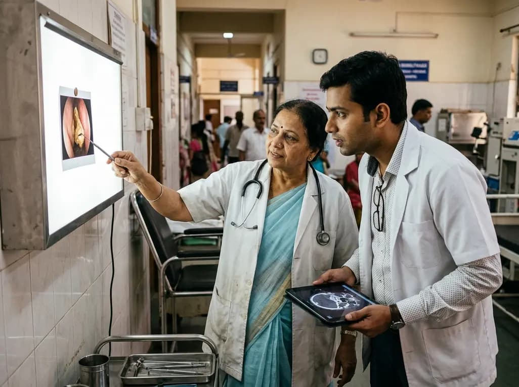
Unilateral nasal symptoms often present substantial diagnostic dilemmas in otorhinolaryngology practice. Although clinicians routinely suspect deviated nasal septum or unilateral rhinosinusitis, rhinolithiasis represents a critical yet underdiagnosed etiology of long-standing nasal obstruction. Recent clinical findings underscore that calcified intranasal concretions foster complex microbial ecosystems, altering sinonasal physiology and predisposing patients to protracted infectious complications.
Rhinolithiasis refers to the progressive deposition of mineral salts around an endogenous or exogenous intranasal nidus. Common exogenous nidi include cotton fibers, beads, and fruit seeds, whereas endogenous sources encompass desiccated mucus, blood clots, or ectopic teeth. Over years, continuous exposure to nasal secretions causes concentric layers of calcium phosphate, calcium carbonate, and magnesium to encrust the inciting material.
Furthermore, this mineralization process progresses very slowly. Patients frequently harbor the foreign mass for decades before experiencing significant functional compromise. As the stone enlarges, it exerts continuous mechanical pressure against adjacent delicate structures. Consequently, this chronic mechanical compression causes mucosal ulceration, localized osteitis, and structural deformation. In severe cases, expanding rhinoliths erode the nasal septum, medial maxillary wall, or hard palate. Therefore, understanding the underlying pathophysiology allows physicians to appreciate why early intervention prevents irreversible sinonasal architectural destruction.
The clinical manifestations of rhinolithiasis frequently mimic those of chronic rhinosinusitis. Most patients complain of persistent, progressive unilateral nasal blockage accompanied by foul-smelling, purulent rhinorrhea. Additionally, individuals may suffer from intermittent epistaxis, facial pain, headache, and severe halitosis. Because the clinical presentation shares extensive overlap with common inflammatory disorders, initial misdiagnosis remains common across primary care settings.
Moreover, patients often receive multiple rounds of empirical oral antibiotics and topical corticosteroids without durable relief. When a patient presents with strictly unilateral sinonasal complaints unresponsive to standard medical regimens, clinicians must suspect a foreign body or calcified concretion. In addition, secondary mucosal hypertrophy and polypoid mucosal degeneration frequently obscure the underlying stone during simple anterior rhinoscopy. Hence, physician vigilance remains paramount, particularly when assessing adult patients who present with decades of unexplained unilateral nasal drainage.
A meticulous diagnostic pathway is essential to confirm rhinolithiasis and assess regional structural complications. First, clinicians must perform rigid nasal endoscopy following mucosal decongestion. Endoscopic examination typically reveals an irregular, gray-to-brown, rock-hard lesion situated along the nasal floor or between the inferior turbinate and nasal septum. Gentle palpation with an instrumental probe confirms the stony, gritty texture characteristic of calcified concretions.
However, rigid endoscopy alone cannot define the full posterior extent or sinus involvement. Therefore, non-contrast computed tomography of the paranasal sinuses serves as the gold standard radiological investigation. Computed tomography scans accurately visualize a hyperdense calcified mass with characteristic central hypodensity corresponding to the organic nidus. Furthermore, coronal and axial views evaluate secondary osteomeatal unit obstruction, bony erosion, and coexisting sinus mucosal thickening. Consequently, combining high-definition endoscopy with thin-slice computed tomography provides crucial anatomical roadmaps for safe surgical planning.
While anatomical obstruction receives extensive attention, the microbiological landscape surrounding rhinolithiasis remains underappreciated. Recent case data reveal that long-standing rhinoliths host complex polymicrobial environments dominated by anaerobic bacteria. Specifically, microbial isolates frequently recover strict anaerobes such as Bacteroides species, alongside facultative pathogens including Streptococcus species and Escherichia coli.
Importantly, this polymicrobial profile mirrors the microbial shifts observed in recalcitrant chronic rhinosinusitis. Mechanical stasis disrupts the physiological mucociliary escalator, resulting in mucus stagnation and decreased tissue oxygen tension. Consequently, this hypoxic niche fosters rapid proliferation of anaerobic organisms that produce putrid sulfur compounds, explaining the characteristic malodor. In addition, colonized rhinoliths act as persistent reservoirs for protective biofilm matrix development. Because standard aerobic swabs frequently fail to detect fastidious anaerobes, clinicians must request dedicated anaerobic cultures during surgical extraction. Ultimately, targeted antimicrobial therapy depends entirely upon accurate microbiological identification.
The definitive curative therapy for rhinolithiasis is complete surgical extraction. Transnasal endoscopic removal represents the standard therapeutic approach, avoiding external facial incisions. For small or mobile rhinoliths, surgeons can perform simple endoscopic grasping and extraction under local anesthesia in an outpatient setting. However, extensive, impacted stones accompanied by severe septal deviation or sinus opacification necessitate general anesthesia.
Furthermore, surgical teams frequently perform comprehensive functional endoscopic sinus surgery concurrently. In complex presentations, procedures such as maxillary antrostomy, total ethmoidectomy, septoplasty, and turbinate reduction effectively restore natural mucociliary drainage pathways. When large stones cause significant mechanical impaction, surgeons carefully fragment the mass using endoscopic forceps, ultrasonic lithotripsy, or holmium lasers before delivering pieces through the anterior nares. Additionally, meticulous intraoperative debridement removes necrotic granulation tissue while protecting underlying bone. Consequently, this combined surgical paradigm completely resolves nasal airway obstruction and eradicates the infectious source.
Postoperative care following rhinolithiasis excision demands systematic attention to prevent recurrent mucosal inflammation and synechia formation. Immediately following surgery, clinicians recommend regular high-volume nasal saline irrigations to facilitate gentle mucosal clearance. Moreover, postoperative antibiotic selection should cover both gram-negative bacilli and anaerobes whenever deep tissue infection or severe rhinosinusitis coexists.
In addition, routine histological evaluation of surrounding granulation tissue remains vital. Chronic local irritation can rarely induce severe dysplasia or mask concurrent inverted papillomas and malignant sinonasal neoplasms. Fortunately, when surgical debridement achieves complete stone removal, long-term patient prognosis remains exceptional. Recurrence remains extremely rare unless an untreated anatomic deformity traps subsequent foreign matter. Therefore, general practitioners and otolaryngologists must maintain high suspicion for rhinolithiasis in any patient suffering from persistent unilateral sinonasal symptoms. Early endoscopic detection prevents long-term sinus disease and restores healthy breathing.
Patients with rhinolithiasis typically present with persistent unilateral nasal obstruction and malodorous purulent rhinorrhea. Furthermore, individuals often report facial pressure, recurrent epistaxis, and chronic halitosis over several years. Because these symptoms closely resemble unilateral chronic rhinosinusitis or foreign body impaction, clinicians must perform a thorough rigid nasal endoscopy. Additionally, computed tomography imaging confirms the calcified intranasal mass, differentiating it from osteomas, fungal concretions, or sinonasal malignancies.
Rhinoliths create prolonged mechanical obstruction within the nasal cavity, impairing normal mucociliary clearance and natural sinus drainage. Consequently, stagnant secretions and persistent tissue hypoxemia produce a favorable microenvironment for anaerobic organisms like Bacteroides alongside aerobic species such as Escherichia coli and Streptococcus. Moreover, the calcified foreign body acts as a permanent nidus for dense biofilm formation. Therefore, long-standing obstruction mirrors the complex microbiologic environment observed in refractory chronic rhinosinusitis.
The definitive management of symptomatic rhinolithiasis requires complete surgical extraction, predominantly through an endoscopic approach under local or general anesthesia. Additionally, surgeons often perform concurrent procedures, including functional endoscopic sinus surgery, septoplasty, or turbinate reduction, to restore normal anatomical ventilation. Following successful stone removal, clinicians should obtain microbiological cultures to direct post-procedure antimicrobial therapy. Consequently, this comprehensive intervention ensures mucosal healing and prevents persistent sinusitis or recurrent chronic infection.
Disclaimer: This content is for informational and educational purposes only and does not constitute medical advice, diagnosis, or treatment. Healthcare professionals should exercise their independent clinical judgment. Refer to the latest local and national guidelines for clinical practice.
References

Read summarized clinical updates, watch expert medical content, and earn CME certifications right from your smartphone.
Open your smartphone camera to scan and install.





Rhinolithiasis is an uncommon etiology of chronic unilateral sinonasal obstruction. A recent case report reveals that long-standing rhinoliths harbor polymicrobial anaerobic pathogens, emphasizing early endoscopic diagnosis, comprehensive surgical debridement, and targeted antimicrobial management.
Today
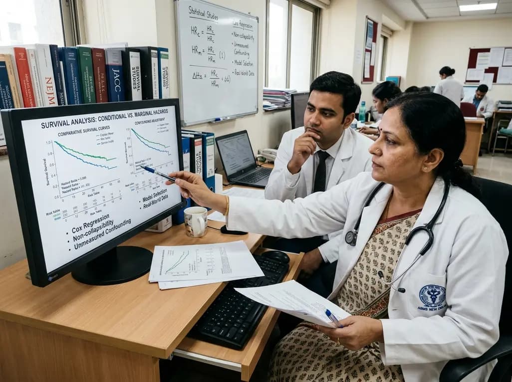
Survival analyses often report hazard ratios, but mathematical non-collapsibility creates crucial differences between conditional and marginal estimates. Clinicians must account for unmeasured confounding and model selection when interpreting trial endpoints and real-world cardiovascular outcomes.
Today

A cross-sectional study of 700 college students highlights significant contraceptive knowledge disparities between sexually active and inactive young adults. The findings demonstrate a critical need for proactive, comprehensive sexual health counseling before sexual debut.
Today
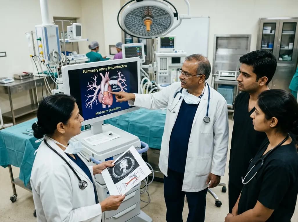
Autologous tissue-engineered vascular graft patches created via in-body tissue architecture show excellent mid-term durability and safety for pulmonary artery reconstruction in congenital heart surgery, particularly for peripheral branch augmentation.
Today

A novel cluster analysis of real-world datasets reveals four distinct CPPD disease phenotypes, validating existing EULAR categories while uncovering previously unrecognized monoarticular and axial presentations to refine patient management.
Today
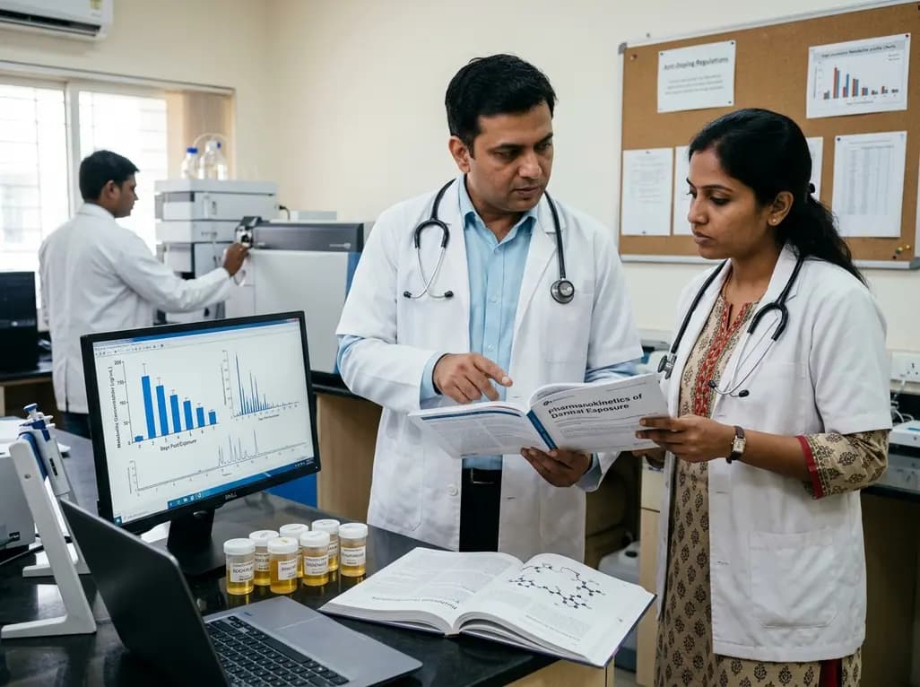
Recent pharmacokinetic research demonstrates that trace dermal exposure to microgram quantities of trenbolone and metenolone produces detectable urinary metabolites for days to over a week, providing critical objective data for evaluating unintentional contamination claims in sports drug testing.
Today
