.webp&w=384&q=75)

Loading, please wait...
Loading, please wait...

Vestibular schwannomas represent benign, slow-growing lesions of the eighth cranial nerve that present major surgical dilemmas. While gross total resection offers definitive oncological cure, neurosurgeons increasingly prioritize neural preservation to maintain patient quality of life. In complex anatomical presentations, subtotal resection provides a safe alternative that prevents devastating cranial nerve deficits. However, leaving behind tissue introduces the risk of tumor recurrence. Recent evidence demonstrates that measuring residual tumor volume serves as a critical prognostic indicator following incomplete resection. Consequently, skull base teams must combine surgical skill with volumetric monitoring to guide secondary interventions effectively.
Historically, surgical teams considered complete microsurgical extirpation the gold standard for all vestibular schwannomas. Complete resection effectively eliminates tumor recurrence, yet it carries substantial risks for large lesions. Specifically, aggressive dissection around the cerebellopontine angle frequently threatens the facial nerve and adjacent brainstem structures. Therefore, modern skull base teams have shifted toward functional preservation over radical tumor eradication. Surgeons deliberately perform subtotal resection when severe arachnoid adherence complicates tumor detachment. This planned subtotal approach protects neurological integrity, especially in patients presenting with borderline facial nerve function. Moreover, clinicians recognize that preserving facial expression and ocular closure profoundly impacts long-term psychosocial well-being. Consequently, partial resection has emerged as an intentional tactic rather than an operative failure. Clinicians nevertheless encounter the challenge of managing residual tissue safely. Because remaining remnants can progress unpredictably, surgical centers must establish robust post-surgical monitoring paradigms. Multidisciplinary teams now blend careful surgical debulking with subsequent observation or adjuvant stereotactic radiotherapy. Thus, contemporary practice balances maximal tumor cytoreduction against the preservation of essential cranial nerves. Furthermore, individualized operative planning ensures that each intervention respects critical patient priorities while controlling immediate mass effect.
Accurate surveillance after subtotal microsurgery depends entirely on precise radiological assessments. Traditional two-dimensional measurements often fail to capture asymmetrical or irregular remnants along the internal acoustic canal. In contrast, advanced three-dimensional volumetric segmentation provides superior spatial accuracy. Radiologists utilize dedicated software platforms to outline contrast-enhancing tissue across high-resolution T1-weighted sequences. This rigorous methodology isolates the exact residual tumor volume remaining after resection. Recent surgical cohorts demonstrate that remnant volume directly correlates with subsequent progression rates. For instance, investigators observed that patients requiring secondary radiation harbored an average residual volume of approximately 37.5 percent. Conversely, patients who maintained stable disease without further intervention exhibited an average remnant burden of only 15.3 percent. Furthermore, volumetric analysis detects subtle three-dimensional shifts long before linear diameters reflect true growth. Early detection of volumetric progression allows clinicians to deliver salvage therapy promptly. As a result, volumetric evaluation transforms post-surgical management from subjective estimation into an objective science. Neurotologists and neurosurgeons can now tailor subsequent follow-up intervals based on concrete volumetric calculations. Additionally, standardized volumetric tracking across longitudinal follow-up visits minimizes interobserver variability between different assessing neuroradiologists.
Preserving the anatomical and functional integrity of the facial nerve remains paramount during skull base surgery. Surgeons evaluate facial outcomes using the standardized House-Brackmann grading scale immediately after surgery and during follow-up. When tumors adhere densely to nerve fibers, pursuing aggressive resection often precipitates permanent paresis. Therefore, operators deliberately halt resection to leave a thin tumor remnant over fragile neurovascular planes. In clinical studies, patients undergoing functional debulking demonstrate favorable House-Brackmann scores at 90 days postoperatively. In addition, this measured approach avoids secondary complications such as corneal exposure keratopathy. However, intentional tumor retention requires surgeons to accept an ongoing oncological responsibility. Residual remnants may eventually resume cellular proliferation, threatening hearing preservation or compressing adjacent structures. Clinicians must weigh the immediate benefits of nerve preservation against the eventual need for secondary radiation. Consequently, open communication with the patient regarding potential salvage therapy is vital. Multidisciplinary teams emphasize that intentional subtotal resection is the first step of a staged management paradigm rather than a standalone curative procedure. Furthermore, thorough baseline electrophysiological monitoring during surgery provides critical objective guidance that assists surgeons in deciding the safest moment to cease resection.
Identifying which patients will experience remnant progression remains a critical goal for multidisciplinary teams. Preoperative tumor volume alone does not accurately predict the eventual necessity of salvage radiotherapy. Instead, the precise percentage of remnant tissue left behind serves as the most dependable predictor. Multivariate regression analyses indicate that larger residual percentages significantly increase the likelihood of secondary therapy. Notably, clinical data demonstrate that patients retaining more than twenty-five percent residual burden face elevated progression risk. In contrast, patients with very small remnants frequently achieve long-term tumor control through conservative surveillance alone. Furthermore, tumor vascularity, proliferation index, and intrinsic biological behavior also contribute to late expansion. Clinicians carefully schedule contrast-enhanced magnetic resonance imaging at standardized intervals to monitor structural changes. When serial imaging confirms measurable growth, stereotactic radiosurgery or fractionated radiotherapy provides reliable rescue therapy. Salvage radiation halts active cell division and stabilizes tumor architecture in most treated individuals. Therefore, identifying high-risk volumetric thresholds enables clinicians to deliver timely salvage therapy before massive recurrence occurs. Moreover, timely radiation prevents the severe neurological morbidity that frequently accompanies secondary salvage microsurgery in an already scarred surgical corridor.
The management of complex skull base tumors demands tight collaboration between diverse medical specialists. Otolaryngologists, neurosurgeons, neuroradiologists, and radiation oncologists must align their treatment strategies to optimize outcomes. Specifically, the multidisciplinary tumor board provides an indispensable forum for reviewing detailed volumetric data. When initial debulking leaves a substantial remnant, team members evaluate whether immediate radiation or watchful waiting is superior. Adjuvant radiotherapy administered shortly after surgery may prevent recurrence, yet upfront irradiation exposes patients to radiation-induced side effects. In contrast, deferred salvage radiosurgery reserves ionizing radiation exclusively for demonstrable progressive disease. Most tertiary skull base centers now prefer proactive surveillance, initiating stereotactic radiosurgery only upon radiographic growth. Additionally, specialized rehabilitation therapists assist patients with vestibular retraining and facial muscle rehabilitation after surgery. Comprehensive care teams also educate patients on recognizing subtle symptoms of cranial nerve deterioration. Consequently, individualized management pathways improve functional independence while delivering robust long-term oncological control for every treated patient. Furthermore, maintaining clear shared documentation between surgical and radiation teams ensures that any radiographic progression triggers rapid clinical reassessment without dangerous diagnostic delay.
Ongoing innovations in neuroimaging and digital analysis promise to refine acoustic schwannoma surveillance further. Automated artificial intelligence segmentation tools are rapidly replacing laborious manual volumetric contouring. Furthermore, these emerging algorithms calculate volumetric growth velocities across serial imaging studies with remarkable precision. Clinicians can identify microscopic volumetric progression months before conventional radiographic assessments confirm tumor recurrence. Similarly, novel biological markers from resected tissue may soon enhance postoperative prognostic models. Combining advanced molecular profiling with three-dimensional volumetric parameters will enable personalized post-surgical surveillance protocols. In the near future, skull base clinicians will calibrate surveillance frequencies according to unique biological and volumetric risk profiles. Thus, integrating computational technologies into routine clinical workflows will enhance long-term patient safety and therapeutic precision. Ultimately, these diagnostic advancements will empower clinical teams to provide timely, minimally invasive care tailored to each individual patient's anatomical condition.
Studies indicate that retaining a residual tumor volume exceeding 25 to 30 percent significantly increases progression risk. In recent clinical cohorts, patients who required adjuvant radiation had an average remnant of 37.5 percent, whereas stable patients averaged 15.3 percent. Higher remnant volumes typically prompt active salvage radiotherapy.
Subtotal resection avoids excessive traction and microvascular injury to delicate facial nerve fibers during dissection. By deliberately leaving a thin tumor remnant over the nerve, surgeons maintain anatomical continuity. Consequently, patients preserve House-Brackmann grade I or II facial function far more effectively than with aggressive complete resection.
Three-dimensional volumetric analysis measures irregular, multiplanar remnants accurately across the entire tumor surface. In contrast, standard linear measurements capture only single dimensions, frequently missing asymmetric growth along the skull base. As a result, 3D segmentation identifies subtle disease progression earlier, guiding timely intervention and treatment decisions.
Disclaimer: This content is for informational and educational purposes only. It is not intended to be a substitute for professional medical advice, diagnosis, or treatment. Always seek the advice of your physician or other qualified health provider with any questions you may have regarding a medical condition. Refer to the latest local and national guidelines for clinical practice.
References
Unterberger A et al. Residual tumor volume guides post-surgical treatment paradigm and need for salvage radiotherapy in vestibular schwannoma management. J Neurooncol. 2026 May 09. doi: 10.1007/s11060-026-05603-6. PMID: 42105194.
Higuchi Y, Nakano S, Aoyagi K, et al. Growth potential of small residual tumors after vestibular schwannoma surgery: comparison between remnants and the natural history of small tumors. J Neurosurg. 2022 Dec;138(6):1658-1665. doi: 10.3171/2022.10.JNS22680.
Tveiten ØV, Link MJ, Neff BA, et al. Salvage radiosurgery following subtotal resection of vestibular schwannomas: does timing influence tumor control? J Neurosurg. 2022 Jul;138(2):418-426. doi: 10.3171/2022.5.JNS22249.

Read summarized clinical updates, watch expert medical content, and earn CME certifications right from your smartphone.
Open your smartphone camera to scan and install.





New clinical research reveals that volumetric measurement of residual tumor volume after subtotal resection of vestibular schwannoma reliably predicts tumor progression and the need for salvage radiotherapy, balancing functional facial nerve preservation with durable disease control.
Today

An Australian cohort of 34,449 adults in an unsubsidized tirzepatide digital weight loss service showed that peer-referred patients achieved significantly higher 6-month adherence (48% vs 42%) and greater weight loss (16.16% vs 14.06%) than matched non-referred peers, highlighting social support in obesity care.
Today
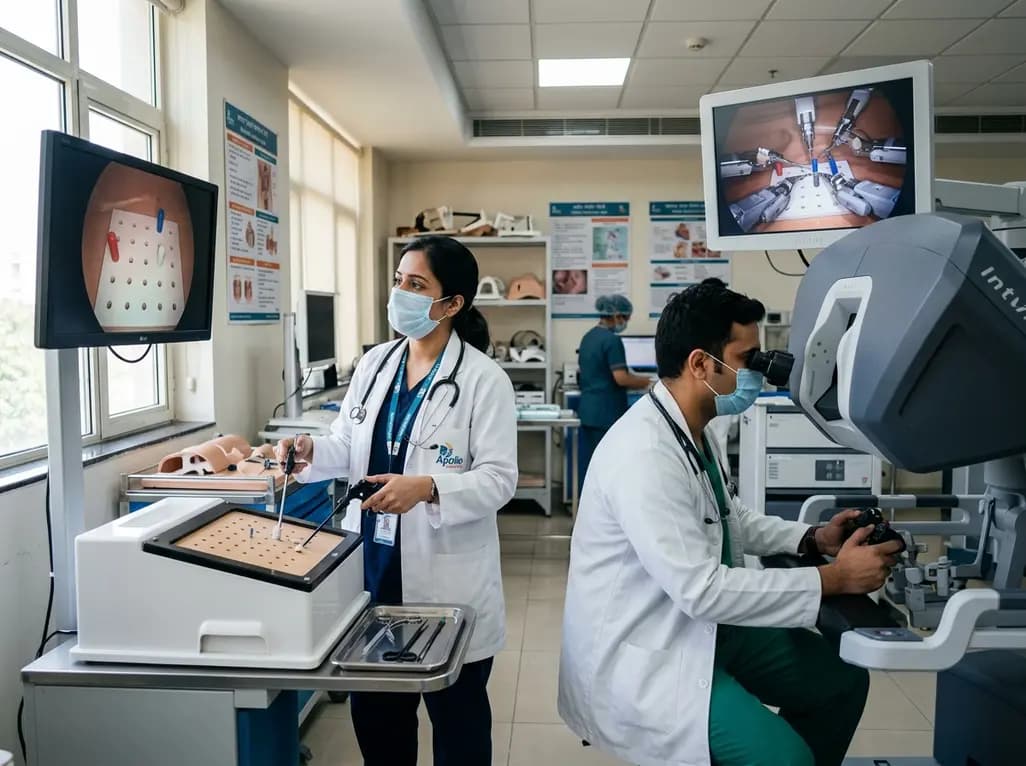
A randomized crossover study demonstrates that conventional and robotic laparoscopy require distinct neuropsychological and psychomotor skills. While trainees master conventional laparoscopy faster, robotic proficiency demands individualized, adaptive curricula tailored to specific learner profiles.
Today

A retrospective study of 109 patients undergoing elective carotid artery stenting evaluated radiation exposure on biplane flat-panel systems. Standardized workflows and dedicated low-dose protocols provide crucial baseline data to establish neurointerventional diagnostic reference levels and optimize patient safety.
Today

A scoping review of 51 studies reveals that spinal manual therapy on the lumbo-pelvic area is commonly used for isolated lower extremity pain, predominantly knee conditions. While thrust manipulation is frequent, reporting deficits highlight the need for standardized technique descriptions to guide practice.
Today

The Supreme Court has reserved its judgment on FSSAI's proposed front-of-pack nutritional warning labels for packaged foods. With debates over compliance timelines, added versus total sugars, and ultra-processed food definitions, this landmark decision carries profound public health implications for India.
Today
