.webp&w=384&q=75)

Loading, please wait...
Loading, please wait...

Elective carotid artery stenting radiation warrants systematic scrutiny as endovascular revascularization increasingly treats extracranial carotid occlusive disease. Interventionalists frequently manage complex cervical pathology in elderly patients with significant vascular comorbidities. Consequently, proceduralists must balance optimal stent deployment against the biological hazards of prolonged ionizing beam exposure. Although endovascular carotid repair provides a proven alternative to endarterectomy, fluoroscopy duration and serial angiography deliver considerable cumulative radiation. Therefore, tertiary centers must establish rigorous benchmarks to track dose metrics and safeguard patient safety. A recent retrospective investigation evaluated radiation dose determinants across 109 consecutive patients undergoing elective stenting for symptomatic carotid stenosis. Notably, the operating team executed all interventions on an advanced biplane flat-panel fluoroscopy system utilizing a dedicated low-dose acquisition protocol. The investigators meticulously documented key dosimetric parameters, specifically dose-area product, total fluoroscopy time, and acquisition frame runs. Furthermore, the researchers analyzed procedural variables alongside demographic factors to isolate genuine drivers of elevated beam exposure. By examining these standardized clinical workflows, clinicians can identify crucial leverage points for radiological quality improvement. Ultimately, understanding dose distribution enables intervention teams to refine technical habits without compromising therapeutic success.
Procedural duration and equipment manipulation directly dictate cumulative patient exposure during endovascular carotid interventions. When operators encounter challenging vascular access, total beam-on time inevitably rises. In this single-center investigation, the researchers identified clear associations between specific procedural maneuvers and overall dose metrics. For instance, repeated balloon dilatations, whether during predilatation or post-stent optimization, substantially increased both fluoroscopy time and cumulative dose-area product. Furthermore, crossing calcified or ulcerated plaques often demanded multiple angiographic roadmaps, which multiplied exposure series. The interventional team tracked fluoroscopy time and the exact count of acquisition runs across all 109 stenting cases. Interestingly, operator experience significantly influenced procedural efficiency and radiation utilization. Experienced neurointerventionalists achieved definitive vessel reconstruction with fewer high-dose cine loops. Conversely, prolonged catheter manipulations during complex aortic arch navigation markedly escalated radiation scatter. In addition, deploying embolic protection filters introduced extra technical steps that occasionally extended total screening periods. However, adherence to a standardized procedural pathway prevented unwarranted fluoroscopy extension during routine maneuvers. These observations demonstrate that technical discipline directly governs radiological safety. Therefore, operators must deliberately minimize unnecessary angiographic series while completing complex intravascular carotid reconstructions.
Patient-specific characteristics and unique vascular anatomy heavily influence endovascular radiation requirements during carotid revascularization. Specifically, elevated body mass index frequently compels modern X-ray generators to augment beam intensity automatically. As a result, obese patients often receive higher dose-area product values despite comparable fluoroscopy durations. Furthermore, severe cervical vascular tortuosity presents formidable mechanical barriers during sheath placement and target lesion engagement. When clinicians navigate unfavorable Type III aortic arches or acutely angulated common carotid bifurcations, catheter positioning demands prolonged fluoroscopic guidance. In addition, extensive vessel wall calcification obscures clear lumography, thereby requiring supplementary oblique acquisition angles. The retrospective study systematically examined these anatomical variables alongside patient age and gender. While demographic features like age showed negligible independent impact, anatomical difficulty consistently correlated with expanded acquisition runs. Moreover, tandem intracranial lesions and contralateral stenosis required comprehensive diagnostic surveys, further increasing cumulative entrance kerma. Nevertheless, standardized low-dose imaging protocols effectively mitigated extreme dose outliers even across technically demanding anatomies. Consequently, neurointerventionalists must anticipate anatomical hurdles during preoperative cross-sectional imaging to plan optimal fluoroscopy angles in advance. Such forward planning curtails unnecessary radiation while executing difficult endovascular carotid procedures.
Establishing robust diagnostic reference levels remains an urgent priority for modern neurointerventional surgery and radiological safety programs. Although international bodies provide broad benchmarks for coronary and peripheral interventions, standardized reference levels for carotid stenting remain sparse. Therefore, dosimetric data from high-volume academic centers offer an essential empirical foundation for prospective multicenter registries. By recording precise dose-area product values and fluoroscopy intervals across 109 standardized procedures, this study delivers valuable reference percentiles. Specifically, tracking median values and seventy-fifth percentile thresholds permits inter-facility comparisons and quality auditing. Furthermore, institutional reference metrics allow catheterization laboratories to detect systemic dose creep and equipment calibration anomalies promptly. When departments identify procedural doses exceeding the seventy-fifth percentile, clinical teams can trigger targeted workflow reviews. In addition, comparative dosimetric analysis highlights variations between individual operators, driving constructive peer education. Establishing national and global reference distributions also protects healthcare personnel from excessive occupational scatter exposure during lengthy neurovascular cases. Accordingly, continuous dosimetric monitoring transforms passive recommendations into actionable institutional benchmarks. High-volume endovascular programs must therefore publish their dosimetric findings to accelerate consensus definition of neurointerventional diagnostic reference levels.
Clinical teams can achieve profound radiation reduction during carotid stenting by implementing structured workflow refinements and technical safeguards. First, interventionalists should routinely configure flat-panel imaging systems to low-pulse fluoroscopy modes, such as four to seven pulses per second. This single technical adjustment drastically reduces cumulative beam energy without degrading necessary spatial resolution for microwire tracking. Second, operators must employ tight physical collimation around the carotid bifurcation, which minimizes stray scatter radiation and sharpens image contrast. Furthermore, biplane flat-panel systems allow simultaneous orthogonal visualization, substantially reducing iodinated contrast volume and extra digital acquisition series. Clinicians should also exploit modern roadmapping capabilities and store digital fluoroscopy loops rather than acquiring repetitive high-dose cine angiograms. Moreover, maintaining adequate distance from the X-ray tube and elevating table heights optimizes geometric magnification while shielding pelvic organs. Interventional teams in India and worldwide must embrace these ALARA principles as mandatory operational standards. In addition, routine multidisciplinary briefings prior to arterial puncture ensure seamless coordination among operators, nurses, and radiographers. Ultimately, deliberate procedural discipline and modern technological protocols preserve patient safety while delivering excellent therapeutic outcomes during endovascular carotid interventions.
Several technical and patient factors drive radiation exposure during carotid revascularization. Specifically, elevated patient body mass index compels imaging systems to increase tube output automatically. Furthermore, complex vascular anatomy, including tortuous aortic arches and calcified bifurcations, requires extended fluoroscopy time and multiple catheter adjustments. In addition, frequent balloon post-dilatations and repeated high-frame-rate digital subtraction angiography runs dramatically expand cumulative dose-area product, underscoring the need for meticulous procedural planning.
Dedicated low-dose protocols utilize advanced digital filtration, optimized detector sensitivity, and reduced pulse rates, typically between four and seven pulses per second. Consequently, these settings preserve spatial resolution while halving continuous beam output. Furthermore, tight X-ray collimation diminishes scatter radiation, which substantially improves vessel contrast. Modern biplane flat-panel systems also integrate sophisticated roadmapping software. Therefore, interventionalists successfully deploy protective filters and carotid stents with superior navigational precision without compromising essential diagnostic visibility.
Diagnostic reference levels provide standardized benchmarks that identify unusually high radiation doses across medical facilities. Because endovascular carotid stenting involves variable anatomical challenges and multiple angiographic runs, establishing reference thresholds prevents unwarranted radiation exposure. Furthermore, reference levels help interventional suites evaluate technical workflows, audit operator performance, and identify faulty imaging hardware. Ultimately, implementing these benchmarks fosters quality improvement, protects patients from deterministic radiation injuries, and reduces long-term occupational radiation exposure for catheterization laboratory staff.
Disclaimer: This content is for informational and educational purposes only... Refer to the latest local and national guidelines for clinical practice.
References

Read summarized clinical updates, watch expert medical content, and earn CME certifications right from your smartphone.
Open your smartphone camera to scan and install.





A retrospective study of 109 patients undergoing elective carotid artery stenting evaluated radiation exposure on biplane flat-panel systems. Standardized workflows and dedicated low-dose protocols provide crucial baseline data to establish neurointerventional diagnostic reference levels and optimize patient safety.
Today
Fatigue affects nearly half of IBD patients even during clinical remission. Discover how a novel 7-week app-based cognitive behavioral therapy intervention aims to transform fatigue management, enhance patient self-efficacy, and reduce disease burden in quiescent Crohn's disease and ulcerative colitis.
Today
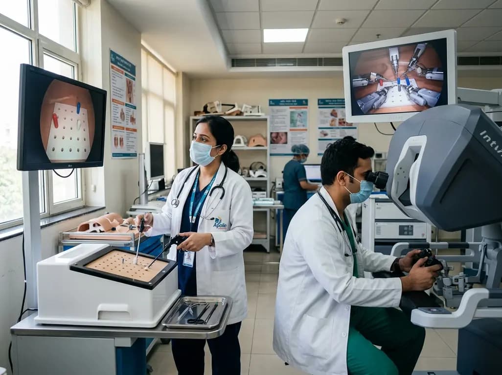
A randomized crossover study demonstrates that conventional and robotic laparoscopy require distinct neuropsychological and psychomotor skills. While trainees master conventional laparoscopy faster, robotic proficiency demands individualized, adaptive curricula tailored to specific learner profiles.
Today

A scoping review of 51 studies reveals that spinal manual therapy on the lumbo-pelvic area is commonly used for isolated lower extremity pain, predominantly knee conditions. While thrust manipulation is frequent, reporting deficits highlight the need for standardized technique descriptions to guide practice.
Today
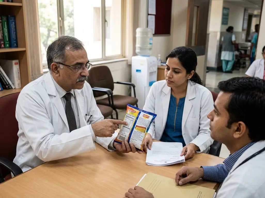
The Supreme Court has reserved its judgment on FSSAI's proposed front-of-pack nutritional warning labels for packaged foods. With debates over compliance timelines, added versus total sugars, and ultra-processed food definitions, this landmark decision carries profound public health implications for India.
Today
