.webp&w=384&q=75)

Loading, please wait...
Loading, please wait...
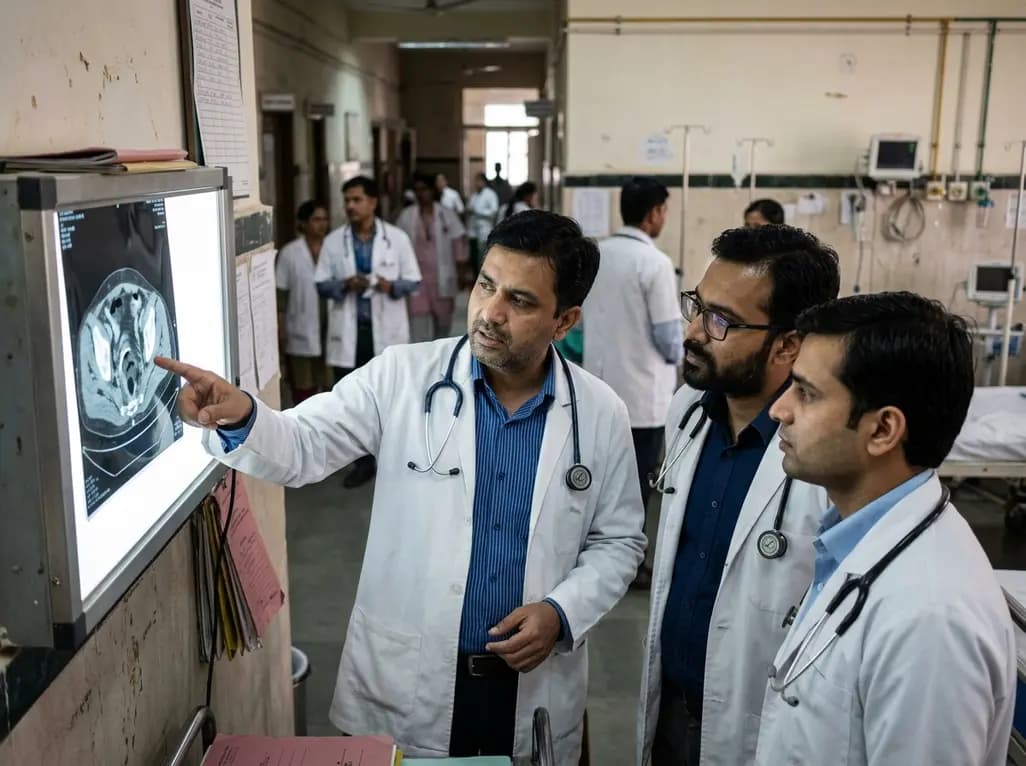
Ingestion of foreign bodies is a relatively common clinical scenario in emergency medicine and gastroenterology. While the majority of ingested objects pass through the gastrointestinal tract without causing mucosal injury or structural damage, sharp rigid objects such as animal bones present a significant hazard. Rectal perforation is an exceptionally rare yet life-threatening manifestation of foreign body ingestion. When a sharp object traverses the entire gastrointestinal tract to reach the distal rectum, local entrapment and delayed transmural erosion can occur. Patients frequently present with nonspecific symptoms, including localized pelvic discomfort or severe anal pain, which can easily be misdiagnosed as uncomplicated perianal disease. Clinical vigilance is necessary because diagnostic delay significantly increases the risk of severe septic complications, tissue necrosis, and extensive abscess formation. Understanding the clinical trajectories, imaging features, and surgical protocols associated with lower gastrointestinal tract foreign body injuries is vital for acute care providers, general surgeons, and gastroenterologists evaluating complex perianal pain.
The gastrointestinal tract possesses several physiological sphincters and anatomical bends where ingested sharp objects are prone to impaction. Although the ileocecal valve and sigmoid colon are the most frequent sites of foreign body entrapment, pointed items such as chicken or meat bones occasionally navigate into the rectal vault. Once located in the distal rectum, forceful sphincter contraction, localized spasm, or manual manipulation can drive the sharp edge directly into the rectal wall. Transmural injury initiates a rapid cascade of localized bacterial contamination from rectal flora, resulting in localized inflammation, micro-perforations, and subsequent abscess formation. Manual attempts by the patient to self-extract or manipulate the impacted object often exacerbate tissue trauma, accelerating full-thickness perforation into adjacent fascial planes. Furthermore, foreign body impaction can compromise regional mucosal perfusion, leading to localized ischemia and necrosis. Recognizing these dynamic pathophysiological mechanisms underscores why early cross-sectional imaging and definitive surgical management are required to prevent widespread pelvic sepsis and complex fistula formation.
Diagnosing lower gastrointestinal foreign body injuries remains remarkably challenging due to nonspecific symptom presentation and absent patient history. Patients rarely recall swallowing bone fragments during meals, particularly when ingestion occurred several days prior to symptom onset. Consequently, severe anal pain or perianal swelling is frequently misattributed to common benign disorders such as thrombosed external hemorrhoids, anal fissures, or simple perianal abscesses. Standard physical examination, including digital rectal examination, may be limited by severe pain or muscle guarding, potentially delaying tactile identification of the impacted foreign body. Laboratory findings typically demonstrate leukocytosis and elevated inflammatory markers, though these findings lack specificity. Non-contrast abdominal and pelvic computed tomography represents the primary diagnostic modality of choice. Computed tomography accurately visualizes radiopaque foreign bodies, identifies precise perforation sites, and evaluates the extent of soft tissue inflammation, free air, or pelvic abscess formation. Utilizing prompt cross-sectional imaging ensures accurate diagnosis, preventing inappropriate conservative management.
Surgical intervention remains the definitive treatment strategy for complex lower rectal perforation caused by sharp foreign bodies. The primary surgical objectives include complete retrieval of the foreign object, thorough drainage of pelvic or anorectal abscesses, and primary repair or diversion of the perforated rectal segment. During surgical exploration, direct visualization enables meticulous assessment of tissue viability and precise identification of transmural defects. In cases featuring localized contamination and viable tissue edges, primary closure of the rectal perforation accompanied by wide drain placement can achieve favorable outcomes. However, extensive abscess formation or gross fecal contamination may necessitate fecal diversion via a temporary loop colostomy to promote bowel rest and tissue healing. Postoperative management relies on broad-spectrum intravenous antimicrobial therapy targeting enteric gram-negative bacilli and anaerobes. Clinical monitoring focuses on resolving local sepsis, controlling pain, and ensuring smooth wound healing. Early surgical consultation and individualized operative management are essential to minimizing long-term anorectal dysfunction.
Failure to promptly recognize and treat lower gastrointestinal perforations can lead to severe postoperative morbidity and life-threatening systemic complications. Distal rectal transmural injuries carry a high risk of spreading infection into the supralevator, ischiorectal, and retrorectal spaces. Uncontrolled infection can progress rapidly to pelvic sepsis, necrotizing fasciitis, systemic inflammatory response syndrome, or septic shock. Furthermore, chronic inflammatory responses surrounding persistent foreign bodies or poorly drained abscesses can result in complex anorectal fistulae, chronic sinus tracts, and persistent fecal incontinence. Patient outcomes heavily depend on the timeliness of surgical intervention, overall systemic health, and the adequacy of initial source control. Broad-spectrum antibiotic coverage must be tailored based on intraoperative culture results to clear resistant pathogen strains. Multidisciplinary care involving general surgeons, gastroenterologists, and infectious disease specialists optimizes clinical recovery. Long-term follow-up examination ensures proper healing of the rectal mucosa, restoration of sphincter integrity, and prevention of recurrent pelvic infectious complications.
Managing sharp foreign body ingestions demands high clinical suspicion whenever patients present with acute, severe anal pain or unexplained perianal suppuration. Physicians should systematically inquire about dietary history, specifically recent consumption of unboned meat or fish dishes, even when patients do not spontaneously report ingestion. Physical manipulation or attempted self-removal of rectal foreign bodies must be strongly discouraged, as manual force significantly increases the risk of deep transmural laceration and severe soft tissue contamination. Early utilization of pelvic computed tomography scanning provides vital anatomical details, guiding operative strategy and surgical approach. Prompt surgical source control, combined with targeted broad-spectrum antimicrobial administration, remains the cornerstone of successful clinical management. Implementing standardized diagnostic pathways for atypical perianal pain ensures rapid identification of foreign body perforations, preventing life-threatening pelvic sepsis and preserving long-term functional anatomy.
Rectal perforation from ingested foreign bodies is challenging to diagnose because patients rarely recall swallowing bone fragments during eating. Additionally, the clinical presentation often mirrors common anorectal conditions like perianal abscesses or fissures. Symptoms such as acute anal pain are non-specific, and initial physical examinations can be limited by patient discomfort, delaying accurate clinical identification without high-resolution computed tomography imaging.
Computed tomography is the definitive imaging modality for evaluating suspected gastrointestinal foreign bodies. It reliably detects radiopaque items like meat or fish bones, identifies the exact site of mucosal perforation, and assesses adjacent soft tissue involvement. Furthermore, computed tomography scans reveal critical secondary complications such as localized extraluminal air, fluid collections, or complex anorectal abscesses, directly guiding surgical planning.
Treatment involves emergency surgical exploration to remove the foreign body, thoroughly drain surrounding anorectal abscesses, and repair the rectal perforation. Depending on the extent of tissue necrosis and fecal contamination, surgeons may perform primary closure with drainage or establish a temporary fecal diversion, such as a colostomy. Adjunctive broad-spectrum intravenous antibiotic therapy is routinely administered to control polymicrobial pelvic infection.
Disclaimer: This content is for informational and educational purposes only and does not constitute medical advice, diagnosis, or treatment. Always seek the advice of a qualified healthcare provider with any questions you may have regarding a medical condition. Refer to the latest local and national guidelines for clinical practice.
References

Read summarized clinical updates, watch expert medical content, and earn CME certifications right from your smartphone.
Open your smartphone camera to scan and install.





A 40-year-old male presented with severe acute anal pain and was found to have a complicated rectal perforation with anorectal abscesses secondary to an ingested meat bone. Prompt imaging and surgical intervention prevented severe morbidity, highlighting key clinical management strategies.
last month

A multicenter study validates a hybrid clinical decision support system combining rule-based logic and machine learning to optimize anticoagulant prescription reviews, reducing alert fatigue and intercepting prescribing errors.
Today

The ClinGen Prenatal Gene Curation Expert Panel evaluated 63 disease relationships across 61 genes, establishing clinical validity for severe fetal phenotypes like hydrops and stillbirth to enhance prenatal genomic interpretation and clinical care.
Today
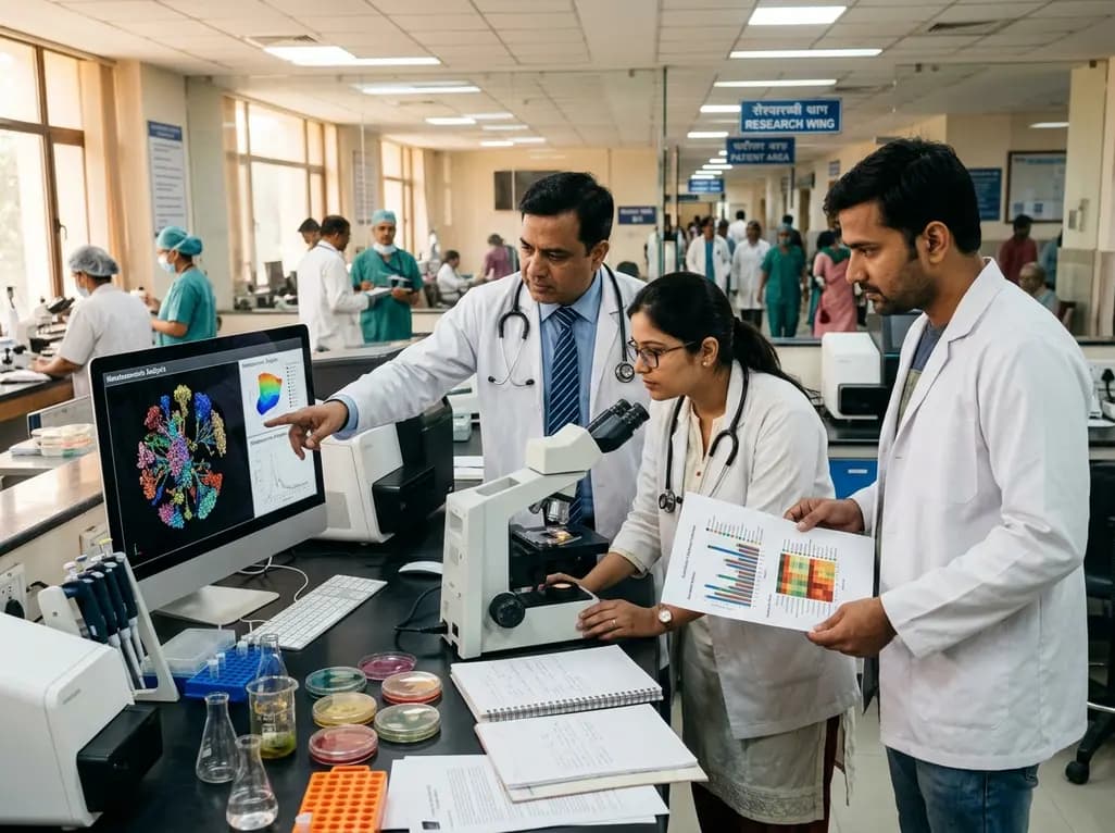
A metataxonomic study reveals distinct gut bacteriome biomarkers in type 2 diabetes, obesity, and cardiovascular complications, identifying specific bacterial shifts that pave the way for precision metabolic medicine.
Today

A psychometric validation study confirms that the Turkish Copenhagen Hip and Groin Outcome Score (HAGOS-T) offers excellent reliability, construct validity, and interpretability for patients with hip osteoarthritis, providing clinicians with a robust tool for functional assessment.
Today

Clinical guidelines rely heavily on isolated biomarkers like IGF-1 and HbA1c. However, portal insulin delivery fundamentally gates hepatic growth hormone sensitivity. This physiological continuum unites type 1 and type 2 diabetes, obesity, cirrhosis, and acromegaly, challenging conventional treatment strategies.
Today
