.webp&w=384&q=75)

Loading, please wait...
Loading, please wait...
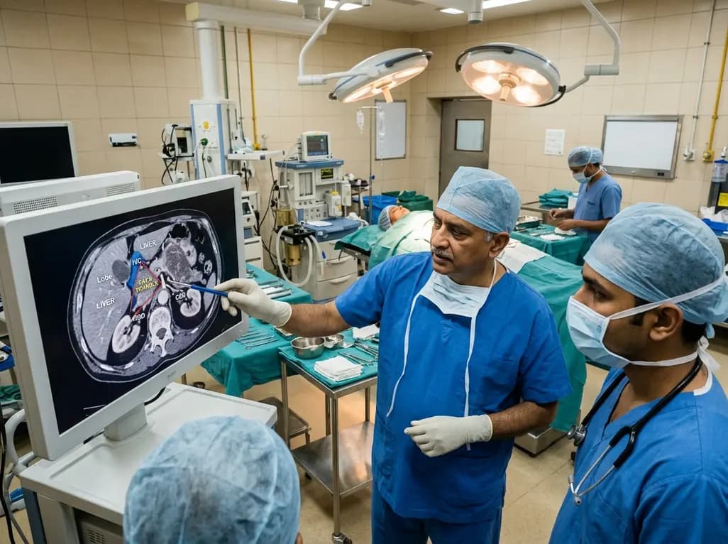
Surgical oncology continually strives to balance radical tumor clearance with patient safety during extensive cytoreductive procedures. In advanced abdominal malignancies, achieving complete cytoreduction represents the cornerstone of prolonged overall survival. However, surgeons frequently encounter occult tumor deposits tucked beneath and behind the liver. To address this technical challenge, researchers at the All India Institute of Medical Sciences (AIIMS) in New Delhi have described Ray's triangle as a critical surgical warning zone during upper abdominal oncologic operations.
During cytoreductive surgery for advanced peritoneal surface malignancies, surgeons must often navigate challenging retrohepatic spaces. The team at AIIMS defined Ray's triangle to delineate an acute danger zone situated at the confluence of the liver, the inferior vena cava, and the right hepatic vein. Although it does not represent an isolated classical anatomical entity, this landmark serves as an indispensable practical surgical boundary.
Specifically, the boundaries encompass the retrohepatic segment of the inferior vena cava, the right hepatic vein trunk, and the posterior surface of the liver parenchyma. Dissection within this restricted space demands extreme precision because aberrant venules and fragile parenchymal bridges traverse the area. Consequently, inadvertent traction or blunt dissection can tear large-caliber venous channels within seconds. Therefore, understanding this regional anatomy enables surgical teams to anticipate major vascular pathways before exerting significant upward or medial traction on the right hepatic lobe. Furthermore, clear visualization prevents catastrophic blood loss during complex peritonectomy procedures.
Peritoneal carcinomatosis arising from epithelial ovarian cancer frequently seeds occult recesses within the right upper quadrant. These hidden sites include Morrison's pouch, the right subdiaphragmatic peritoneum, the right subhepatic space, and the bare area of the liver. Preoperative computed tomography scans often fail to capture microscopic or sheet-like tumor involvement in these secluded locations. Therefore, surgeons cannot rely solely on gross imaging findings to guide resection limits.
To inspect and clear these hidden anatomical zones thoroughly, the oncology surgeon must perform complete mobilisation of the liver. The surgical team initiates this maneuver by incising the right triangular and coronary ligaments through a midline laparotomy. Subsequently, progressive dissection detaches the hepatic attachments from the anterior surface of the retrohepatic vena cava. By elevating the right hepatic lobe medially, the operating team exposes all occult surfaces for direct visual inspection. Consequently, the surgical team can palpate and resect suspicious diaphragmatic implants that would otherwise cause early peritoneal recurrence.
The landmark study from the Department of Surgical Oncology at Dr BRA-IRCH, AIIMS, reviewed 205 patients undergoing extensive cytoreduction with liver mobilisation between January 2014 and December 2025. The cohort comprised individuals treated with either upfront cytoreductive surgery or interval debulking following neoadjuvant systemic chemotherapy. Notably, the operating teams achieved complete or near-complete cytoreduction in 194 patients, representing an impressive 94.6 percent of the study cohort.
In addition, 178 patients attained complete cytoreduction with no macroscopic residual tumor whatsoever. Only 16 patients retained minimal residual nodules measuring less than 2.5 millimeters. However, the researchers recorded right hepatic vein lacerations in nine patients, yielding an overall vascular complication rate of 4.4 percent. Interestingly, seven of these nine vascular injuries transpired during the first 100 operations. In contrast, only two injuries occurred throughout the subsequent 105 cases, reflecting a declining trend from 7.0 percent to 1.9 percent as surgical familiarity evolved. While this decrease lacked formal statistical significance, it clearly demonstrated the vital role of surgical experience.
Major vascular compromise during upper abdominal cytoreduction introduces immediate morbidity and extends postoperative recovery. Patients who experienced intraoperative right hepatic vein injury in the AIIMS cohort exhibited significantly higher blood loss and required prolonged intensive care management. Therefore, establishing a systematic approach to the hepato-caval junction remains an urgent surgical priority for oncologists.
Surgeons must strictly avoid blind finger dissection and excessive manual traction when delivering the liver into the operative field. Instead, sharp dissection under direct illuminated vision allows early identification of the right hepatic vein trunk. Moreover, surgeons should gently sweep intervening fibroareolar planes away from the retrohepatic cava using fine bipolar electrosurgical instruments. If dense desmoplastic reactions or post-chemotherapy fibrosis obscure tissue planes, the team should pause further dissection. Indeed, the AIIMS researchers appropriately aborted retrohepatic clearance in two patients because severe cicatrix rendered planes unsafe, successfully avoiding fatal vascular hemorrhage.
Achieving a complete macroscopic clearance score represents the single most significant modifiable prognostic factor in advanced ovarian malignancy. Leaving residual macroscopic tumor behind compromises systemic chemotherapy efficacy and shortens progression-free survival intervals. Consequently, modern gynecologic oncology practice mandates assertive upper abdominal cytoreductive techniques rather than conservative intra-abdominal debulking.
Nevertheless, aggressive surgical efforts must never compromise patient safety. The identification of Ray's triangle provides surgical oncology fellows and practicing specialists with a standardized framework for complex abdominal surgery. By formalizing this hepato-caval danger zone, surgical trainers can teach safe liver mobilisation step by step. Furthermore, multidisciplinary surgical teams can plan vascular control strategies prior to touching high-risk retrohepatic margins. Ultimately, this standardized anatomical methodology ensures that patients with extensive peritoneal disease gain the oncologic benefits of complete resection while remaining protected from disastrous vascular complications.
Q1: What exactly is Ray's triangle in abdominal cancer surgery?
Ray's triangle is a surgical danger area located at the junction of the liver parenchyma, the inferior vena cava, and the right hepatic vein. AIIMS surgical oncologists defined this space to warn surgeons about extreme vascular hazards during liver mobilisation. Dissection within this triangle requires meticulous technique to prevent severe hemorrhage.
Q2: Why is complete liver mobilisation necessary during ovarian cancer debulking?
Ovarian cancer frequently deposits metastatic implants on the diaphragm, retrohepatic bare area, and subhepatic retroperitoneal spaces. Preoperative radiological scans frequently miss these occult deposits. Mobilising the liver allows the surgical team to inspect these concealed recesses directly and excise every visible tumor deposit to achieve complete cytoreductive clearance.
Q3: How can surgeons avoid major venous injuries around Ray's triangle?
Surgeons should avoid blunt dissection, forceful liver retraction, and blind tissue division. They must visually delineate the retrohepatic inferior vena cava and identify the right hepatic vein before dividing deep peritoneal ligaments. When post-chemotherapy fibrosis obliterates safe surgical planes, surgeons should prudently halt dissection to preserve vascular integrity.
Disclaimer: This content is for informational and educational purposes only. It does not constitute medical advice or replace professional judgment. Refer to the latest local and national guidelines for clinical practice.
References

Read summarized clinical updates, watch expert medical content, and earn CME certifications right from your smartphone.
Open your smartphone camera to scan and install.





Researchers at AIIMS New Delhi have identified Ray's triangle, a high-risk surgical zone between the liver and inferior vena cava. Recognising this danger area allows surgical oncologists to achieve complete cytoreduction in advanced abdominal cancers while substantially lowering the risk of life-threatening hemorrhage.
Today

Subacute thyroiditis presents diagnostic dilemmas when differentiating it from malignancy or chronic thyroiditis. Discover how 2D shear wave elastography provides quantitative tissue stiffness metrics that enhance diagnostic accuracy and assist in tracking disease resolution during therapy.
Today

An updated systematic review and meta-analysis of 7,875 patients reveals that routine microaxial left ventricular support during nonemergent high-risk PCI does not reduce mortality or ischemic complications, while significantly increasing major bleeding events.
Yesterday
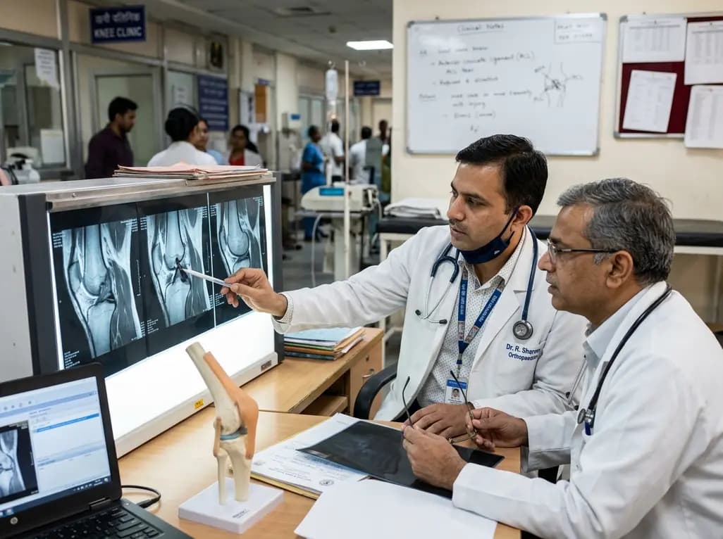
The FIRM-P framework redefines anterior cruciate ligament instability management by separating objective knee phenotypes from patient context and integrating arthrogenic muscle inhibition as a dynamic modifier to optimize individual surgical timing and outcomes.
Today
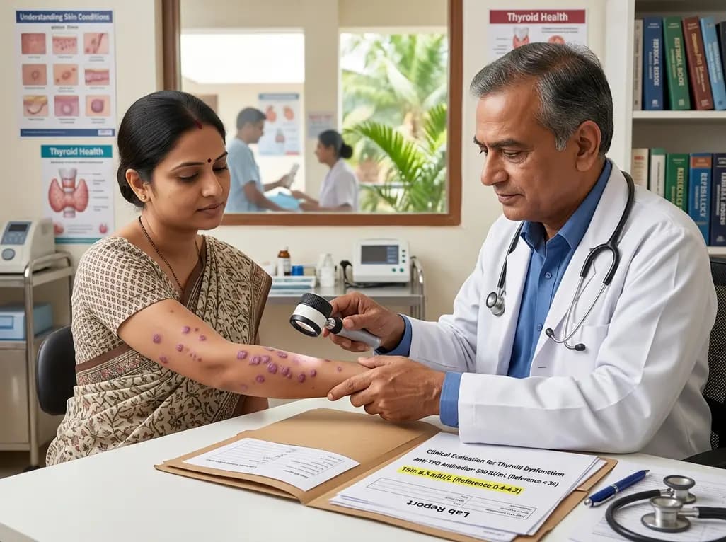
A case-control study demonstrates a significant association between lichen planus and thyroid disorders, with elevated TSH and anti-TPO antibody levels across cutaneous and follicular subtypes, emphasizing the critical value of routine thyroid screening.
Today
