.webp&w=384&q=75)

Loading, please wait...
Loading, please wait...

Current cancer diagnostics primarily rely on serum biomarkers, tissue biopsies, and radiological imaging. However, these tools often lack the sensitivity required for early detection. Traditional biopsies are invasive and cannot easily capture real-time tumor dynamics. Consequently, the medical community is shifting focus toward Raman spectroscopy in CTCs as a cornerstone of modern liquid biopsy. Circulating tumor cells (CTCs) are shed from primary tumors into the bloodstream. These cells provide a minimally invasive window into the tumor biology, allowing for serial sampling throughout the treatment journey.
Raman imaging is a label-free optical technique that identifies molecular fingerprints with exceptional chemical specificity. Specifically, this technology enables the non-destructive analysis of rare cells. Furthermore, recent advances in single-cell Raman spectroscopy allow clinicians to observe intrinsic cellular characteristics without external staining. Surface-Enhanced Raman Scattering (SERS) and Tip-Enhanced Raman Scattering (TERS) have significantly boosted the sensitivity of these platforms. Therefore, researchers can now detect rare CTCs even in early-stage cancer when cell concentrations are extremely low. Notably, these biochemical signatures help in assessing tumor heterogeneity and predicting therapeutic resistance.
Integrating Raman spectroscopy in CTCs into standardized clinical workflows remains a primary goal for oncology. Transitioning from laboratory analysis to routine clinical imaging requires addressing several technical hurdles. These include the need for high-throughput processing and automated data interpretation. Moreover, standardized protocols for sample preparation are essential to ensure reproducibility across different diagnostic centers. Despite these challenges, the ability to monitor real-time tumor evolution through blood samples represents a paradigm shift. Ultimately, this integration could lead to personalized treatment strategies and improved survival rates for cancer patients.
Raman spectroscopy provides a unique biochemical "fingerprint" for each cell. Unlike fluorescence imaging, it does not require external labels or dyes. This allows for a more accurate and non-destructive analysis of rare circulating tumor cells.
Monitoring CTCs allows doctors to track how a tumor changes over time. It helps in early diagnosis, assessing the risk of metastasis, and determining if a specific chemotherapy or immunotherapy is working effectively.
SERS (Surface-Enhanced Raman Scattering) and TERS (Tip-Enhanced Raman Scattering) are advanced techniques that use metallic nanostructures to amplify Raman signals. This amplification is crucial for detecting individual cancer cells in complex blood samples.
Disclaimer: This content is for informational and educational purposes only. It does not constitute medical advice or a professional relationship. Refer to the latest local and national guidelines for clinical practice.
References
Zhang J et al. From spectroscopic analysis to clinical imaging: emerging applications and persistent challenges of raman spectroscopy in CTCs-based liquid biopsies. Ann Med. 2026 Dec undefined. doi: 10.1080/07853890.2026.2634462. PMID: 41746879.
Li D et al. Surface-enhanced Raman spectroscopy for label-free cancer liquid biopsy: from fundamentals to clinical analysis of biofluid. Front Bioeng Biotechnol. 2026 Jan 12. doi: 10.3389/fbioe.2025.1324567.
Depciuch J et al. Analysis of liquid biopsy by Raman spectroscopy to facilitate prediction of response to immunotherapy in non-small-cell lung cancer (NSCLC) patients. Spectrochim Acta A Mol Biomol Spectrosc. 2025 May 05. doi: 10.1016/j.saa.2024.125000.

Read summarized clinical updates, watch expert medical content, and earn CME certifications right from your smartphone.
Open your smartphone camera to scan and install.





This review explores how Raman spectroscopy and CTC-based liquid biopsies are advancing early cancer diagnosis and real-time therapeutic monitoring....
7 months ago
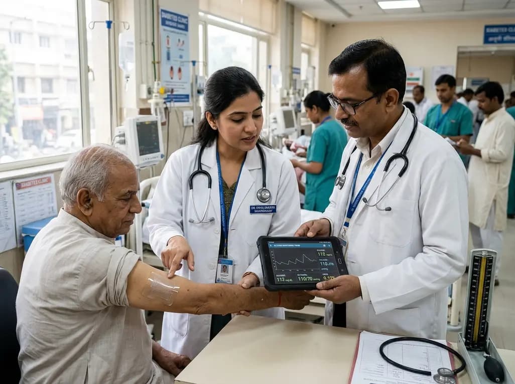
A breakthrough ultrathin multimodal sensor decouples physical hemodynamic monitoring from disposable electrochemical calibration, achieving long-term wearable glucose prediction with 81.36% accuracy and a 9.12% MARD. This offers a robust, drift-free solution for outpatient metabolic surveillance.
Today
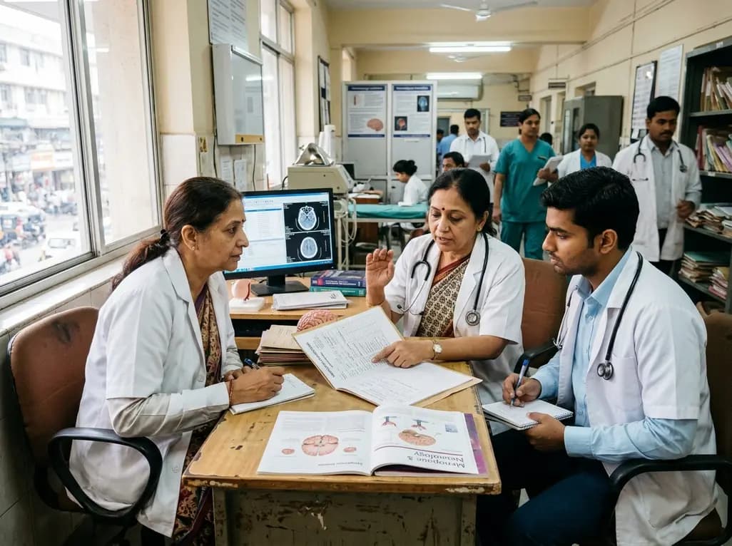
A clinical review of menopause and neurology highlights the neurobiological impacts of midlife hormonal shifts on cognition, epilepsy, migraine, multiple sclerosis, and stroke, emphasizing safe hormone therapy prescribing.
Today
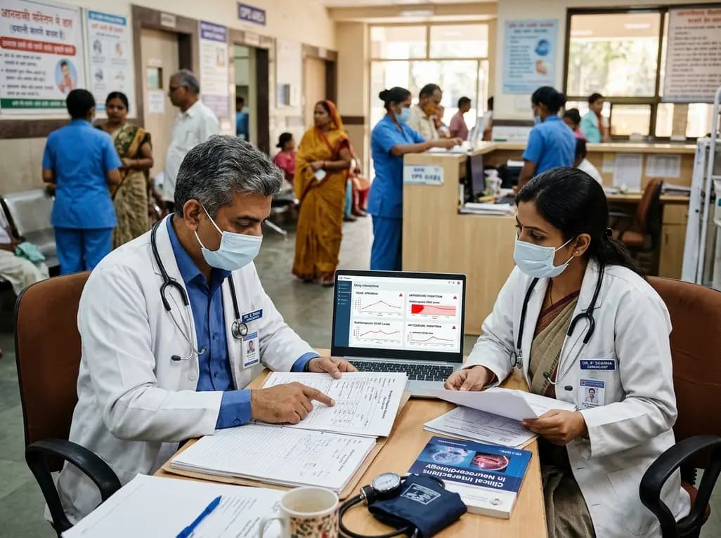
Co-administering antiseizure medications and DOACs frequently causes pharmacokinetic interactions, subtherapeutic drug levels, and thromboembolic failures. This review highlights enzyme induction risks, unpredictable outcomes with levetiracetam, and the vital role of therapeutic drug monitoring.
Today
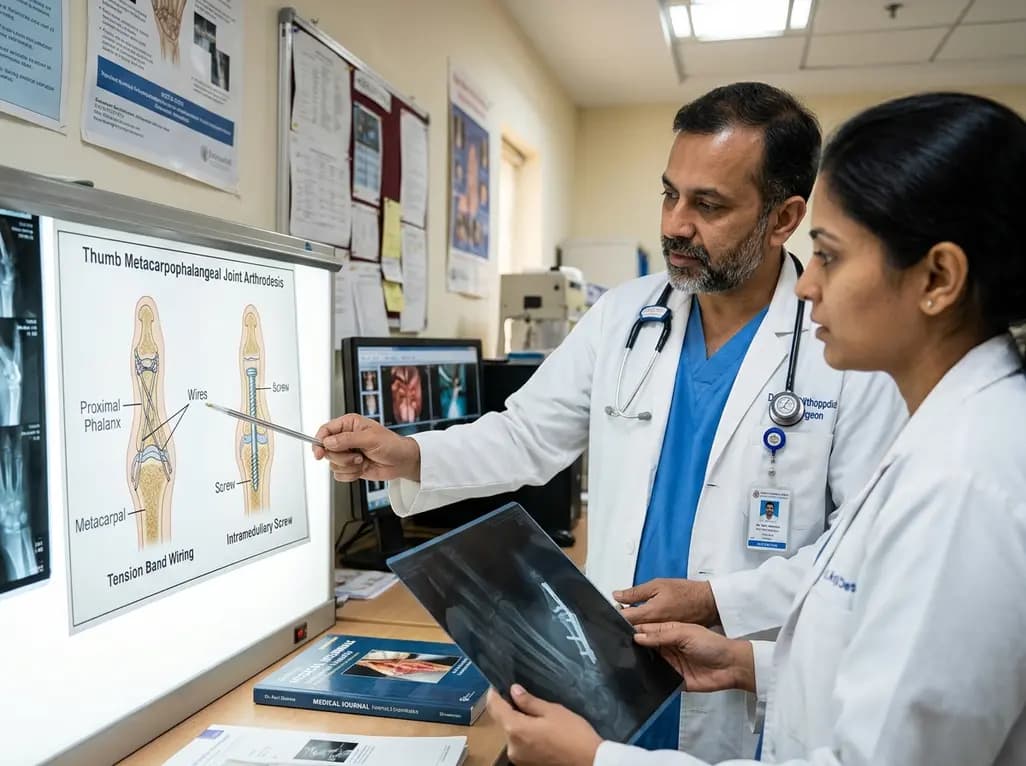
A comparative review of tension band wiring and intramedullary screw fixation in thumb metacarpophalangeal arthrodesis, evaluating union rates, hardware complications, and clinical outcomes.
Today

Following regulatory scrutiny from FSSAI regarding promotional representations and lean muscle preservation claims, Dr. Reddy's Laboratories and Nestlé Health Science are currently reviewing the branding and marketing strategy for their joint nutritional supplement Celevida GLP+.
Today
