.webp&w=384&q=75)

Loading, please wait...
Loading, please wait...

Glioblastoma is the most aggressive primary central nervous system malignancy. Even after complete surgical resection and chemoradiotherapy, tumor recurrence remains inevitable. Clinicians routinely monitor patients using serial neuroimaging. However, the manifestation of relapse varies considerably among individuals. Understanding every unique pattern of radiologic progression in glioblastoma provides vital prognostic insight. Recent evidence applying multi-state mathematical models now reveals how distinct recurrence phenotypes evolve longitudinally, offering clinicians a rigorous framework to anticipate clinical trajectories and refine secondary therapeutic interventions.
Standard response criteria often simplify glioblastoma recurrence into a binary assessment of disease progression. However, clinical presentations encompass diverse neuroimaging manifestations. A landmark longitudinal study of 119 glioblastoma patients identified four distinct radiologic progression patterns. These patterns comprise complete progression, classic-T1 progression, non-responder recurrence, and non-local dissemination.
The complete recurrence phenotype represents a multifaceted radiological group. Specifically, it includes T2-circumscribed lesions, diffuse T2 infiltration, and lesions showing transient contrast loss followed by flare-up. Conversely, classic-T1 progression manifests as localized, sharply defined contrast-enhancing margins surrounding the primary resection cavity. Meanwhile, non-responder status describes rapid, continuous tumor expansion immediately post-surgery, reflecting intense upfront therapeutic resistance. Finally, non-local progression features distant parenchymal relapse far from operative margins.
Consequently, these imaging archetypes illuminate diverse biological processes. For example, non-responder tumors display accelerated proliferation and aggressive invasion. In contrast, patients who develop complete or classic-T1 patterns often maintain temporary radiological stability. Therefore, identifying these distinct progression phenotypes empowers neuro-oncology teams to characterize disease dynamics accurately and select individualized salvage therapies.
Traditional clinical research heavily employs Kaplan-Meier curves and Cox proportional hazards regression to evaluate cancer survival. Although these statistical tools establish foundational benchmarks, they compress complex clinical pathways into isolated static endpoints. Specifically, conventional models treat disease progression as a single intermediate checkpoint, completely ignoring subsequent transitions between different recurrence stages.
To capture disease evolution accurately, investigators built a unidirectional multi-state model. This sophisticated architecture integrates six discrete health states and 14 distinct transitions. The modeled pathway starts at surgical resection, proceeds across the four specified radiologic recurrence states, and concludes at death. As a result, this dynamic framework systematically evaluates how patients transition across sequential post-recurrence milestones.
Furthermore, multi-state modeling computes time-dependent transition hazards that standard regression formulas overlook. Rather than providing an isolated median survival figure, the model determines real-time probabilities for entering each progression state. Neuro-oncology teams can therefore assess individual prognostic trajectories with unprecedented precision. Consequently, clinicians can anticipate critical turning points in disease progression and provide personalized, timely guidance to patients.
Longitudinal evaluation reveals striking temporal variations in recurrence risks across the postoperative timeline. Notably, patients display significant differences in both progression-free survival and overall survival depending on their radiologic phenotype. During the initial six months post-surgery, the cumulative hazard for the non-responder pattern rises sharply. The probability of entering this refractory state peaks at five months, reaching twenty percent.
In contrast, the clinical hazard profile shifts considerably between 6 and 24 months postoperatively. During this window, transition probabilities for classic-T1 and complete progression rise steadily. Specifically, classic-T1 recurrence achieves a peak probability of 12.7 percent between 10 and 12 months. Furthermore, complete progression demonstrates a delayed peak probability of 8.7 percent at 16 months post-surgery. Meanwhile, non-local progression maintains comparatively low, stable transition probabilities throughout follow-up.
Accordingly, these chronological hazard waves carry vital implications for routine clinical surveillance. Neuro-oncologists can tailor magnetic resonance imaging frequency based on expected temporal risks. For example, intense surveillance during the first six months detects aggressive non-responders promptly. Subsequently, sustained monitoring across the second year successfully identifies late-onset classic-T1 and complete recurrences.
Modern neuro-oncology emphasizes molecular profiling to classify gliomas and predict therapeutic responsiveness. To evaluate whether underlying genetic drivers dictate radiographic progression patterns, researchers examined 11 glioma-related genes across 69 profiled patients. Interestingly, the investigation demonstrated no statistically significant differences in key gene alterations across the different radiologic progression phenotypes.
This pivotal finding underscores that genomic mutations alone do not determine macroscopic recurrence patterns. Although tumors share common driver alterations, localized microenvironmental factors heavily dictate imaging phenotypes. Specifically, regional hypoxia gradients, blood-brain barrier disruption, and extracellular matrix remodeling drive distinct radiologic features. For instance, vigorous neoangiogenesis induces focal contrast enhancement in classic-T1 tumors, whereas infiltrative migration produces diffuse non-enhancing T2 changes.
Consequently, clinicians cannot rely exclusively on initial molecular diagnostics to forecast patterns of relapse. While baseline genetics guide initial chemoradiotherapy decisions, longitudinal imaging captures real-time physiological adaptations. Therefore, integrating genomic data with multi-state imaging surveillance provides a comprehensive assessment of glioblastoma biology. This multimodal perspective helps multidisciplinary tumor boards anticipate disease evolution more accurately.
Differentiating radiologic progression patterns enables clinicians to design personalized salvage regimens rather than utilizing uniform salvage strategies. For instance, patients presenting with classic-T1 progression around 10 to 12 months often harbor localized, accessible lesions. In these selected cases, repeat surgical resection or stereotactic radiosurgery can achieve meaningful local tumor control while preserving neurological function.
Conversely, patients displaying the aggressive non-responder phenotype within five months gain minimal benefit from repeat surgery. Because these rapidly recurring tumors exhibit profound chemoresistance, clinicians should prioritize systemic therapy modification or immediate trial enrollment. Additionally, anti-angiogenic agents such as bevacizumab may help manage severe peritumoral edema, reduce steroid requirements, and stabilize acute neurological deficits.
Furthermore, patients who develop diffuse T2 or non-local dissemination require therapies that target widespread parenchymal invasion. Focal re-irradiation often proves ineffective and carries unacceptable neurotoxic risks for non-local relapse. Instead, tumor-treating fields, metronomic systemic regimens, or novel targeted agents offer wider coverage. Ultimately, aligning salvage therapy with specific radiologic progression archetypes optimizes clinical outcomes and prevents ineffective interventions.
Conventional Cox models assess static survival endpoints from a fixed baseline, overlooking intermediate disease stages. In contrast, a multi-state model captures progressive shifts across distinct recurrence states. Consequently, it quantifies continuous transition probabilities over time, offering clinicians detailed prognostic timelines and facilitating adaptive therapeutic decision-making throughout patient follow-up.
Although glioblastoma harbors significant genomic heterogeneity, specific driver mutations frequently persist across divergent radiologic recurrence patterns. Therefore, regional microenvironmental conditions, vascular permeability changes, and peritumoral infiltration may drive phenotypic imaging variation more heavily than isolated mutations alone. Comprehensive multi-omics analyses remain necessary to uncover subtle epigenetic mechanisms governing these pathways.
Early non-responder progression typically manifests within six months post-surgery, portending aggressive disease biology and poor outcomes. Multidisciplinary tumor boards should rapidly re-evaluate imaging to exclude pseudoprogression. Subsequently, clinicians must consider re-resection, enrollment in novel clinical trials, targeted systemic agents, or altered fractionated re-irradiation to mitigate rapid clinical deterioration.
Disclaimer: This content is for informational and educational purposes only... Refer to the latest local and national guidelines for clinical practice.
References

Read summarized clinical updates, watch expert medical content, and earn CME certifications right from your smartphone.
Open your smartphone camera to scan and install.





A longitudinal multi-state model of 119 glioblastoma patients reveals distinct radiologic progression patterns—complete, classic-T1, non-responder, and non-local—with significant survival differences despite similar gene alterations, defining critical chronological hazard peaks to optimize salvage therapy.
Today

Survival analyses often report hazard ratios, but mathematical non-collapsibility creates crucial differences between conditional and marginal estimates. Clinicians must account for unmeasured confounding and model selection when interpreting trial endpoints and real-world cardiovascular outcomes.
Today

A cross-sectional study of 700 college students highlights significant contraceptive knowledge disparities between sexually active and inactive young adults. The findings demonstrate a critical need for proactive, comprehensive sexual health counseling before sexual debut.
Yesterday
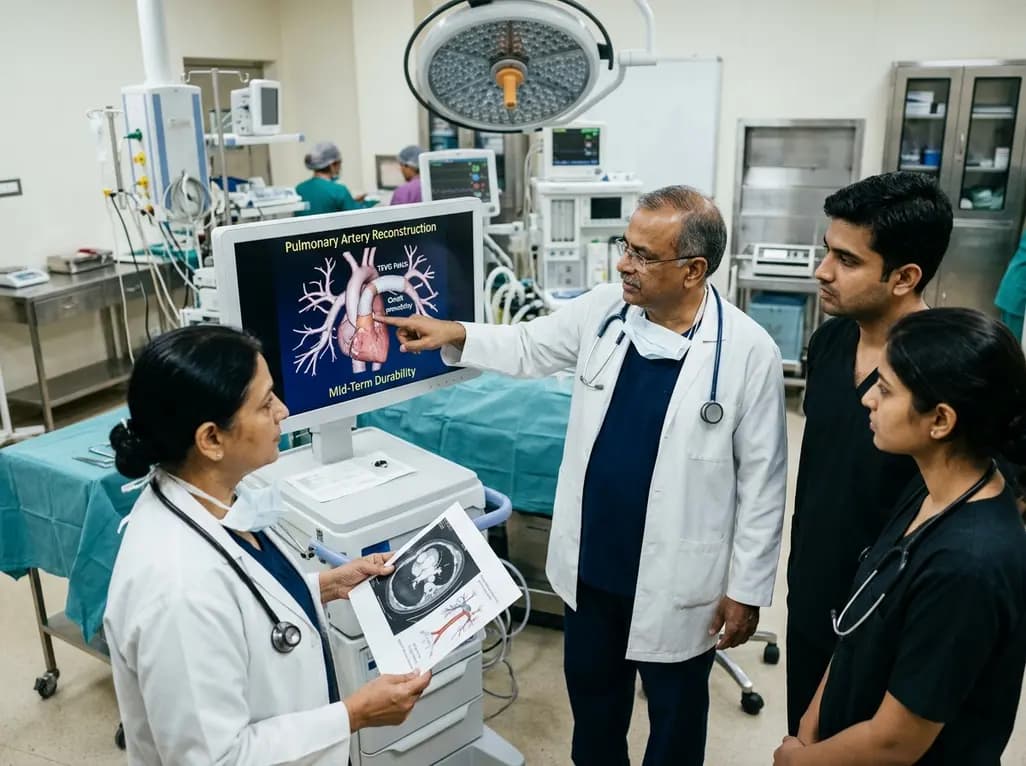
Autologous tissue-engineered vascular graft patches created via in-body tissue architecture show excellent mid-term durability and safety for pulmonary artery reconstruction in congenital heart surgery, particularly for peripheral branch augmentation.
Today

A novel cluster analysis of real-world datasets reveals four distinct CPPD disease phenotypes, validating existing EULAR categories while uncovering previously unrecognized monoarticular and axial presentations to refine patient management.
Today

Recent pharmacokinetic research demonstrates that trace dermal exposure to microgram quantities of trenbolone and metenolone produces detectable urinary metabolites for days to over a week, providing critical objective data for evaluating unintentional contamination claims in sports drug testing.
Today
