.webp&w=384&q=75)

Loading, please wait...
Loading, please wait...
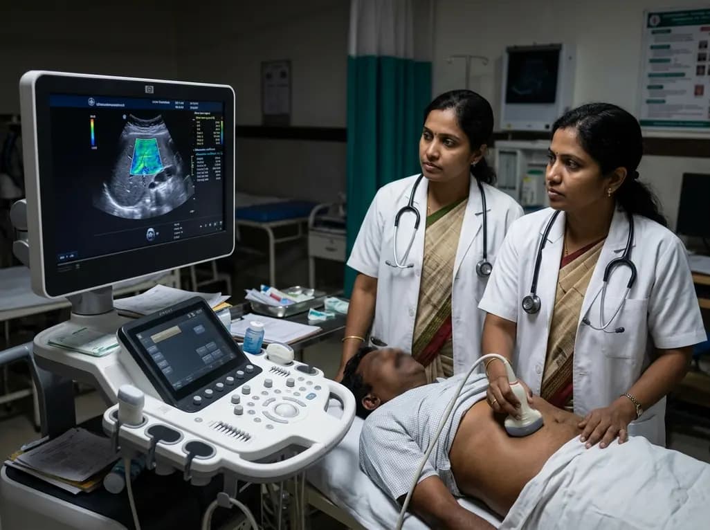
Metabolic dysfunction-associated steatotic liver disease represents a growing global health challenge. Consequently, clinicians need accurate, non-invasive diagnostic tools to evaluate hepatic parenchyma. Standard B-mode ultrasound provides qualitative assessment, but it lacks sensitivity for mild steatosis. To address these clinical limitations, quantitative ultrasound liver grading has emerged as a transformative diagnostic paradigm. This advanced methodology measures acoustic tissue properties directly, thereby eliminating operator subjectivity. Among novel techniques, acoustic energy attenuation measurements have demonstrated high diagnostic precision. Ultrasound attenuation reflects how acoustic waves lose energy while traveling through heterogeneous biological structures. When hepatocytes accumulate intracellular lipids, hepatic acoustic properties change measurably. As a result, ultrasound waves undergo frequency-dependent attenuation that correlates directly with fat concentration. Hepatologists now utilize quantitative ultrasound liver grading to grade steatosis across diverse clinical cohorts. Furthermore, this approach provides reproducible metrics that guide longitudinal patient care.
Evaluating hepatic microarchitecture requires an understanding of distinct acoustic wave physics. Compression-wave attenuation measures the decrease in amplitude of longitudinal ultrasound waves propagating through soft tissue. As longitudinal waves traverse lipid-laden hepatocytes, thermal absorption and scattering mechanisms dissipate acoustic energy. Therefore, higher degrees of macrovesicular steatosis cause greater compression-wave attenuation. In contrast, shear-wave attenuation quantifies the energy loss of transversely propagating acoustic waves. Shear waves generate transverse particle displacements, which interrogate tissue viscoelastic properties rather than bulk compressibility alone. Consequently, shear-wave attenuation captures viscous damping within the hepatic extracellular matrix. When metabolic dysfunction induces both lipid accumulation and inflammatory remodeling, tissue viscosity increases measurably. Thus, shear-wave attenuation provides complementary biophysical data that longitudinal compression waves cannot capture independently. Ultimately, integrating both attenuation modalities offers a comprehensive biophysical profile of disease progression in patients with chronic metabolic liver conditions.
A prospective clinical study investigated the diagnostic performance of compression-wave attenuation and shear-wave attenuation head-to-head. Researchers evaluated fifty-five adult participants, including thirteen healthy volunteers and forty-two patients with biopsy-proven steatotic liver disease. The investigators captured both attenuation parameters simultaneously during the same ultrasound acquisition protocol. Consequently, this design eliminated inter-scan temporal variability and ensured identical physiological conditions. The cohort encompassed the complete clinical spectrum, spanning mild steatosis to advanced steatohepatitis. Researchers compared the ultrasound metrics directly against magnetic resonance imaging proton density fat fraction and histological biopsies. Both attenuation parameters demonstrated robust diagnostic capabilities across all stages. Specifically, compression-wave attenuation and shear-wave attenuation correlated strongly with magnetic resonance imaging measurements. Furthermore, area under the receiver operating characteristic curve analyses confirmed excellent diagnostic discrimination between healthy controls and disease cohorts. Thus, head-to-head evaluation validated their complementary diagnostic roles.
Correlating non-invasive imaging biomarkers with reference standards remains essential for clinical validation. In this prospective investigation, both attenuation metrics showed high statistical correlation with proton density fat fraction measurements. Specifically, compression-wave attenuation achieved a correlation coefficient of 0.71, while shear-wave attenuation achieved 0.77. These strong correlations demonstrate that both ultrasound biomarkers reliably quantify intrahepatic fat content across varying degrees of adiposity. Moreover, multivariate analyses revealed that combining compression and shear wave parameters improved overall diagnostic accuracy. When clinicians differentiate simple steatosis from progressive steatohepatitis, histopathological grading remains the historical gold standard. However, liver biopsy carries procedural risks, sampling errors, and significant inter-observer variability. In contrast, quantitative ultrasound interrogates a substantially larger hepatic volume non-invasively. Additionally, shear-wave attenuation reflects inflammatory changes accurately. Therefore, multi-parametric ultrasound evaluation bridges the gap between conventional ultrasound screening and high-cost magnetic resonance imaging.
Implementing quantitative attenuation imaging into routine outpatient workflows provides substantial clinical advantages. Metabolic liver disease represents a silent pandemic, frequently coexisting with type 2 diabetes, obesity, and cardiovascular disorders. Consequently, primary care physicians and hepatologists need point-of-care tools to stratify patient risk rapidly. Quantitative ultrasound examinations require minimal extra scanning time during routine abdominal sonography. Additionally, quantitative metrics allow physicians to track therapeutic responses objectively during lifestyle interventions or pharmacotherapy. Because quantitative attenuation measurements yield numeric values, clinicians avoid subjective visual grading discrepancies between sonographers. Furthermore, early identification of steatohepatitis enables targeted interventions before irreversible fibrotic remodeling develops. In resource-constrained healthcare environments, quantitative ultrasound offers a cost-effective alternative to serial magnetic resonance imaging examinations. Thus, integrating compression and shear wave attenuation measurements into clinical algorithms significantly enhances risk stratification and personalized management strategies for metabolic liver disease.
Technological development continues to expand the utility of quantitative acoustic measurements in hepatology. Modern ultrasound manufacturers are currently integrating automated quality indicators to assist sonographers during real-time image acquisition. Moreover, deep learning algorithms can process raw radiofrequency data alongside attenuation metrics to refine fat quantification algorithms. Future multicenter trials will establish standardized cutoff values across diverse patient demographics and ultrasound hardware platforms. In addition, ongoing research explores how body mass index and thoracic wall thickness affect attenuation measurements. Standardizing technical protocols across vendors will facilitate widespread clinical adoption in community hospitals and specialized tertiary centers. Ultimately, combining attenuation coefficients with shear-wave elastography and dispersion imaging will create comprehensive point-of-care multiparametric liver assessments. As non-invasive diagnostic capabilities continue to evolve, quantitative acoustic imaging will play a central role in guiding metabolic disease management globally.
Compression-wave attenuation measures energy loss from longitudinal acoustic waves as they travel through liver tissue, reflecting intracellular fat accumulation. In contrast, shear-wave attenuation assesses transverse acoustic waves, capturing tissue viscoelasticity and viscous damping. While compression attenuation directly reflects volumetric fat distribution, shear-wave attenuation provides complementary insight into tissue viscosity and microstructural inflammatory alterations across various disease stages.
Magnetic resonance imaging proton density fat fraction represents the non-invasive reference standard for hepatic fat quantification. However, it remains costly, resource-intensive, and less accessible for routine screening. Quantitative ultrasound attenuation metrics correlate strongly with proton density fat fraction while offering rapid, cost-effective, point-of-care evaluation. Consequently, quantitative ultrasound enables broad population screening and frequent therapeutic monitoring in everyday clinical practice.
Metabolic steatohepatitis represents the progressive, inflammatory phenotype of metabolic liver disease that carries substantial risk for cirrhosis, liver failure, and hepatocellular carcinoma. Accurate non-invasive staging identifies patients requiring intensive lifestyle interventions or emerging pharmacotherapies. Furthermore, reliable quantitative monitoring eliminates the risks of invasive liver biopsies, facilitating safe longitudinal follow-up and timely clinical decision-making in high-risk metabolic patients.
Disclaimer: This content is for informational and educational purposes only. It is not intended to provide medical advice or to be a substitute for professional medical advice, diagnosis, or treatment. Always seek the advice of your physician or other qualified health provider with any questions you may have regarding a medical condition. Refer to the latest local and national guidelines for clinical practice.
References

Read summarized clinical updates, watch expert medical content, and earn CME certifications right from your smartphone.
Open your smartphone camera to scan and install.





Prospective study demonstrates that ultrasound compression-wave and shear-wave attenuation correlate strongly with MRI-PDFF and histopathology, offering complementary non-invasive metrics for grading metabolic dysfunction-associated steatotic liver disease.
last month
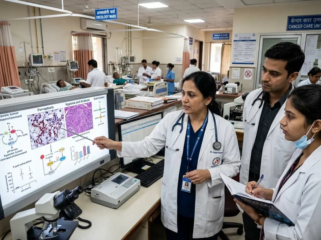
Targeting ferroptosis provides a compelling strategy to dismantle hypoxia- and acidosis-driven immunosuppression in solid tumors, enhancing response to modern immunotherapy.
Today

Findings from Project Viva indicate that sustained postpartum weight retention, rather than excessive gestational weight gain alone, directly worsens women's mid-life cardiovascular health, degrading blood pressure and glycemic parameters decades after delivery.
Today

A large UK Biobank study reveals that plasma proteomic signatures capture preclinical organ damage and improve multiorgan risk prediction across early CKM syndrome stages 0 to 2 beyond traditional PREVENT clinical models.
Today

A retrospective CBCT study reveals that superior TMJ space asymmetry correlates moderately with occipital condyle-atlas-axis asymmetry in unilateral TMJ osteoarthritis, highlighting an imaging correlate between jaw biomechanics and upper cervical alignment rather than a standalone diagnostic marker.
Today

A Bayesian multilevel meta-analysis reveals that aerobic training combined with moderate carbohydrate restriction modestly lowers HbA1c in type 2 diabetes. However, sparse data and very low certainty leave incremental benefits over exercise or diet alone unproven, highlighting the need for individualized care.
Today
