

Loading, please wait...
Loading, please wait...

Managing pediatric osteoarticular infections presents unique clinical challenges, especially in immunocompromised children and neonates. These vulnerable patients frequently exhibit attenuated systemic signs of musculoskeletal infection. Consequently, clinicians encounter diagnostic delays that risk permanent joint destruction or limb deformities. Furthermore, recent retrospective evidence emphasizes that multimodal management provides the best opportunity for clinical resolution. Understanding these complex clinical patterns empowers pediatricians, orthopedicians, and infectious disease specialists to deliver timely interventions.
Neonates and immunocompromised children possess distinct anatomical and immunological vulnerabilities that alter disease progression. Specifically, transphyseal blood vessels in neonates allow metaphysical infections to spread directly across growth plates into the adjacent joint space. Consequently, acute osteomyelitis rapidly induces concurrent septic arthritis in vulnerable infants. Moreover, children with primary or secondary immunodeficiencies cannot organize an effective cellular immune response at infected skeletal sites. In addition, immunosuppressive medications, chemotherapy, and underlying hematological disorders severely blunt local neutrophil recruitment. Therefore, destructive osseous pathology progresses silently without overt warmth or localized erythema. Clinicians must also remember that indwelling central lines and recurrent invasive procedures increase risk for hematogenous bacterial seeding. As a result, transient bacteremia easily colonizes hypervascular metaphyseal zones in growing bones. Because conventional clinical signs remain obscured, early identification demands continuous vigilance. Thus, collaborative assessment between pediatricians and orthopedic surgeons provides the safest strategy to prevent catastrophic growth arrest.
The clinical signs of bone and joint sepsis in fragile pediatric patients often deviate from classic presentations. For example, prominent systemic fevers, intense erythema, and marked localized tenderness may be completely missing. In a recent retrospective cohort, one-third of confirmed patients, predominantly neonates, remained afebrile throughout their clinical admission. Nevertheless, every single patient demonstrated reduced active limb mobility or marked pseudoparalysis during physical examination. Furthermore, localized swelling occurred in nearly seventy-eight percent of cases, offering a reliable focal finding. Healthcare workers frequently confuse newborn limb immobility with obstetric brachial palsy or minor birth trauma. Consequently, critical treatment windows close before clinicians suspect true infectious pathology. Moreover, older immunosuppressed children receiving steroids may report minimal pain despite advanced bone destruction. Clinicians must maintain a heightened index of suspicion whenever an infant resists passive motion or cries during routine diapering. Additionally, systematic physical evaluation of all four limbs remains mandatory in vulnerable infants. Identifying subtle functional deficits prevents extensive joint cartilage breakdown.
Standard hematological tests often yield deceptive results in immunocompromised pediatric cohorts. Notably, peripheral white blood cell counts and absolute neutrophil counts fluctuate unpredictably or remain completely normal. Baseline immunosuppression, myelosuppressive chemotherapy, and primary marrow failures prevent standard inflammatory leukocytosis. In striking contrast, serum C-reactive protein proves uniquely sensitive, demonstrating significant elevation across all confirmed cases in clinical series. Therefore, C-reactive protein provides an essential biochemical indicator to support clinical suspicion. Furthermore, sequential monitoring of C-reactive protein levels serves as an effective objective tool for tracking therapeutic recovery. Clinicians also evaluate erythrocyte sedimentation rate and procalcitonin, although these markers demonstrate variable kinetics in compromised hosts. In addition, physicians must never dismiss potential skeletal sepsis simply because leukocyte counts appear unremarkable. Instead, clinicians should pair elevated inflammatory markers with early imaging studies like ultrasound or magnetic resonance imaging. Prompt magnetic resonance imaging detects subperiosteal fluid collections before cortical bone erosion occurs. Accordingly, combining C-reactive protein testing with advanced imaging establishes a swift, decisive diagnostic pathway.
The microbiological spectrum in immunocompromised pediatric bone infections expands far beyond standard community pathogens. Although Staphylococcus aureus remains the primary organism, atypical pathogens cause a substantial portion of these cases. Specifically, Gram-negative bacilli like Pseudomonas aeruginosa and Stenotrophomonas maltophilia frequently infect vulnerable pediatric patients. Furthermore, clinicians increasingly encounter opportunistic fungal isolates like Candida tropicalis and atypical Mycobacteria in compromised children. Even environmental organisms such as Bacillus species can produce severe musculoskeletal damage in fragile neonates. Because opportunistic microbes resist conventional empiric beta-lactams, routine antimicrobial regimens frequently prove inadequate. Therefore, treating teams must secure operative aspirates, synovial fluid, and tissue biopsies before administering broad-spectrum antibiotics. In addition, diagnostic microbiology laboratories should utilize expanded fungal media, mycobacterial cultures, and molecular sequencing techniques. Identifying the exact offending organism enables prompt transition from empirical coverage to pathogen-directed therapy. Consequently, infectious disease consultation remains indispensable to optimize antimicrobial dosing and overcome complex multidrug resistance.
Achieving successful infection control in vulnerable pediatric hosts requires a coordinated, multimodal management strategy. Notably, surgical intervention serves as a primary pillar of definitive treatment, with over eighty percent of patients requiring operative procedures. Early arthrotomy or arthroscopic joint washout rapidly decompresses intra-articular pressure, preserving delicate epiphyseal perfusion and averting avascular necrosis. Simultaneously, surgeons thoroughly debride necrotic bone, evacuate subperiosteal abscesses, and obtain sterile operative cultures. Alongside timely surgery, targeted intravenous antimicrobial regimens eliminate systemic bacteremia and sterilize deep bony architecture. Furthermore, multidisciplinary teams must integrate immune-directed therapies whenever host defenses remain severely compromised. Specifically, modifying immunosuppressive drugs, administering intravenous immunoglobulins, or applying colony-stimulating factors accelerates tissue clearance. In addition, pediatric physical therapists initiate protected mobilization early to prevent persistent joint contractures. Despite notable biological vulnerabilities, clinical series demonstrate complete infection resolution in nearly seventy-eight percent of cases. Thus, seamless collaboration among pediatricians, orthopedic surgeons, and infectious disease specialists optimizes long-term functional recovery.
Neonates rarely exhibit classic signs like high fever or obvious local warmth. Instead, clinicians must look for reduced active limb movement, pseudoparalysis, and localized swelling. Affected infants frequently cry during diaper changes or show extreme irritability upon gentle passive joint manipulation. Therefore, any unexplained limb immobility or localized fullness in a neonate warrants immediate diagnostic evaluation for osteoarticular infection, even if the infant remains completely afebrile and clinically stable.
Immunocompromised children and neonates often cannot mount a robust leukocytosis due to immature bone marrow reserves, chemotherapy, or primary immunodeficiencies. Consequently, total white cell and absolute neutrophil counts often stay misleadingly normal. In contrast, hepatocytes continue synthesizing C-reactive protein in response to interleukin-6 release during tissue invasion. This acute-phase reactant demonstrates superior sensitivity, consistently rising in active skeletal infections and providing clinicians with a dependable biochemical marker to guide therapy.
Surgeons must intervene urgently when imaging demonstrates joint effusion, subperiosteal abscesses, or failure of medical therapy after forty-eight hours. Prompt surgical drainage decompresses elevated intra-articular pressure, which rapidly compromises local vascular supply and causes permanent chondral destruction. In addition, surgical exploration yields vital microbiological specimens, allowing targeted antibiotic selection against fastidious pathogens. Therefore, early operative debridement paired with targeted medical treatment remains an indispensable component of successful multimodal infection control.
Disclaimer: This content is for informational and educational purposes only and should not be taken as professional medical advice. Refer to the latest local and national guidelines for clinical practice.
References

Read summarized clinical updates, watch expert medical content, and earn CME certifications right from your smartphone.
Open your smartphone camera to scan and install.





A retrospective study evaluates pediatric osteoarticular infections in immunocompromised children and neonates, emphasizing diagnostic challenges, elevated CRP, diverse microbiology, and the necessity of multimodal surgical and medical management to achieve favorable functional outcomes.
Today

Microdosing, fractional dosing, and split dosing of GLP-1 receptor agonists have emerged as popular off-label strategies to manage gastrointestinal side effects. However, these methods carry risks of dosing inaccuracy, peptide degradation, and lack regulatory endorsement.
Today
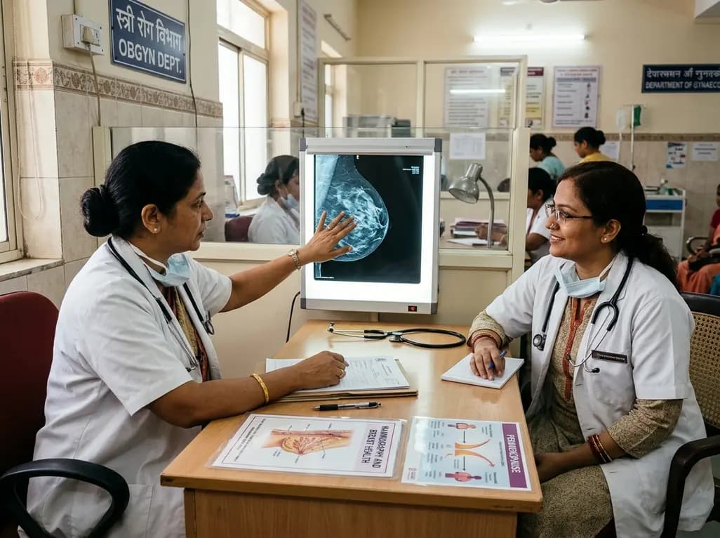
Perimenopause brings erratic hormonal surges and metabolic shifts that significantly affect breast tissue. This comprehensive clinical review examines benign breast conditions, cancer risk stratification, mammographic density challenges, and endocrine therapy transitions in midlife women.
Today

The VOCAL-AHF study reveals that most acute heart failure patients across reduced and non-reduced ejection fraction categories are eligible for in-hospital GDMT initiation. Notably, nearly two-thirds of HFrEF patients qualify for quadruple therapy, while non-reduced EF patients show high eligibility for dual therapy.
Today

A novel fluorine-functionalized near-infrared copper fluorescent probe selectively illuminates lung tissue, revealing critical links between hyperglycemia, ER stress, and pulmonary malignancy for improved early diagnosis.
Yesterday
