.webp&w=384&q=75)

Loading, please wait...
Loading, please wait...
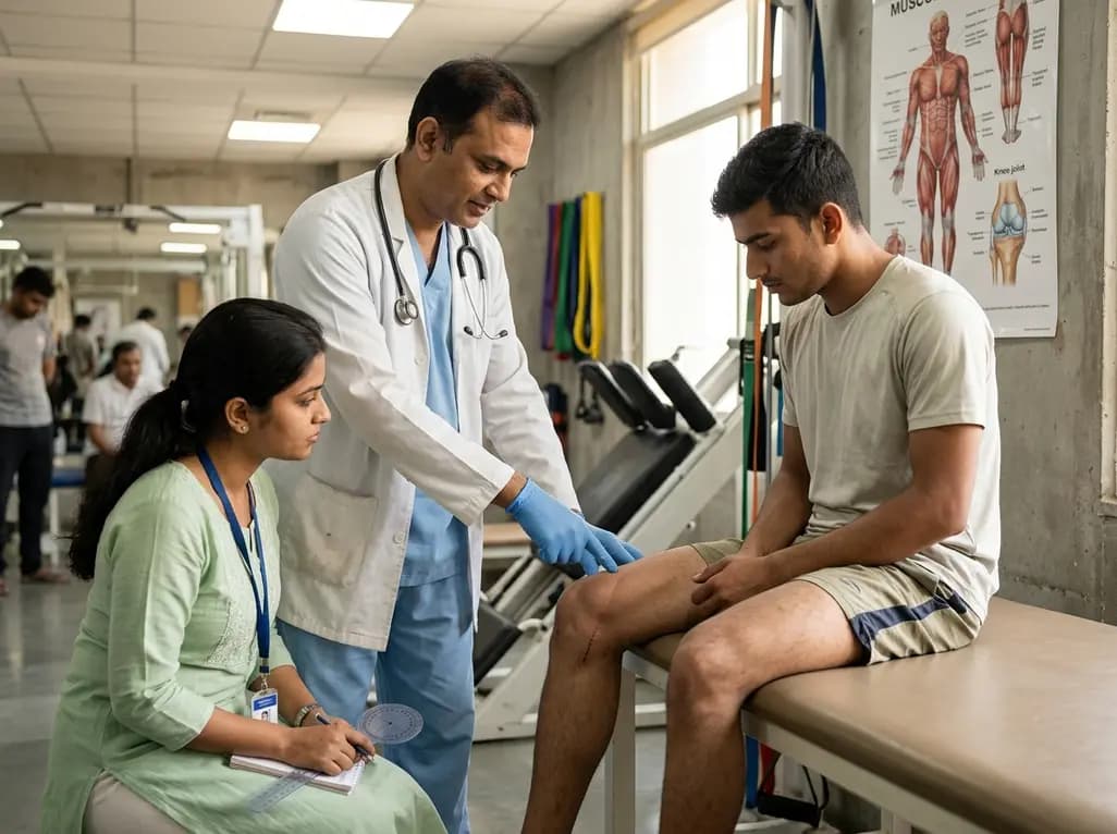
Persistent quadriceps muscle weakness and shrinkage remain substantial clinical challenges following joint trauma. Specifically, quadriceps atrophy after ACL injury delays recovery and increases the risk of reinjury among active individuals. For decades, clinicians debate whether whole-muscle wasting stems from true muscle fibre loss or a reduction in individual fibre dimensions. Furthermore, specialists frequently question whether knee joint trauma triggers peripheral neuromuscular junction degradation. Recent structural analyses provide definitive evidence regarding how anterior cruciate ligament trauma impacts skeletal muscle morphology. Histological evaluations demonstrate that whole-muscle wasting occurs due to rapid reductions in individual myofibre cross-sectional area rather than the loss of overall myofibre count. Consequently, understanding these cellular mechanisms provides valuable direction for designing postoperative exercise regimens.
Whole-muscle histological assessments reveal crucial facts about structural remodelling in the vastus lateralis. Following joint trauma, individual fibre cross-sectional area drops significantly within seven days across both sexes. Moreover, this cellular shrinkage persists during subsequent recovery intervals. Importantly, the total number of skeletal muscle fibres remains completely preserved throughout the acute post-injury timeline. Therefore, whole-muscle diameter reduction reflects individual myofibre atrophy rather than cell death or irreversible hypoplasia. This distinction carries immense therapeutic value for orthopedic specialists and physical therapists. Because the baseline architecture of the muscle remains structurally intact, muscle mass is fully capable of recovery when stimulated by targeted hypertrophic interventions.
Clinical researchers historically hypothesized that early muscle wasting might stem from peripheral denervation at the neuromuscular junction. However, electrophysiological and morphological studies show that compound muscle action potentials remain stable after acute anterior cruciate ligament tears. Furthermore, histological analysis of vastus lateralis motor endplates confirms that neuromuscular junctions maintain complete structural and functional integrity. Motor nerve terminals remain firmly attached to their post-synaptic receptors without evidence of axon degeneration. Consequently, muscle shrinkage is not preceded by overt peripheral denervation. Instead, central inhibitory pathways and local arthrogenic reflexes decrease voluntary activation. Thus, muscle wasting develops downstream of altered neural drive rather than primary terminal nerve destruction.
Joint trauma also drives notable shifts in skeletal muscle metabolic phenotype. Specifically, all Type II fast-twitch subtypes undergo rapid shrinkage after joint disruption. Additionally, the vastus lateralis displays a marked phenotypic transition toward Type IIb glycolytic fibres. This structural shift closely mirrors the physiological adaptations observed in classic models of limb unloading and joint disuse. Electromyographic evaluations during locomotion confirm reduced muscle firing amplitude and shortened stance durations on the injured extremity. Consequently, decreased mechanical loading accelerates fast-twitch fibre shrinkage and reduces overall force production capacity. Because Type II fibres generate rapid joint-stabilizing torque, reversing this glycolytic disuse shift represents a primary rehabilitation objective.
The primary barrier to restoring muscle volume is arthrogenic muscle inhibition. After an acute joint injury, intra-articular effusion, pain, and mechanoreceptor disruption trigger a reflexive shutdown of quadriceps alpha motor neurons. Consequently, patients cannot volitionally recruit high-threshold motor units during standard resistance training. Because the neuromuscular junction remains structurally sound, clinicians can employ adjunctive neuromuscular electrical stimulation to bypass central inhibition. Furthermore, blood flow restriction training creates localized metabolic stress, which stimulates muscle protein synthesis without excessive joint loading. In addition, implementing focal joint cryotherapy before exercise dampens inhibitory spinal reflex arcs. Therefore, multimodal strategies help restore quadriceps activation and reverse myofibre shrinkage.
Current post-injury rehabilitation regimens often fall short of completely resolving muscle deficits before return-to-sport clearance. Because quadriceps atrophy after ACL injury is driven by myofibre shrinkage rather than cell death, targeted hypertrophic exercise is highly viable. Clinicians should incorporate progressive overload protocols that specifically challenge Type II fast-twitch fibres. Moreover, combining eccentric loading with early neuromuscular activation restores force-generating capacity and rate of torque development. Addressing bilateral limb asymmetries early prevents long-term compensatory movement patterns. Ultimately, structured preoperative and postoperative therapy programs that focus on myofibre hypertrophy can accelerate functional recovery and lower the risk of secondary knee osteoarthritis.
No, ACL injury does not cause permanent muscle fibre loss. Research demonstrates that total myofibre number remains fully preserved in the vastus lateralis after trauma. Instead, whole-muscle atrophy results from a decrease in individual muscle fibre cross-sectional area. Because the original fibres remain present, muscle size can be restored with targeted rehabilitation.
Quadriceps weakness occurs primarily due to arthrogenic muscle inhibition rather than peripheral nerve damage. Intra-articular swelling, pain, and altered sensory signals trigger reflex pathways in the spinal cord that suppress alpha motor neuron excitability. Consequently, the brain cannot fully activate the quadriceps muscle, leading to rapid disuse-mediated fibre atrophy.
Effective modalities include neuromuscular electrical stimulation, blood flow restriction training, and progressive resistance exercise. Electrical stimulation directly depolarizes motor axons to overcome central inhibition, while blood flow restriction promotes muscle hypertrophy using lighter joint-friendly loads. Combining these tools with early cryotherapy helps restore quadriceps volume and dynamic joint stability.
Disclaimer: This content is for informational and educational purposes only. It is not intended to be a substitute for professional medical advice, diagnosis, or treatment. Always seek the advice of your physician or other qualified health provider with any questions you may have regarding a medical condition. Refer to the latest local and national guidelines for clinical practice.
References
Stoneback L et al. Structural determinants of quadriceps atrophy following ACL injury: Evidence for fibre atrophy without fibre loss or overt peripheral denervation. J Physiol. 2026 Aug 23. doi: 10.1113/JP291436. PMID: 42633494.
Graham MC, Thompson KL, Hawk GS, Fry CS, Noehren B. Muscle fiber cross-sectional area is associated with quadriceps strength and rate of torque development after ACL injury. J Strength Cond Res. 2024;38(6):e273-e279.
Rice DA, McNair PJ. Quadriceps arthrogenic muscle inhibition: neural mechanisms and treatment perspectives. Semin Arthritis Rheum. 2010;40(3):250-266.

Read summarized clinical updates, watch expert medical content, and earn CME certifications right from your smartphone.
Open your smartphone camera to scan and install.





New evidence reveals that quadriceps atrophy after ACL injury is driven by decreased muscle fibre cross-sectional area rather than permanent fibre loss or denervation, opening avenues for targeted rehabilitation.
last month

A systematic review of 41 preclinical studies reveals that baicalin dampens systemic inflammation, inhibits NF-κB and NLRP3 pathways, and improves organ preservation in sepsis models. However, substantial study heterogeneity and a complete lack of human clinical trials highlight the need for further translational research.
Today
Discover the crucial distinctions between mesonephric and mesonephric-like proliferations of the female genital tract. Learn the key histomorphologic, immunohistochemical, and molecular differences essential for accurate diagnosis and clinical management.
Today

A large UK Biobank study reveals that plasma proteomic signatures capture preclinical organ damage and improve multiorgan risk prediction across early CKM syndrome stages 0 to 2 beyond traditional PREVENT clinical models.
Today
A narrative review investigates whether the articularis genus muscle functions as an independent anatomical entity or blends with the vastus intermedius. We evaluate its morphology, role in retracting the suprapatellar bursa, and direct clinical significance in anterior knee pain and arthroplasty.
Today

A Bayesian multilevel meta-analysis reveals that aerobic training combined with moderate carbohydrate restriction modestly lowers HbA1c in type 2 diabetes. However, sparse data and very low certainty leave incremental benefits over exercise or diet alone unproven, highlighting the need for individualized care.
Today
