.webp&w=384&q=75)

Loading, please wait...
Loading, please wait...

Pulmonary Arteriovenous Malformations (PAVMs) are rare but critical vascular anomalies that create direct shunts between pulmonary arteries and veins. These shunts bypass the capillary bed's filtration system, allowing thrombi to enter the systemic circulation. Consequently, this mechanism often leads to a pulmonary arteriovenous malformation stroke, a serious complication that clinicians must recognize early to prevent permanent disability. Furthermore, the absence of specific biomarkers makes diagnosis challenging, often requiring advanced imaging for confirmation.
A 67-year-old male with no prior medical history recently presented with recurrent cerebral embolism within three months. This occurred despite therapeutic anticoagulation. Therefore, doctors performed a contrast-enhanced chest CT, which revealed an underlying PAVM. Because the patient had no other risk factors, the medical team attributed the paradoxical embolism to this shunt. Specifically, the shunt allowed venous clots to bypass the lungs and enter the cerebral vasculature, bypassing the body's natural pulmonary filter.
Interventional radiology remains the gold standard for managing symptomatic PAVMs. Percutaneous transcatheter embolization involves using coils or vascular plugs to occlude the feeding artery effectively. Moreover, the procedure demonstrates high efficacy with minimal invasiveness compared to surgical alternatives. In this specific case, the patient underwent successful embolization and remained symptom-free at a 4-year follow-up. Additionally, timely intervention prevents recurrent embolic events by closing the direct communication between the pulmonary and systemic circuits. However, physicians must carefully assess the risk of hemorrhagic transformation during the post-procedural period.
Ultimately, this case underscores the importance of considering PAVMs in unexplained cerebral embolism cases. Because diagnosis is often delayed, maintaining a high index of suspicion is vital. Furthermore, embolization provides a definitive solution that anticoagulation alone cannot achieve in these patients. Early identification and transcatheter management are essential to improve patient outcomes and prevent disabling neurologic events.
Contrast-enhanced chest computed tomography (CT) and pulmonary angiography are the primary tools for diagnosing and mapping the angioarchitecture of PAVMs. These modalities help identify the feeding artery size, which guides treatment decisions.
PAVMs create a right-to-left shunt that bypasses the lungs' capillary filter. Consequently, paradoxical emboli—such as blood clots, air, or bacteria—travel directly from the venous system to the brain, bypassing the lung’s natural filtration mechanism.
Disclaimer: This content is for informational and educational purposes only. It does not constitute professional medical advice, diagnosis, or treatment. Always seek the advice of your physician or other qualified healthcare provider with any questions you may have regarding a medical condition. Refer to the latest local and national guidelines for clinical practice.
References
Qin X et al. Percutaneous Transcatheter Embolization for Recurrent Cerebral Embolism Due to Pulmonary Arteriovenous Malformation. Am J Case Rep. 2026 Jun 07. doi: 10.12659/AJCR.952379. PMID: 42251485.
Ribeiro RP, et al. Recurrent Stroke Due to Pulmonary Arteriovenous Malformation: Diagnostic Challenges and Management. Cureus. 2025 Dec 3;17(12):e98393.
Jiang X, et al. Pulmonary multifocal arteriovenous malformations lead to ischemic stroke in young adults: a case report and literature review. Cardiovasc Diagn Ther. 2024.

Read summarized clinical updates, watch expert medical content, and earn CME certifications right from your smartphone.
Open your smartphone camera to scan and install.





Expert review of a case where percutaneous transcatheter embolization successfully treated recurrent cerebral embolism caused by an undiagnosed PAVM....
4 months ago

A retrospective cohort study reveals that frailty quadruples the odds of pressure injuries in hospitalized older adults. Integrating the Clinical Frailty Scale with comprehensive geriatric care is crucial for identifying vulnerability early and implementing targeted multidisciplinary prevention.
Today
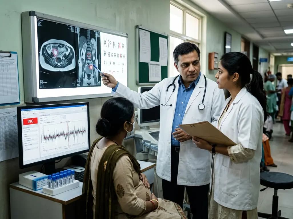
Occult maternal malignancy accounts for 3.82% of persistent unreportable NIPT results with genomic noise. Multiple chromosomal aberrations, notably on chromosome 18, warrant thorough oncological evaluation and whole-body MRI to ensure timely maternal diagnosis while preserving fetal care.
Today

This article reviews the forensic and clinical complexities of acute myocarditis arising on left ventricular hypertrophy in children, highlighting early diagnostic red flags and multidisciplinary postmortem autopsy protocols.
Today
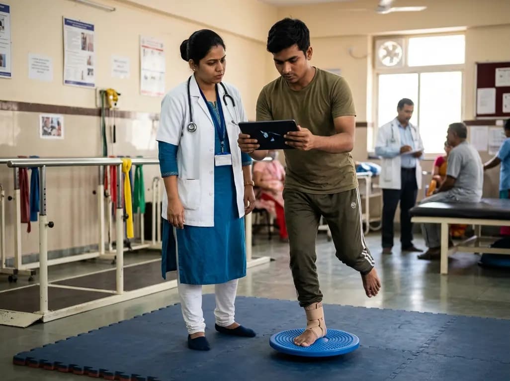
A randomized controlled trial demonstrates that an eight-week dual-task training intervention significantly improves static and dynamic postural control over standard balance exercises in individuals with chronic ankle instability, addressing critical neurocognitive deficits during athletic and daily activities.
Today
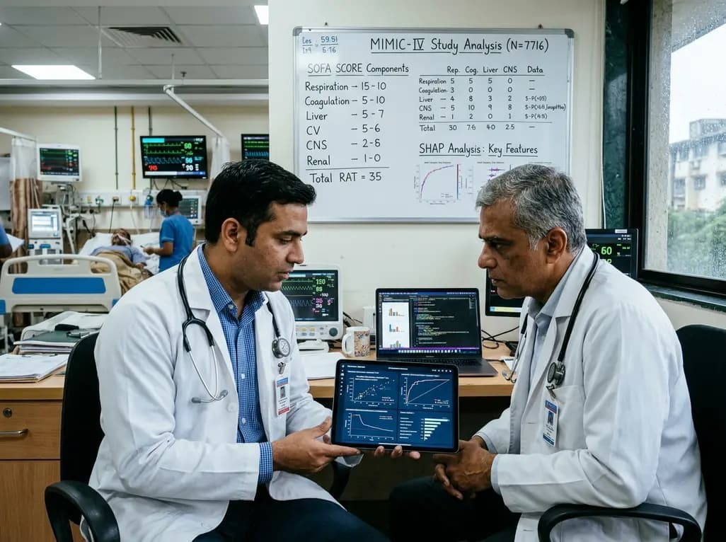
A retrospective cohort study of 7,716 ICU patients from the MIMIC-IV database demonstrates that Gradient Boosting Decision Trees significantly outperform traditional SOFA scores in predicting in-hospital mortality among patients with sepsis and diabetes, with SHAP analysis providing clinical interpretability.
Today
