

Loading, please wait...
Loading, please wait...

Mechanical thrombectomy represents the established standard of care for patients who experience acute ischemic stroke secondary to anterior circulation large vessel occlusion. Clinicians routinely evaluate neuroimaging to assess core infarct size, tissue viability, and collateral flow before intervention. However, subtle baseline parenchymal changes often pose diagnostic dilemmas for acute stroke teams. In particular, the presence of prethrombectomy hemorrhagic infarction on sensitive MRI sequences raises crucial questions regarding tissue vulnerability. Historically, clinicians regarded petechial hemorrhagic transformation as an incidental or benign phenomenon. Recent multicenter evidence challenges this conventional paradigm by highlighting its profound prognostic implications.
Emergency clinicians frequently detect subtle petechial bleeding within ischemic brain parenchyma during acute stroke evaluations. Radiologists classify hemorrhagic transformation into petechial hemorrhagic infarction and discrete parenchymal hematoma using validated European Cooperative Acute Stroke Study criteria. Petechial changes, termed HI type 1 and type 2, historically carried a reputation as benign epiphenomena of spontaneous collateral reperfusion. In contrast, hematomas with significant space-occupying mass effect clearly drive secondary neurological decline.
Nevertheless, modern imaging protocols utilizing gradient-recalled echo or susceptibility-weighted imaging detect early petechiae with unprecedented sensitivity. When prethrombectomy hemorrhagic infarction appears before endovascular recanalization, it signals substantial microvascular compromise rather than benign tissue viability. Consequently, treating teams must look beyond simple core volume measurements. Identifying subtle petechiae before groin puncture helps clinicians anticipate procedural risks more effectively. While these petechial findings do not exclude patients from undergoing life-saving revascularization, they establish an essential baseline of tissue fragility. Thus, recognizing early hemorrhagic transformation allows acute stroke physicians to temper prognostic expectations and prepare for intensive post-procedural vigilance.
Understanding the pathophysiological cascade of hemorrhagic transformation explains why early petechial changes carry serious prognostic weight. Ischemia triggers rapid failure of endothelial ion pumps, cellular swelling, and progressive degradation of the vascular basal lamina. In addition, matrix metalloproteinases rapidly break down extracellular matrix proteins that normally reinforce capillary walls. Consequently, severe disruption of the blood-brain barrier occurs even before therapeutic recanalization takes place.
Furthermore, restored arterial flow delivers high hydrostatic pressures into structurally compromised downstream capillary beds. When interventionalists successfully retrieve the occluding thrombus, this rapid reperfusion exposes fragile microvessels to shear stress. If subtle petechiae already exist before procedure initiation, the microvasculature has already failed at multiple microscopic sites. Therefore, sudden recanalization may accelerate blood extravasation and convert microscopic petechiae into destructive confluent hematomas. In addition, reperfusion prompts intense neuroinflammation, leukocyte infiltration, and free radical generation. These destructive downstream processes amplify localized tissue injury within ischemic territories. Thus, pretreatment petechial hemorrhage reflects a primed biological state of severe reperfusion vulnerability rather than harmless collateral leakage.
A landmark multicenter retrospective study recently evaluated 479 consecutive acute ischemic stroke patients undergoing mechanical thrombectomy across academic stroke centers. All enrolled participants received standardized pretreatment T2*-weighted magnetic resonance imaging before endovascular groin puncture. Investigators identified pretreatment petechial hemorrhagic infarction in 5.2% of the overall cohort, demonstrating exceptional inter-rater diagnostic consistency.
Importantly, multivariable analyses revealed that baseline hemorrhagic infarction independently predicted adverse clinical outcomes. Patients displaying pretreatment petechial hemorrhage experienced a sixfold higher adjusted odds of 90-day functional dependence compared to those without baseline hemorrhage. Both frequentist and Bayesian statistical frameworks confirmed this robust association after adjusting for age, initial stroke severity, comorbidities, and baseline infarct volume. Moreover, the presence of baseline petechiae demonstrated strong posterior probabilities of association with post-procedural parenchymal hematoma and symptomatic intracerebral hemorrhage. Specifically, the frequentist analysis showed an adjusted odds ratio of 6.02 for developing symptomatic hemorrhage after thrombectomy. Consequently, these statistical findings provide compelling empirical evidence that early petechial bleeding identifies patients at high risk for catastrophic post-recanalization transformation.
Interventional neurologists and neurosurgeons must interpret these findings carefully without withholding indicated endovascular therapy. Current international guidelines strongly support prompt mechanical thrombectomy for eligible large vessel occlusions. Persistent arterial occlusion almost invariably causes severe permanent disability or death. Therefore, identifying prethrombectomy petechial bleeding should never serve as an automatic contraindication to reperfusion therapy.
Instead, neurointerventional teams should incorporate this radiographic finding into comprehensive procedural risk stratification models. When interventionalists identify pretreatment petechial hemorrhage on MRI, they can adjust intraprocedural techniques and anticipate post-recanalization management challenges. For instance, operators might exercise heightened caution during device passes to avoid unnecessary vessel trauma. Additionally, clinicians should re-evaluate the risk-benefit balance of adjunctive intra-arterial thrombolytics or aggressive glycoprotein IIb/IIIa inhibitors during the intervention. Although chemical thrombolysis can improve microvascular clearing, it may exacerbate bleeding in patients with compromised basal membranes. Thus, detecting early petechiae guides thoughtful therapeutic restraint regarding secondary pharmacotherapy while maintaining rapid primary mechanical recanalization efforts.
Optimal post-thrombectomy critical care plays a paramount role in protecting patients with baseline hemorrhagic infarction. Following successful recanalization, maintaining strict hemodynamic control directly prevents excessive hydrostatic perfusion into damaged brain parenchyma. Contemporary clinical trials suggest that intensive blood pressure targets prevent hematoma expansion after endovascular therapy. Consequently, neurocritical care teams must implement precise continuous arterial line monitoring.
Furthermore, clinicians should avoid permissive hypertension once vessel patency is achieved, particularly among individuals with confirmed baseline microvascular breakdown. Titrating intravenous antihypertensive agents helps stabilize capillary perfusion pressure while avoiding catastrophic parenchymal hematoma formation. In addition, critical care teams should obtain urgent repeat neuroimaging within 24 hours to detect early parenchymal hematoma evolution. If severe hemorrhagic transformation occurs, clinicians must promptly correct coagulopathies and maintain close neurosurgical consultation. Similarly, managing glycemic levels, hyperthermia, and systemic oxygenation prevents secondary neurovascular degradation. Therefore, proactive post-procedural protocols provide an indispensable defense against clinical deterioration in these vulnerable patients.
The recognition of pretreatment petechiae as a marker of reperfusion vulnerability opens promising avenues for translational stroke research. Historically, clinical trials evaluating neuroprotective agents struggled to demonstrate efficacy in heterogeneous stroke populations. However, stratifying participants by baseline blood-brain barrier disruption may identify a subgroup that derives maximal benefit from targeted vascular stabilization.
Novel therapeutic candidates aiming to inhibit matrix metalloproteinases, scavenge free radicals, or reinforce endothelial junctions could prevent reperfusion injury. If future clinical trials specifically enroll patients exhibiting prethrombectomy microvascular breakdown, researchers might observe pronounced treatment effects. Furthermore, advanced artificial intelligence algorithms may improve the automated detection of subtle petechiae on acute gradient echo sequences. Integrating automated lesion quantification into emergency workflows will accelerate real-time clinical decisions. Ultimately, evolving from binary treatment paradigms toward personalized neurovascular profiling will enhance patient safety across comprehensive stroke systems.
No, detecting pretreatment petechial hemorrhage does not constitute a contraindication to mechanical thrombectomy. Large vessel occlusions carry devastating morbidity and mortality if left untreated, and mechanical recanalization remains the most effective proven therapy. However, identifying early petechiae alerts clinicians to heightened microvascular fragility. Consequently, stroke teams should proceed with thrombectomy while avoiding unnecessary adjunctive antithrombotics and instituting rigorous post-procedural blood pressure control to prevent catastrophic secondary parenchymal hematomas.
Hemorrhagic infarction consists of small, patchy petechial hemorrhages confined within infarcted tissue without generating significant mass effect or structural midline shift. In contrast, parenchymal hematoma represents a confluent, dense blood collection that exerts substantial mass effect, compresses surrounding brain structures, and markedly increases intracranial pressure. While petechiae indicate underlying blood-brain barrier damage, confluent hematomas cause direct mechanical tissue destruction and drive severe early neurological deterioration in acute stroke patients.
Gradient-recalled echo and susceptibility-weighted MRI sequences exhibit extraordinary sensitivity to magnetic field distortions caused by paramagnetic blood degradation products, particularly deoxyhemoglobin and hemosiderin. Non-contrast head CT frequently misses microscopic petechial hemorrhages because subtle blood extravasation blends into surrounding hypodense tissue edema. Consequently, sensitive baseline MRI protocols reliably visualize early microvascular leakage and subtle petechiae hours before conventional computed tomography can identify visible hemorrhagic transformation in evolving ischemic stroke territories.
Disclaimer: This content is for informational and educational purposes only. It is not intended to be a substitute for professional medical advice, diagnosis, or treatment. Always seek the advice of your physician or another qualified health provider with any questions you may have regarding a medical condition. Refer to the latest local and national guidelines for clinical practice.
References

Read summarized clinical updates, watch expert medical content, and earn CME certifications right from your smartphone.
Open your smartphone camera to scan and install.





A multicenter cohort study shows that pretreatment hemorrhagic infarction detected on baseline MRI significantly increases the risk of 90-day functional dependence and severe hemorrhagic transformation following mechanical thrombectomy for acute large vessel occlusion stroke.
Today
A large retrospective cohort study demonstrates that explainable machine learning models, notably XGBoost, accurately predict hospital admission at emergency triage. By analyzing routine triage parameters, hospitals can streamline bed allocation, decrease boarding times, and alleviate acute care overcrowding.
Today

A new quality-improvement initiative reveals that universal maternal mental health screening in the NICU increased screening rates from 29.5% to 95.0%, with nearly 60% of screened mothers requiring psychosocial referrals, highlighting the urgent need for embedded mental health resources.
Today
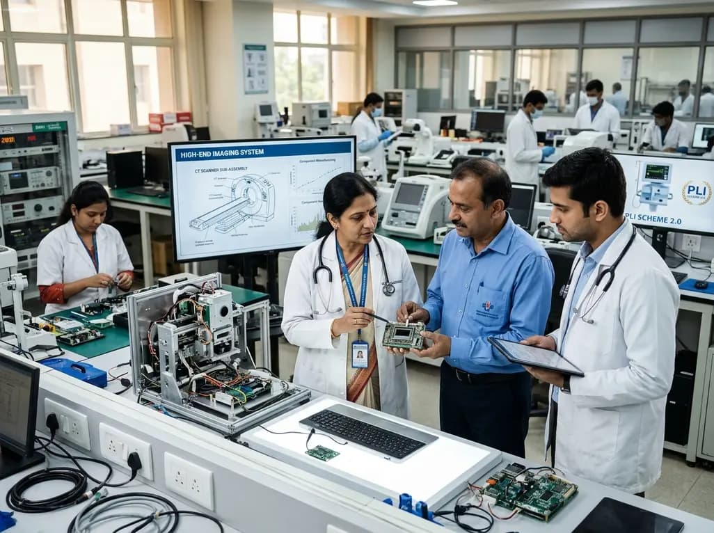
The Department of Pharmaceuticals has extended the medical devices PLI scheme to 2035-36 with a Rs 2,700 crore outlay. Featuring relaxed investment barriers and eliminated sales thresholds, the revised framework targets deep manufacturing, high-end diagnostics, critical MedTech components, and supply resilience.
Today
A metabolomic study reveals that aging delays glutathione synthesis and gamma-glutamylcysteine accumulation following muscle injury. Although precursor supplementation targets this enzymatic bottleneck, in vitro tests show no immediate effect on myogenesis, highlighting the complexity of muscle repair in older adults.
Today

A systematic review evaluates psychological interventions for smoking cessation in diabetes, showing short-term success but highlighting challenges in long-term abstinence.
Today
