

Loading, please wait...
Loading, please wait...

Accurately determining brain aneurysm rupture risk remains one of the most difficult challenges in modern neurovascular practice. Intracranial aneurysms affect roughly three percent of the general population worldwide. Although subarachnoid hemorrhage causes severe morbidity, prophylactic interventions carry distinct procedural risks. Consequently, clinicians must balance treatment hazards against natural history rupture rates. Traditional clinical scores provide baseline estimates. However, these scoring rubrics often fail to capture individual aneurysm biology. Recent advances in high-resolution magnetic resonance imaging and machine learning offer promising solutions. By evaluating vessel wall enhancement and subtle tissue textures, advanced predictive models transform clinical decision-making.
For decades, neurosurgeons have relied on demographic parameters and luminal dimensions to guide clinical care. The PHASES score serves as the primary tool for estimating rupture likelihood. This clinical metric incorporates patient population, hypertension, age, aneurysm size, prior subarachnoid hemorrhage, and aneurysm site. Nevertheless, clinical scoring systems display notable limitations in real-world practice. For instance, many subarachnoid hemorrhages arise from aneurysms smaller than seven millimeters. These diminutive lesions often carry deceptively low PHASES scores. In a prospective study by Sagues and colleagues, the standalone clinical model achieved an area under the curve of only 0.62. Furthermore, it demonstrated a modest 70% accuracy, 44% sensitivity, and 79% specificity. Therefore, conventional scores clearly lack the sensitivity required to detect every unstable lesion. Morphological metrics help address this clinical deficit. Incorporating geometric parameters like size ratio significantly improves diagnostic performance. Specifically, combining PHASES with size ratio increased the area under the curve to 0.79. While geometric parameters reveal flow disturbances, they treat the arterial wall as an inert vessel. True clinical precision requires direct insight into the biological vitality of the aneurysm wall itself.
High-resolution magnetic resonance vessel wall imaging has emerged as a transformative modality in neurovascular evaluation. Unlike digital subtraction angiography, vessel wall MRI directly visualizes the structural layers of the arterial pouch. Aneurysm wall enhancement following intravenous gadolinium serves as a valuable surrogate marker for tissue inflammation. Histopathological analyses demonstrate that wall enhancement correlates with macrophage infiltration, neovascularization, and mural degradation. When hemodynamic stress triggers endothelial injury, inflammatory cascades induce proteolytic destruction of elastin and collagen. Consequently, the aneurysmal dome thins, predisposing the structural pouch to rupture. In the cohort evaluated by Sagues and co-authors, incorporating 3D aneurysm wall enhancement metrics elevated predictive performance to an area under the curve of 0.82. Additionally, this enhanced model achieved an impressive 81% accuracy and 86% specificity in detecting symptomatic presentation. Wall enhancement reflects an active, unstable pathological process rather than static morphology. Moreover, circumferential enhancement demonstrates a much higher instability risk than focal enhancement. Therefore, high-resolution vessel wall imaging provides functional biological insights that help clinicians identify rupture-prone lesions with greater confidence. Furthermore, non-contrast sequences often fail to differentiate quiescent lesions from inflamed pouches nearing structural exhaustion. By tracking contrast retention across three-dimensional maps, neurovascular specialists gain an objective readout of localized tissue stress.
Multivariable risk modeling allows clinicians to evaluate the incremental diagnostic yield of complementary imaging biomarkers. In their clinical investigation, the researchers systematically tested four hierarchical tiers of predictive models. First, the baseline clinical tier evaluated PHASES variables alone, confirming that clinical history offers limited predictive capability. Subsequently, the second model introduced morphological complexity, specifically the ratio of aneurysm height to parent vessel diameter. This morphological integration yielded an immediate improvement in diagnostic sensitivity, rising from 44% to 77%. Next, the third tier incorporated quantified three-dimensional aneurysm wall enhancement. Consequently, this addition notably strengthened diagnostic specificity to 86%, while achieving an overall accuracy of 81%. As a result, these sequential improvements demonstrate that structural morphology and mural inflammation deliver non-redundant diagnostic information. Morphology reflects chronic hemodynamic shear stress, whereas contrast enhancement captures active biological breakdown. In addition, symptomatic presentation strongly correlates with inflammatory remodeling within the wall matrix. Patients experiencing sentinel headaches or cranial neuropathies frequently display marked wall enhancement on magnetic resonance imaging. Thus, integrating geometric parameters with contrast-enhanced imaging creates a robust, highly reliable decision-making framework. Moreover, combining these parameters mitigates false-positive findings associated with non-inflammatory wall thickening. Clinicians can thereby distinguish between benign vascular remodeling and high-risk aneurysmal expansion.
Radiomics represents the latest frontier in quantitative neurovascular analysis. This computational technique extracts hundreds of mathematical features from medical images, exposing spatial pixel heterogeneity invisible to the naked human eye. In the study by Sagues and colleagues, the best-performing model combined patient age with wall radiomic features. Remarkably, this advanced computational model achieved an area under the curve of 0.87. Furthermore, it demonstrated 76% accuracy, 88% sensitivity, and 72% specificity in identifying unstable aneurysms. Radiomics quantifies microstructural tissue variations, signal gradients, and complex textural disorganization within the aneurysm wall. Consequently, these computational signatures identify vulnerable lesions before gross morphological expansion occurs. Furthermore, radiomic textures reflect advanced biological degradation, including intramural microthrombosis and inflammatory cellular density. By automating high-dimensional feature extraction, radiomic algorithms minimize the inter-observer variability common in manual assessments. Machine learning models synthesize these multidimensional patterns into reliable risk classifications. Therefore, integrating radiomics into neuroimaging analysis provides unmatched diagnostic sensitivity, ensuring clinicians promptly detect highly fragile aneurysms. In contrast to subjective visual inspection, quantitative texture analysis detects subtle intra-wall contrast variations. Consequently, this algorithmic assessment standardizes vulnerability scoring across diverse clinical presentations. Clinicians can thus identify high-risk lesions that appear deceptively unremarkable on standard angiograms.
Translating multimodal prediction models into daily practice will significantly refine neurointerventional triage. Currently, clinicians often rely on maximum aneurysm diameter to justify preventive coiling or microsurgical clipping. However, relying strictly on size creates a notable clinical paradox. While large aneurysms carry higher relative rupture rates, most subarachnoid hemorrhages occur from smaller lesions. High-resolution vessel wall imaging directly resolves this clinical dilemma. Specifically, identifying wall enhancement or aggressive radiomic signatures helps justify proactive treatment in small, high-risk aneurysms. Conversely, documenting wall stability can reassure patients with low-risk lesions, avoiding costly procedural complications. In addition, vessel wall imaging clarifies which aneurysm has destabilized in patients with multiple intracranial lesions. Nevertheless, broad adoption requires standardizing imaging protocols and feature extraction pipelines across diverse hospital centers. Future longitudinal investigations should evaluate whether medical therapies, including statin therapy, can dampen mural inflammation and reverse enhancement. Ultimately, combining patient demographics, hemodynamics, and radiomics will establish a personalized paradigm for intracranial aneurysm management. Furthermore, automated software tools embedded directly into picture archiving and communication systems could deliver immediate risk calculations. This seamless point-of-care integration will empower neurosurgeons and neurologists during urgent triage consultations.
The PHASES score predicts intracranial aneurysm rupture using clinical and anatomical factors like age, hypertension, and size. However, it relies heavily on population averages and aneurysm dimensions. Because many small aneurysms rupture despite low PHASES scores, it lacks adequate sensitivity to assess individual biological wall stability reliably.
High-resolution vessel wall MRI directly visualizes the arterial pouch rather than just the lumen. Following contrast administration, intense wall enhancement indicates active inflammation, macrophage infiltration, and neovascularization. These pathological processes degrade the structural collagen matrix, serving as measurable imaging biomarkers of imminent aneurysm instability and potential rupture.
Radiomics extracts hundreds of quantitative mathematical features from cross-sectional imaging that exceed visual human perception. By analyzing complex signal intensity variations and spatial heterogeneity within the aneurysm wall, radiomic algorithms identify subtle structural degradation. This computational approach achieved an impressive 0.87 AUC, significantly enhancing diagnostic sensitivity.
Disclaimer: This content is for informational and educational purposes only and does not constitute medical advice, diagnosis, or treatment. Healthcare professionals should exercise their independent clinical judgment when reviewing medical literature. Patients should consult a qualified physician regarding any medical condition or treatment options. Refer to the latest local and national guidelines for clinical practice.
References

Read summarized clinical updates, watch expert medical content, and earn CME certifications right from your smartphone.
Open your smartphone camera to scan and install.





High-resolution vessel wall MRI and radiomics significantly improve brain aneurysm rupture risk assessment compared to conventional clinical metrics alone. Combining morphological data, wall enhancement, and radiomic features delivers superior predictive accuracy for clinical decision-making.
Today

A 60-year-old woman presented with subacute cerebellar ataxia, dysautonomia, and hyperreflexia. Despite negative commercial antibody panels, tissue-based assays and phenotypic evolution to blepharospasm guided diagnostic resolution through specialized neuroimmunology testing.
Today

A prospective study reveals significant first-trimester trace-element alterations in pregnant women with epilepsy. Lower serum selenium independently predicted subsequent seizure recurrence despite prior remission, highlighting the vital clinical role of micronutrient profiling during antenatal care.
Today
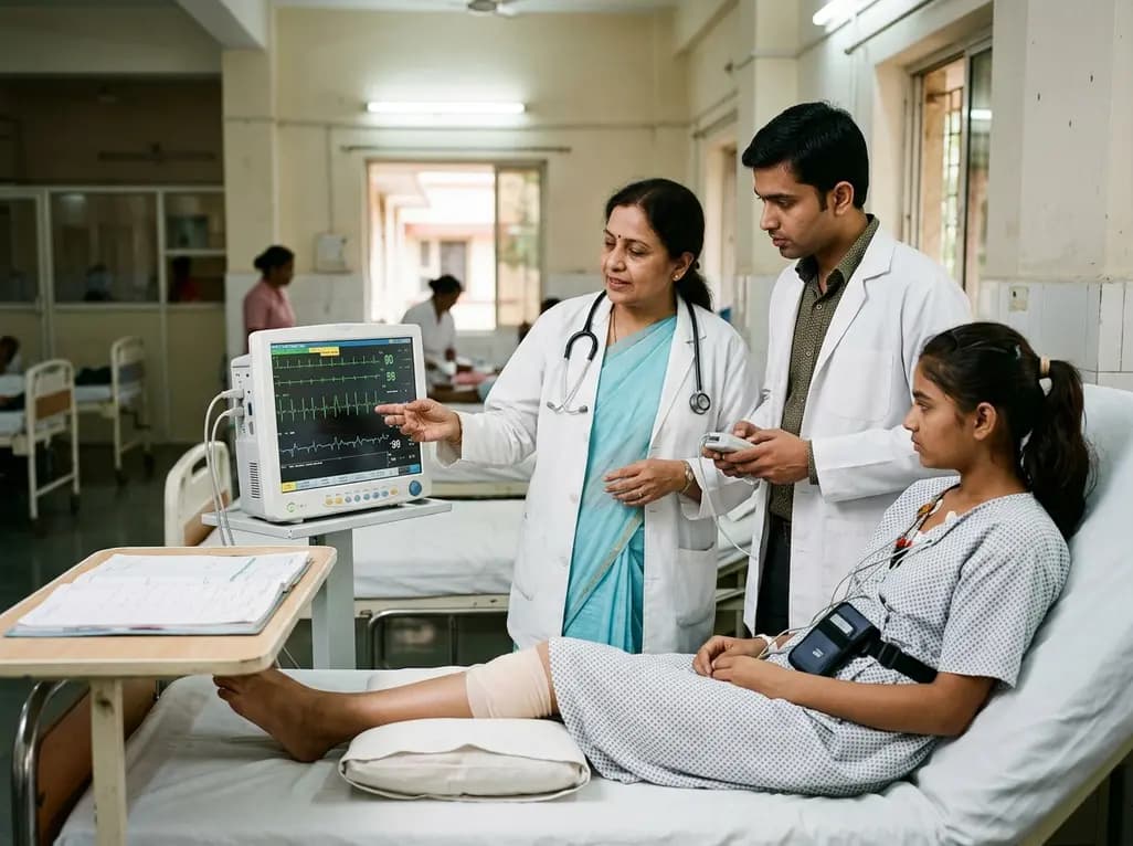
A 15-year-old patient presented with knee arthralgia and acute rheumatic fever, exhibiting complex arrhythmias including AV blocks and non-sustained ventricular tachycardia despite lacking severe valvulitis. This highlights the importance of extended Holter monitoring in pediatric rheumatic carditis.
Today
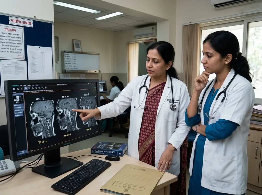
An eight-year longitudinal imaging follow-up reveals severe temporomandibular joint remodeling in nodulosis-arthropathy-osteolysis (NAO) syndrome. Despite extensive osseous destruction, the patient maintained functional jaw opening without TMJ interventions, highlighting the role of conservative management.
Today

Discover essential clinical insights into AGLP1-type amyloidosis, a rare localized complication of glucagon-like peptide-1 analog injections. Learn how histopathology and mass spectrometry differentiate benign iatrogenic amyloidomas from systemic disease, preventing misdiagnosis and unwarranted therapies.
Today
