.webp&w=384&q=75)

Loading, please wait...
Loading, please wait...

Postoperative rehabilitation remains an essential component of comprehensive breast cancer survivorship care. While physical therapy after standard mastectomy reliably restores mobility, clinicians historically hesitated to prescribe intensive regimens following reconstructive procedures. Concerns regarding flap compromise, wound breakdown, and implant displacement often led to prolonged immobilization. However, a rigorous systematic review now highlights the clinical value of breast reconstruction physical therapy. This comprehensive analysis clarifies how early mobilization protocols accelerate functional recovery, reduce upper-extremity disability, and preserve surgical safety in post-mastectomy patients.
Historically, reconstructive surgeons enforced prolonged immobilization for four weeks or longer after tissue reconstruction. Surgeons worried that premature movement might shear microvascular pedicles or cause prosthetic displacement. Consequently, patients frequently developed severe glenohumeral stiffness, pectoral tightness, and chronic myofascial pain. Furthermore, secondary shoulder impairments significantly delayed return to work and reduced daily independence. Recent surgical paradigms, however, prioritize rapid functional restoration through structured movement protocols. A systematic review synthesis evaluated fifteen clinical studies including 1,020 patients undergoing either implant-based or autologous reconstruction. Importantly, structured physiotherapy delivered consistent improvements in shoulder range of motion and overall upper-limb function. In addition, quality-of-life scores showed marked enhancement among active cohorts compared to immobilized controls. Clinicians observed these functional gains without any accompanying surge in surgical site complications or reconstructive failures. Therefore, structured physical therapy represents an empowering and safe standard of care rather than an operative hazard. Early mobilization successfully mitigates chronic post-mastectomy morbidity. Moreover, proactive guidance instills confidence and dispels patient anxieties during early convalescence.
The timing of rehabilitation initiation remains a critical clinical question for surgical and physiatric teams. Traditional management relied on prolonged shoulder splinting to protect underlying muscular and vascular repairs. In contrast, the systematic review demonstrated that introducing rehabilitation within two weeks postoperatively yields superior functional recovery. Patients engaging in early exercise achieved accelerated shoulder flexion and abduction compared to those subjected to traditional four-week immobilization. Moreover, early gentle movement reduced regional muscular guarding and mitigated myofascial tightness across the anterior chest wall. Fortunately, earlier mobilization did not increase the incidence of hematoma, seroma formation, or wound dehiscence. Similarly, drain duration and infectious complications remained equivalent between early and delayed rehabilitation groups. Thus, clinical evidence strongly challenges prolonged immobilization schedules. By initiating progressive, supervised range-of-motion routines inside the initial two-week recovery window, clinicians substantially hasten upper-extremity recovery. Consequently, patients regain independence in activities of daily living much sooner. Furthermore, timely therapy prevents permanent capsular adhesions without threatening reconstructed tissue flaps or synthetic prostheses.
Rehabilitation needs differ significantly based on the reconstructive approach chosen for each individual patient. Autologous procedures, such as deep inferior epigastric perforator flaps or latissimus dorsi transfers, entail extensive donor-site tissue harvesting. Consequently, these patients experience complex biomechanical deficits affecting both donor and recipient sites. Latissimus dorsi flaps, for example, directly compromise shoulder adduction and internal rotation strength. Therefore, autologous rehabilitation protocols require targeted trunk mobilization, core stabilization, and gradual latissimus compensation training. Conversely, implant-based techniques primarily involve subpectoral or prepectoral prosthetic placement with acellular dermal matrices. In subpectoral cases, pectoral muscle elevation triggers severe postoperative muscle spasm and pectoralis tightness. Consequently, implant-based physical therapy focuses on pectoral stretching, scapular retraction, and gentle capsular accommodation. Clinicians must recognize these distinct anatomical considerations when designing postoperative exercise plans. Tailoring physical therapy to the exact reconstructive modality ensures targeted recovery and minimizes muscular compensation patterns. In addition, individualized therapy prevents secondary cervical strain. Ultimately, recognizing these procedural nuances enables therapists to optimize long-term kinematic recovery.
Physiotherapists utilize various manual techniques to relieve postoperative discomfort and restore soft-tissue suppleness. Myofascial release, scar massage, and lymphatic drainage represent standard tools in breast cancer rehabilitation. The systematic review evaluated the clinical efficacy of these manual modalities alongside alternative therapies. Specifically, researchers investigated whether adding acupuncture, meditation, or electrotherapy enhanced physical recovery. Interestingly, massage-based interventions demonstrated clear benefits for tissue pliability and subjective comfort, but adjunctive modalities provided limited additional value. Alternative techniques failed to produce statistically significant improvements in objective shoulder range of motion. Therefore, clinical resources should prioritize direct kinesitherapy, progressive stretching, and targeted soft-tissue mobilization. Furthermore, specialized therapists must monitor tissue compliance around incisions and radiation-treated fields. Active physical movement consistently drives neuromuscular re-education far more effectively than passive or alternative therapies. Clinicians should thus center therapeutic regimens on active functional exercise rather than auxiliary modalities. Moreover, evidence indicates that active patient engagement produces superior functional resilience. Consequently, structured exercise remains the true foundation of restorative physical care.
Translating clinical evidence into daily surgical oncology workflows requires multidisciplinary coordination between surgeons, oncologists, and physical therapists. Before hospital discharge, clinicians should provide clear postural education and gentle distal extremity exercises. During the initial two weeks, patients can safely perform elbow, wrist, and controlled shoulder pendulum movements. Once surgical drains are removed and surgical wound healing stabilizes, therapists introduce active-assisted flexion and abduction. Importantly, movements should stay below ninety degrees of shoulder elevation until tissue integration progresses satisfactorily. Furthermore, therapists must continuously monitor for signs of seroma, erythema, flap perfusion compromise, or lymphedema. Gradually, therapists advance patients toward full active range of motion and resistance training over eight to twelve weeks. Educating patients about normal healing sensations also reduces kinesiophobia and promotes home exercise adherence. Through structured collaboration, surgical teams can achieve excellent reconstructive outcomes while preserving complete upper-limb function. In conclusion, multidisciplinary care bridges the gap between surgical oncologic success and lifelong functional well-being. Therefore, early rehabilitation pathways should become standard practice in modern breast reconstruction centers.
Most patients can safely begin gentle range-of-motion exercises within the first two weeks following surgery. Early mobilization protocols accelerate shoulder flexion and abduction recovery without increasing wound complications, hematomas, or seromas. However, clinicians must tailor the exact start date to the reconstruction type, drain status, and initial incision healing. Supervised programs ensure movements progress gradually from protected distal exercises to full overhead stretches.
Rehabilitation protocols differ because each surgical approach disrupts different anatomical structures. Autologous flaps involve donor-site morbidity, requiring targeted core stabilization, donor-muscle re-education, and spine mobility work alongside shoulder exercises. Conversely, implant-based reconstructions often cause significant pectoralis major tension and chest tightness. Therefore, implant therapy focuses predominantly on pectoralis stretching, scapular kinematics, and posture correction to prevent capsular stiffness and shoulder restriction.
Current clinical evidence demonstrates that structured, early physical therapy does not increase the risk of seroma formation, flap compromise, or implant loss. Although arm movement slightly elevates lymphatic drainage volume, it does not delay overall wound healing or increase reconstructive complications. Patients who initiate guided physical therapy within two weeks experience faster recovery of shoulder mobility while maintaining surgical safety under proper professional supervision.
Disclaimer: This content is for informational and educational purposes only and should not be considered medical advice. Healthcare professionals should exercise their independent clinical judgment when applying this information. Refer to the latest local and national guidelines for clinical practice.
References
Glynou SP et al. Postoperative Physical Therapy After Breast Reconstruction: A Systematic Review. Aesthet Surg J. 2026 Oct 03. doi: undefined. PMID: 42827398.
Yang A, Sokol H, Nealon K, et al. Early free range-of-motion upper limb exercises after mastectomy and immediate implant-based reconstruction are safe and beneficial: a randomized trial. Ann Surg Oncol. 2020;27(12):4750-4759.
De Groef A, Van Kampen M, Vervloesem N, et al. Effectiveness of Postoperative Physical Therapy for Upper-Limb Impairments After Breast Cancer Treatment: A Systematic Review. Arch Phys Med Rehabil. 2015;96(6):1140-1153.
Soran A, Gimbel M, Kurupati S, et al. Exercise after Breast Reconstruction Surgery: Evaluating Current Trends and Practices of U.S. Plastic Surgeons. Plast Reconstr Surg Glob Open. 2021;9(10):e3871.

Read summarized clinical updates, watch expert medical content, and earn CME certifications right from your smartphone.
Open your smartphone camera to scan and install.





A systematic review evaluates the safety and efficacy of postoperative physical therapy after breast reconstruction. Early rehabilitation within two weeks accelerates range of motion without increasing complications, highlighting key distinctions between autologous and implant-based procedures.
Today
Plasma calprotectin correlates strongly with acute phase reactants, joint counts, and autoantibody load in rheumatoid arthritis. Evaluating calprotectin alongside CRP identifies aggressive inflammatory phenotypes, providing clinicians with actionable insights to refine treatment and monitoring strategies.
Today

A pharmacometric and machine learning study reveals that specific vaginal bacteria alter cervical antiretroviral exposure, highlighting the need for precision HIV PrEP strategies.
Today
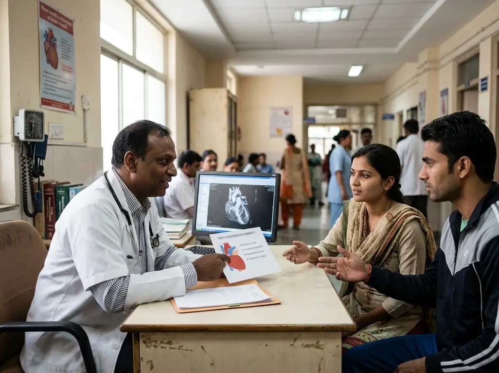
New clinical evidence demonstrates a shift away from strict exercise restrictions in genetic heart disease. While patients with channelopathies engage in more vigorous activity than those with cardiomyopathies, shared decision-making and individualized risk assessments ensure safe, beneficial exercise participation.
Today
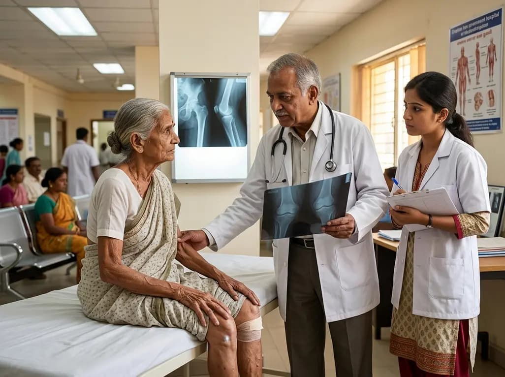
Sarcopenia significantly impairs functional independence and amplifies fracture risks in patients with osteoarthritis and osteoporosis. Learn about modern diagnostic strategies, GLP-1 therapy considerations, and evidence-based non-pharmacological management across multidisciplinary clinical settings.
Today

Recent research defines ferro-aging as a conserved iron-lipid axis driving organ decline across primates. Age-related iron dyshomeostasis stimulates ACSL4-mediated lipid peroxidation, promoting cellular senescence. Notably, vitamin C directly inhibits ACSL4, suppressing phospholipid oxidation and multi-organ decay.
Today
