.webp&w=384&q=75)

Loading, please wait...
Loading, please wait...

Clinicians traditionally perform a postoperative CT brain biopsy to screen for asymptomatic hemorrhages following stereotactic brain biopsy (SBB). However, the clinical utility of this routine practice remains a subject of intense debate. While SBB is generally a safe diagnostic procedure, reported hemorrhage rates vary significantly between 1% and 60%. Recent evidence suggests that a selective, symptom-driven strategy could potentially replace routine imaging without compromising patient safety.
A comprehensive retrospective study of 753 biopsies recently evaluated the factors associated with post-biopsy hemorrhage. Specifically, the researchers detected blood on postoperative imaging in 42% of cases. Despite this high detection rate, the findings altered clinical management in only 5% of all cases. Furthermore, most management changes occurred in patients who were already symptomatic. These results indicate that the positive predictive value of routine CT for detecting new or expanding hemorrhage is only 17%.
Moreover, certain risk factors clearly correlate with an increased likelihood of hemorrhage. For example, detecting blood through the biopsy needle intraoperatively or having a preoperative intralesional hemorrhage significantly increases risk. Additionally, patients with platelet counts between 100 and 150 × 10⁹/L face nearly double the risk of bleeding. Consequently, stable patients without these specific risk factors may not require routine scans. Therefore, a more targeted approach could reduce unnecessary radiation exposure and healthcare costs.
Adopting a selective imaging approach requires careful clinical monitoring. Physicians should prioritize the postoperative CT brain biopsy for individuals who exhibit new or worsening neurological symptoms, such as altered mental status or motor deficits. Furthermore, patients with a known bleeding diathesis or significant intraoperative complications definitely require imaging. Otherwise, for asymptomatic patients, the final decision to perform a CT study remains at the treating physician's discretion. This shift in practice ensures high-quality care while optimizing hospital resources.
No, routine imaging may not be warranted for all patients. Evidence suggests that a symptom-driven approach is safe for stable, asymptomatic patients who had no intraoperative complications.
Key risk factors include intraoperative bleeding through the biopsy needle, preoperative intralesional hemorrhage, and platelet counts below 150 × 10⁹/L.
Studies indicate that a symptom-driven approach does not compromise safety, as clinically significant hemorrhages typically present with neurological changes that prompt imaging regardless.
Disclaimer: This content is for informational and educational purposes only. It does not constitute professional medical advice, diagnosis, or treatment. Always seek the advice of your physician or other qualified healthcare provider with any questions you may have regarding a medical condition. Refer to the latest local and national guidelines for clinical practice.
References
Abi Faraj C et al. Reevaluating routine imaging: clinical utility of postoperative CT after stereotactic brain biopsy. J Neurosurg. 2026 Apr 10. doi: 10.3171/2025.11.JNS251196. PMID: 41962153.
Elhag A et al. To scan or not to scan? A retrospective cohort study analysing the efficacy of routine post-operative CT after brain biopsy. Acta Neurochir. 2024;166(1):1-8. doi: 10.1007/s00701-024-06180-1.
Blumrich L et al. Routine postoperative computed tomography scan after craniotomy: systematic review and evidence-based recommendations. Neurosurg Rev. 2021;44(5):2523–2531. doi: 10.1007/s10143-021-01473-w.
"
Read summarized clinical updates, watch expert medical content, and earn CME certifications right from your smartphone.
Open your smartphone camera to scan and install.





New research suggests routine CT after brain biopsy may be unnecessary for stable patients, favoring a symptom-driven approach to imaging....
6 months ago

Following regulatory scrutiny from FSSAI regarding promotional representations and lean muscle preservation claims, Dr. Reddy's Laboratories and Nestlé Health Science are currently reviewing the branding and marketing strategy for their joint nutritional supplement Celevida GLP+.
Today
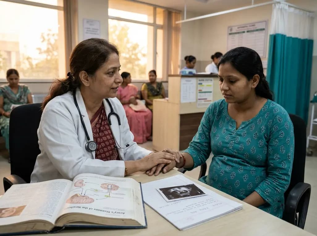
A comprehensive scoping review synthesises evidence linking prenatal maternal mental health to long-term offspring neurodevelopment, evaluating neuroendocrine, inflammatory, and epigenetic pathways while highlighting clinical screening and future research priorities.
Today
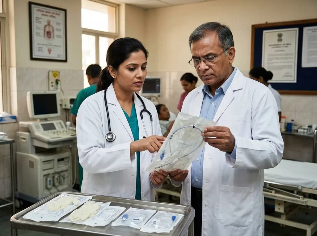
The Directorate General of Health Services directs Indian states to halt the hazardous reuse and reprocessing of single-use medical devices. Discover regulatory implications under the Drugs and Cosmetics Act, critical infection risks, legal liabilities, and operational changes required across clinical specialties.
Today
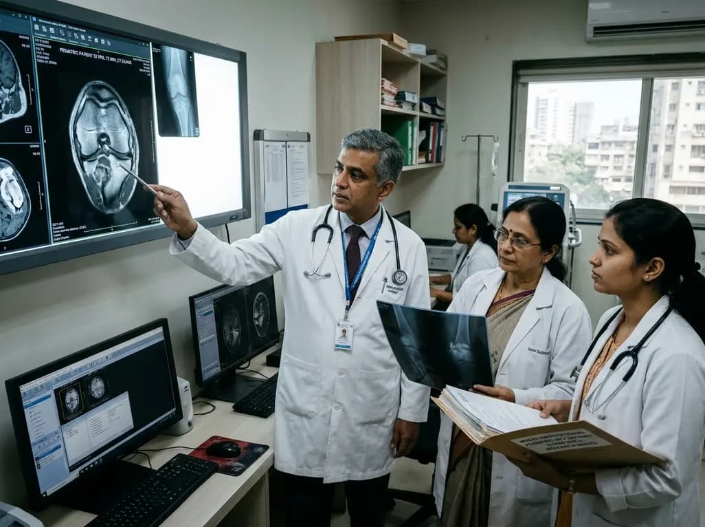
A 25-year multi-institutional cohort reveals high recurrence rates in pediatric giant cell tumor of bone, particularly in patients with open physes and bone marrow edema on preoperative MRI. Early surveillance and careful balance between growth plate preservation and tumor clearance are essential.
Today

India is advancing preventive healthcare by integrating genomics, artificial intelligence, and quantum technologies. With the SANGAM programme sequencing one million Indian genomes, clinicians can unlock personalized therapies, detect cardiometabolic risks early, and curb the burden of diabetes and heart disease.
Today
