.webp&w=384&q=75)

Loading, please wait...
Loading, please wait...
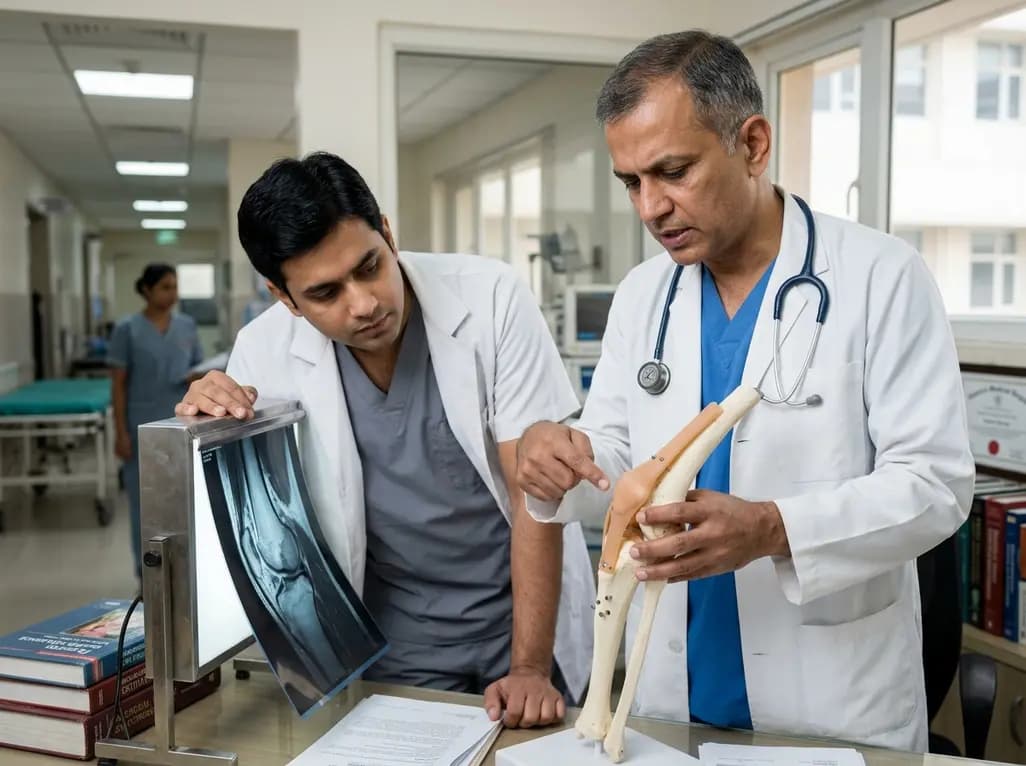
Orthopedic surgeons frequently face diagnostic and technical challenges when treating complex rotatory and lateral knee instability. Isolated injuries to the lateral structures are uncommon, and concomitant disruption of adjacent stabilizing elements can severely compromise surgical outcomes. Specifically, performing a posterolateral corner reconstruction without addressing underlying proximal tibiofibular joint laxity often leads to persistent joint dysfunction and premature graft failure. Surgeons must therefore evaluate all static and dynamic stabilizers to restore proper biomechanics.
The posterolateral aspect of the knee relies on a delicate interplay between several static and dynamic structures. Primary stabilizers include the fibular collateral ligament, the popliteus tendon, and the popliteofibular ligament. These structures collectively resist varus forces, external rotation, and posterior tibial translation. However, the proximal tibiofibular joint acts as an essential foundation for these ligamentous insertions. Disruption of the anterior and posterior superior tibiofibular ligaments destabilizes the fibular head itself. Consequently, any graft anchored to an unstable fibular head experiences altered vector mechanics and excessive stress. When surgeons perform posterolateral corner reconstruction, they must recognize that fibular hypermobility compromises graft tensioning. Unaddressed proximal tibiofibular joint instability leads to subtle micro-motion, which prevents solid graft integration and causes persistent lateral pain. Therefore, restoring stability to both the posterolateral structures and the proximal tibiofibular joint is vital for durable knee kinematics. Modern cadaveric investigations emphasize that simultaneous stabilization creates a reliable structural base, which protects soft tissue constructs during postoperative rehabilitation and early mobilization.
Evaluating complex posterolateral knee injuries requires a high index of suspicion during initial clinical assessment. Clinicians often miss proximal tibiofibular joint disruption because its symptoms closely mimic isolated posterolateral corner tears or lateral meniscal pathology. Standard magnetic resonance imaging protocols may fail to demonstrate subtle ligamentous laxity at the superior tibiofibular articulation. Therefore, targeted physical examination maneuvers are indispensable. Surgeons should systematically evaluate varus stress laxity, rotational instability at thirty degrees of knee flexion, and direct anteroposterior translation of the fibular head relative to the tibia. Comparing laxity with the uninjured contralateral side provides crucial diagnostic clarity. Additionally, weight-bearing radiographs and stress views help identify subtle joint subluxation that unweighted imaging misses. Missing a proximal tibiofibular joint injury prior to surgery frequently results in residual instability after isolated reconstruction. As a result, patients continue to report persistent lateral knee discomfort, mechanical clicking, and subjective giving way during pivot activities. Integrating rigorous clinical examination with advanced dynamic stress imaging ensures comprehensive identification of all compromised lateral stabilizing structures.
Simultaneous surgical stabilization of the posterolateral corner and proximal tibiofibular joint demands meticulous anatomical knowledge and precise tunnel creation. Surgeons utilize dual soft-tissue tendon autografts to reconstruct the native ligamentous anatomy accurately. Standard surgical exposure involves careful identification and neurolysis of the common peroneal nerve to prevent iatrogenic nerve injury during drilling. Anatomical landmarks dictate the precise entry points for femoral, fibular, and tibial tunnels. A transverse fibular head tunnel facilitates reconstruction of both the fibular collateral ligament and the anterior proximal tibiofibular ligament. Furthermore, passing a graft through dedicated tibial sockets restores the popliteofibular ligament and the posterior tibiofibular capsule. Maintaining safe bone bridge margins between drilled tunnels minimizes the risk of intraoperative fracture. Careful graft passage ensures that reconstructed vectors replicate natural motion without causing mechanical impingement. By anchoring grafts across both the tibiofibular joint and the femorofibular axis, surgeons re-establish comprehensive static stability. This dual-reconstruction strategy eliminates abnormal translational forces and restores physiological stress distribution across the lateral column.
A critical component of successful complex knee reconstruction is the implementation of a sequential fixation strategy. Fixing all graft limbs at a single flexion angle often results in over-constraining the joint or leaving dynamic laxity. In contrast, independent graft tensioning allows surgeons to secure each construct at its specific physiological angle. For instance, the fibular collateral ligament is typically tensioned near full extension to optimize varus stability. Conversely, the popliteus tendon and popliteofibular components are tensioned at higher angles of knee flexion to control tibial external rotation effectively. Securing the proximal tibiofibular joint ligaments first provides a rigid, stable fibular foundation. This rigid base enhances the mechanical reliability of subsequent posterolateral corner constructs. Biomechanical testing demonstrates that independent tensioning avoids graft over-tensioning, which could otherwise limit knee flexion or cause premature joint degeneration. Controlled tensioning aligns with known joint kinematics, promoting uniform load distribution across soft tissues. Consequently, this refined fixation sequence reduces peak mechanical strain on individual grafts during functional rehabilitation.
Managing complex multiligament knee injuries requires holistic surgical planning to avoid high failure rates. In clinical practice, unrecognised lateral instability remains a primary cause of primary cruciate ligament graft failure. When anterior or posterior cruciate ligament reconstructions are performed alongside an inadequately stabilized posterolateral corner, excessive rotational and varus forces degrade the cruciate grafts over time. Adding proximal tibiofibular joint stabilization to the surgical plan addresses a previously underappreciated biomechanical vulnerability. This strategy is particularly valuable in high-demand athletes and patients with chronic, high-energy knee dislocations. Furthermore, establishing a reproducible surgical technique using widely available autografts makes this procedure practical for orthopedic specialists. Early biomechanical results show that anatomic dual stabilization restores qualitative knee stability across standard clinical stress maneuvers. While long-term patient-reported outcome measures are still being gathered, this technical approach establishes a clear framework. Orthopedic surgeons treating severe lateral instability can adopt these anatomical principles to optimize patient outcomes and reduce revision surgery rates.
The proximal tibiofibular joint provides the structural anchor for key lateral stabilizers, including the fibular collateral ligament and biceps femoris. If the proximal tibiofibular joint remains hypermobile, any newly reconstructed posterolateral corner graft will lack a firm base. This persistent instability leads to abnormal graft stress, altered knee kinematics, residual pain, and high rates of premature surgical graft failure.
Independent graft tensioning allows surgeons to fix each reconstructed ligament at its specific biomechanical angle of knee flexion. For example, fixing the fibular collateral ligament in extension and the popliteus complex in flexion prevents joint over-constraining. This precise sequential strategy ensures optimal load sharing across soft tissue grafts, restores natural kinematics, and minimizes postoperative joint stiffness during early physical therapy.
Comprehensive evaluation requires combining physical examination with specialized diagnostic imaging. Surgeons evaluate varus stress laxity, rotational movement using the dial test, and manual translation of the fibular head relative to the tibia. Dynamic weight-bearing radiographs, stress radiographs, and high-resolution magnetic resonance imaging help visualize subtle ligamentous disruption and joint subluxation, ensuring accurate surgical planning for combined soft tissue injuries.
Disclaimer: This content is for informational and educational purposes only and does not constitute medical advice, diagnosis, or treatment. Refer to the latest local and national guidelines for clinical practice.
References

Read summarized clinical updates, watch expert medical content, and earn CME certifications right from your smartphone.
Open your smartphone camera to scan and install.





Concomitant PLC and PTFJ injuries create complex knee instability. This study presents an anatomic dual-reconstruction technique using tendon autografts and sequential tensioning to provide a stable fibular base, optimize load distribution, and improve posterolateral knee stability.
last month
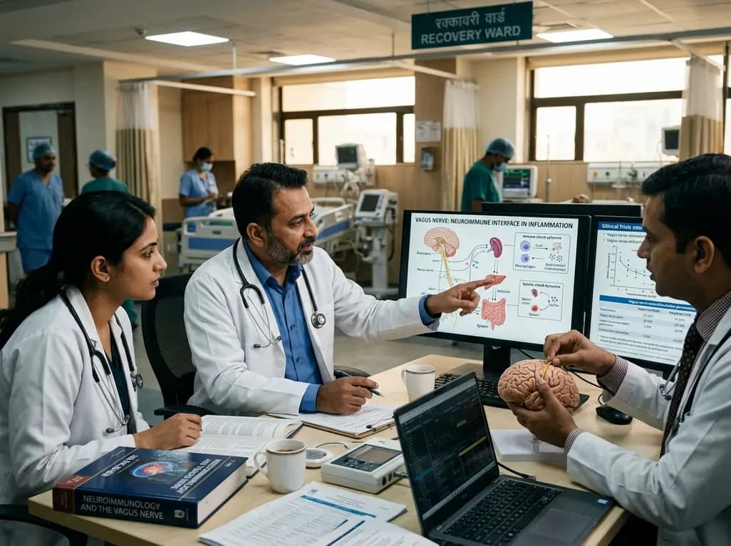
A review examines how vagal neuroimmune circuits link the brain and immune system, sensing cytokine surges and dampening peripheral inflammation. Bioelectronic medicine harnesses these pathways to provide targeted, non-pharmacological therapies for chronic inflammatory and autoimmune diseases.
Today
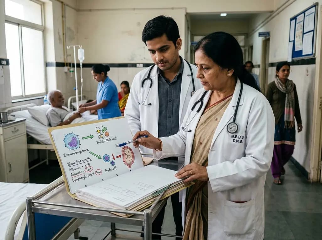
Explore the crucial link between dietary protein and immunocompetence. This review examines how low-protein nutrition shifts metabolic resources away from pathogen defense toward early reproduction, compromising host survival and immunity against severe bacterial infections.
Today

A post hoc analysis of the PARM-T2D study shows pemafibrate exerts baseline-dependent effects on LDL-C and particle size in type 2 diabetes and hypertriglyceridemia. It lowers triglycerides and remnants while favorably remodeling atherogenic LDL particles based on initial lipid control status.
Today

A longitudinal study reveals that patients undergoing staged bilateral hip arthroscopy experience progressive contralateral labral deterioration during surgical intervals. However, structural degradation does not compromise 2-year postoperative clinical outcomes or patient-reported satisfaction scores.
Today
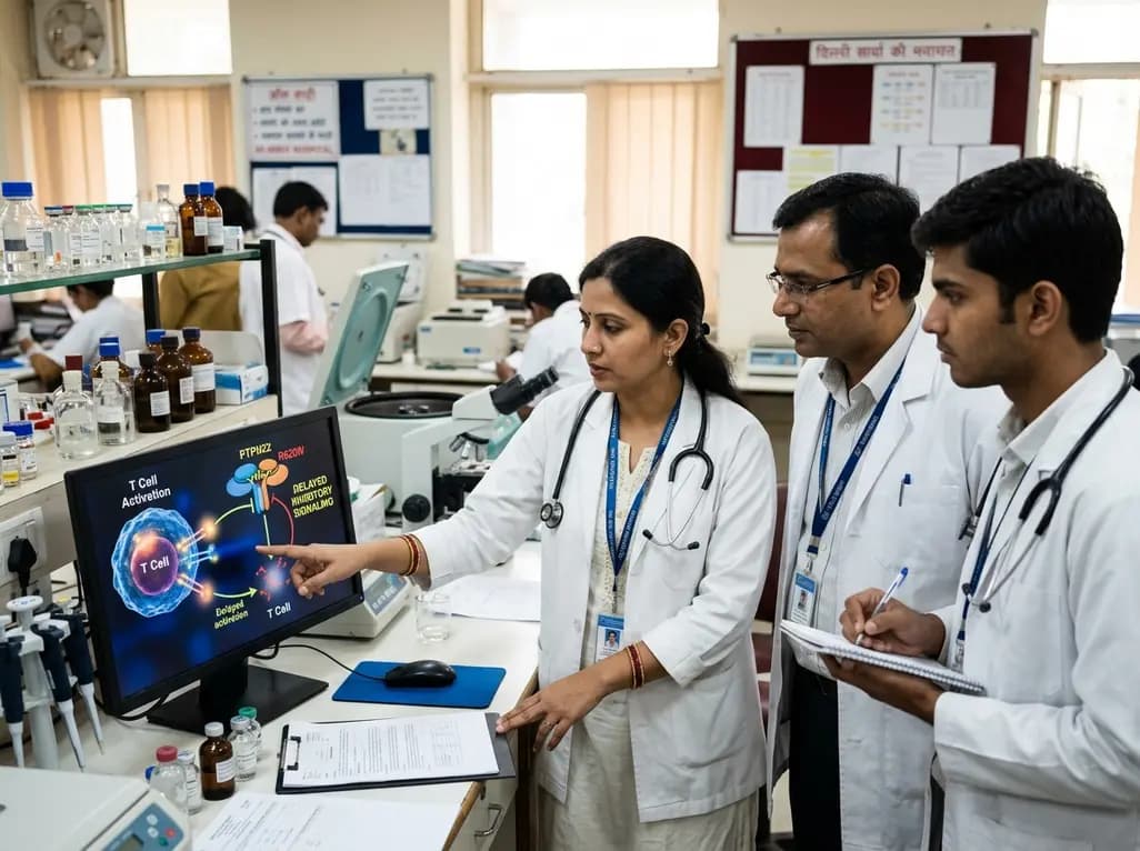
New immunological research reveals how PTPN22 forms a delayed inhibitory signaling complex to restrain T cell activation, explaining how the autoimmune-linked R620W variant disrupts feedback control and promotes persistent inflammation in conditions like rheumatoid arthritis and type 1 diabetes.
Today
