.webp&w=384&q=75)

Loading, please wait...
Loading, please wait...

Pediatric liver transplantation successfully restores long-term health in infants facing end-stage liver disease, particularly biliary atresia. However, postoperative immunological disturbances occasionally trigger devastating hematologic complications. Post-transplant autoimmune hemolytic anemia represents an uncommon complication that affects approximately one to three percent of pediatric liver allograft recipients. Although rare, this autoimmune phenomenon carries high morbidity and mortality when diagnosis is delayed. Clinicians typically diagnose the condition several months after transplantation, often when immunosuppression is being tailored. In particular, calcineurin inhibitors like tacrolimus disrupt regulatory T-cell homeostasis and can allow autoreactive B-cell clones to escape peripheral tolerance. Consequently, these unregulated B cells generate pathogenic autoantibodies directed against recipient erythrocytes. The resulting direct antiglobulin test positivity heralds accelerated intravascular and extravascular red cell destruction. Most alarming, severe hemolysis produces profound systemic downstream effects that extend far beyond simple anemia. Recent clinical evidence demonstrates that intense immune hemolysis creates a potent prothrombotic state. Therefore, physicians caring for pediatric transplant recipients must maintain a high index of suspicion. Early recognition of abnormal hematologic indices prevents irreversible tissue ischemia, allograft failure, and fatal systemic vascular occlusion.
Children with post-transplant hemolysis often exhibit nonspecific symptoms initially, including pallor, fatigue, irritability, and poor oral intake. However, physical examination frequently reveals scleral icterus, dark urine, and unexpected allograft hepatomegaly. In a representative case of a ten-month-old female transplanted for biliary atresia, severe hemolytic crisis emerged five months postoperatively. Laboratory testing revealed a precipitous drop in hemoglobin to 7.5 g/dL alongside marked elevations in lactate dehydrogenase to 1751 IU/L. Furthermore, clinicians detected profound hyperbilirubinemia, marked consumptive thrombocytopenia, and a prolonged prothrombin time. A strongly positive direct antiglobulin test confirmed the diagnosis of warm autoimmune hemolysis. Concurrently, coagulation profiling revealed a critical depletion of Protein S activity to 48.1 percent and a marked rise in D-dimer levels to 3.78 mg/L. These laboratory aberrations reflect acute systemic thrombophilia and widespread fibrin turnover. Therefore, clinicians must not treat post-transplant cytopenias as isolated benign drug reactions. Instead, medical teams should immediately order comprehensive hemolysis panels and thorough coagulation studies. Additionally, any sudden drop in platelets during active hemolysis demands urgent vascular imaging to exclude occult thrombotic complications.
The sudden co-occurrence of portal vein thrombosis during acute hemolytic crisis highlights a dangerous, hemolysis-driven hypercoagulable cascade. Intravascular erythrocyte destruction releases substantial quantities of cell-free hemoglobin and procoagulant erythrocyte microparticles into the circulation. Consequently, cell-free hemoglobin scavenges local nitric oxide, which impairs physiological vasodilation and triggers platelet hyperactivation. Furthermore, erythrocyte microparticles expose negatively charged phosphatidylserine on their outer membrane leaflet. This membrane alteration actively promotes assembly of the tenase and prothrombinase complexes, vastly accelerating thrombin generation. At the same time, ongoing inflammation and microvascular injury consume natural anticoagulant factors, producing a marked drop in Protein S activity. Reduced Protein S impairs the activated protein C pathway, leaving the splanchnic circulation vulnerable to thrombogenesis. Moreover, altered portal hemodynamics, surgical anastomotic geometry, and postoperative vessel narrowing further accentuate local shear stress. When turbulent portal inflow encounters a hypercoagulable circulation, platelet aggregation accelerates rapidly within the portal trunk. Thus, post-transplant hemolysis generates a biological storm that transforms small anatomical vulnerabilities into extensive portal vein thrombosis. Recognizing this molecular link enables teams to initiate preventive anticoagulant measures before complete vascular occlusion occurs.
Successfully resolving autoimmune hemolysis complicated by vascular thrombosis requires an aggressive, dual-pronged therapeutic strategy. Clinicians must simultaneously quell autoantibody synthesis and dissolve the intravascular thrombus without provoking fatal hemorrhage. In practice, medical teams immediately reduce or modify tacrolimus dosing to alleviate calcineurin inhibitor-mediated immune dysregulation. Concurrently, physicians initiate high-dose intravenous methylprednisolone at 4 mg/kg per day to halt reticuloendothelial clearance of opsonized erythrocytes. Furthermore, adding rituximab provides targeted B-cell depletion, eliminating the clonal lymphocyte population producing pathogenic autoantibodies. In conjunction with aggressive immunosuppression, therapeutic systemic anticoagulation with unfractionated or low-molecular-weight heparin is indispensable. Clinicians must carefully monitor anti-factor Xa levels or activated partial thromboplastin time to balance clot resolution against post-surgical bleeding. In documented clinical experience, this prompt multimodality regimen produced complete portal vein recanalization on Doppler ultrasound within five days. Moreover, the patient attained sustained direct antiglobulin test negativity and full hematologic recovery. Therefore, combining pulse corticosteroids, monoclonal anti-CD20 therapy, and titrated heparin safely rescues both the vascular conduit and the hepatic graft.
Preventing devastating vascular sequelae in pediatric liver recipients demands rigorous, standardized postoperative surveillance protocols. Transplant programs must educate clinicians and families to recognize subtle signs of early hemolysis and impending portal hypertension. Specifically, whenever a pediatric recipient develops unexplained anemia or jaundice, teams must perform an urgent direct antiglobulin test. Additionally, clinicians should systematically order complete coagulation profiles, including D-dimer levels and Protein S activity, during any acute hemolytic event. Bedside color-flow Doppler ultrasonography represents the non-invasive gold standard for assessing portal vascular patency. Ultrasonographers should evaluate the portal vein diameter, flow velocity, and flow direction to detect early thrombus or turbulent flow patterns. If ultrasound findings appear equivocal, contrast-enhanced computed tomography provides definitive anatomic delineation of the portal venous axis. Furthermore, multidisciplinary collaboration between pediatric hepatologists, hematologists, transplant surgeons, and interventional radiologists ensures rapid therapeutic escalation. Establishing institutional care pathways that mandate immediate vascular imaging at the onset of hemolysis significantly reduces graft loss. Ultimately, proactive vigilance and protocolized emergency interventions ensure optimal long-term survival for high-risk pediatric liver transplant recipients.
Autoimmune hemolysis releases cell-free hemoglobin and procoagulant erythrocyte microparticles directly into the bloodstream. Free hemoglobin rapidly scavenges endothelial nitric oxide, which impairs vasodilation and accelerates platelet activation. Concurrently, erythrocyte microparticles expose phosphatidylserine, triggering massive thrombin generation. In addition, severe systemic inflammation and consumption critically deplete Protein S activity. These combined hemolytic alterations create a severe thrombophilic state that promotes acute portal vein thrombosis within surgically reconstructed vessels.
Clinicians must implement an aggressive, combined medical strategy targeting both autoantibody production and vascular occlusion. Management begins by reducing tacrolimus dosages to alleviate immune dysregulation while administering high-dose intravenous methylprednisolone at 4 mg/kg daily. Furthermore, early rituximab therapy provides targeted B-cell depletion to achieve durable immunologic remission. Simultaneously, therapeutic anticoagulation with intravenous heparin is essential to dissolve portal thrombi, provided teams closely monitor bleeding parameters.
Whenever clinicians suspect post-transplant autoimmune hemolysis, they must immediately obtain a direct antiglobulin test, complete blood count, reticulocyte count, and serum lactate dehydrogenase. In addition, assessing total bilirubin and haptoglobin confirms active hemolysis. Because hemolysis induces thrombophilia, physicians must systematically evaluate coagulation profiles, including D-dimer and Protein S levels. Finally, urgent color-flow Doppler ultrasonography of the liver is mandatory to assess portal vein patency and detect early vascular thrombosis.
Disclaimer: This content is for informational and educational purposes only and is not intended as medical advice, diagnosis, or treatment. It does not replace professional clinical judgment. Healthcare professionals must evaluate each clinical situation individually, and patients should always consult their physician or a qualified healthcare provider regarding any health condition or treatment decisions. Guidelines and practices vary across healthcare systems; clinicians must exercise clinical discretion and follow local regulations and institutional protocols. The author and publisher are not liable for any consequences resulting from the application of information contained in this material. Refer to the latest local and national guidelines for clinical practice.
References

Read summarized clinical updates, watch expert medical content, and earn CME certifications right from your smartphone.
Open your smartphone camera to scan and install.





Post-transplant autoimmune hemolytic anemia is a rare pediatric complication that can trigger acute portal vein thrombosis via hypercoagulable pathways. Rapid diagnosis with Doppler ultrasound, systemic anticoagulation, and targeted immunotherapy can achieve graft salvage and complete hematologic remission.
Today
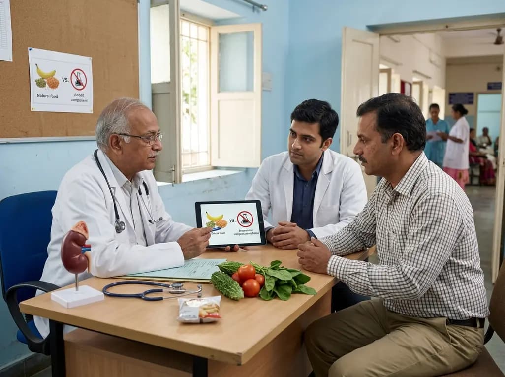
Modern renal nutrition is shifting toward additive-aware dietary guidance. This paradigm differentiates between intrinsic nutrients in whole foods and hyper-bioavailable inorganic additives, offering practical strategies to mitigate additive stacking and toxicity without restricting nutritious whole foods.
Today

A multiparametric MRI approach combining quantitative T2 shading, apparent diffusion coefficient (ADC) measurements, and morphological signs provides superior diagnostic accuracy over subjective evaluation to differentiate ovarian endometriomas from benign hemorrhagic cysts.
Today

A landmark case report reveals a donor heart beating for a century in a 63-year-old recipient, challenging conventional age limits in cardiac transplantation and offering critical lessons on organ longevity, donor selection, and long-term graft survival.
Today

A new randomized crossover trial reveals that a hinged ulnar gutter orthosis significantly improves hand function and weight-bearing tolerance compared to standard volar splints in patients with degenerative triangular fibrocartilage complex lesions, supporting functionally permissive orthotic designs.
Today

New research reveals that M1 macrophage-derived exosomes aggravate diabetic nephropathy by transferring WTAP to stabilize S1PR2 mRNA. Silencing WTAP in these vesicles attenuates endothelial injury and renal fibrosis, pointing toward innovative nanomedicine therapies.
Yesterday
