.webp&w=384&q=75)

Loading, please wait...
Loading, please wait...
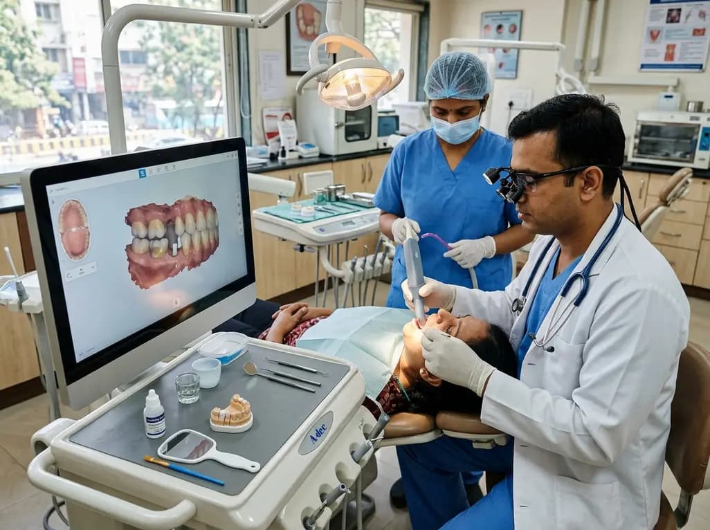
Digital impressions have rapidly transformed contemporary restorative dentistry, yet capturing deep endodontic cavities directly remains a demanding clinical challenge. Restoring severely damaged endodontically treated teeth frequently necessitates custom post and core restorations, which clinicians increasingly fabricate using digital chairside workflows. However, the narrow, steep geometry of prepared root canals often restricts optical sensor penetration, leading to digital artifacts and surface distortions. A recent in vitro investigation evaluated how radicular morphological parameters and surface conditioning influence post space scanning trueness using the TRIOS 5 intraoral scanner. By systematically assessing post space opening diameter, preparation depth, taper, adjacent tooth presence, and the application of liquid scanning-aid material, the researchers provided critical insights into optimizing digital post space acquisition. Consequently, these findings provide dental professionals with evidence-based strategies to improve intraoral scanning reliability in prosthodontic practice.
Intraoral scanning systems rely on structured light or optical confocal microscopy to construct detailed three-dimensional virtual models. While these scanners demonstrate exceptional precision on supragingival tooth preparations, narrow root canals present substantial illumination and triangulation barriers. Specifically, the steep axial walls of endodontic post spaces scatter optical rays, generating deep shadows where scanner sensors fail to capture data accurately. Moreover, surface reflections from moist or highly translucent dentinal walls can produce data voids and stitching errors during image reconstruction. Traditionally, clinicians bypassed these optical hurdles by fabricating indirect resin patterns or taking conventional elastomeric impressions. Although analog techniques offer proven accuracy, they introduce laboratory turnaround times and increase patient discomfort. Direct digital impressions eliminate elastomer distortion and streamline the post and core workflow; however, clinicians must understand the physical constraints of light transmission within narrow root canals before eliminating conventional methods entirely.
The accuracy of digital post space acquisition depends heavily on four fundamental anatomical and preparation variables. First, canal opening diameter plays a pivotal role, as wider coronal orifices allow significantly greater light transmission into the deeper canal segments. Second, preparation depth directly dictates optical access; deeper canal preparations inevitably increase the distance from the scanner tip to the apical seat. In the evaluated study, preparations extending to 10.0 mm showed substantially higher deviation values than those limited to 8.0 mm. Third, preparation taper directly influences the scanner line-of-sight, with wider tapers of 0.08 mm/mm providing vastly superior scan fidelity compared to narrow 0.04 mm/mm tapers. Fourth, the presence of adjacent teeth restricts the operational angulation of the scanner wand, thereby exacerbating optical shadowing. When bilateral adjacent teeth were present, scanning deviation increased significantly. Consequently, canal geometry and surrounding anatomical structures interact synergistically to influence post space scanning trueness during clinical procedures.
To counteract light reflection and scattering within dark, confined canal spaces, investigators assessed the utility of liquid scanning-aid material. Intraoral scanning-aid liquids create an ultra-thin, uniform matte coating across the prepared dentin surfaces before scanning. This specialized layer significantly suppresses specular glare and promotes diffuse light reflectance, thereby allowing intraoral optical sensors to register steep canal walls with high fidelity. Without applying the scanning-aid liquid, root mean square error (RMSE) values ranged broadly between 50.0 and 804.5 μm, indicating substantial dimensional discrepancies across different preparations. In contrast, after applying the liquid scanning aid, median RMSE values dropped markedly to a range between 46.0 and 360.0 μm. This significant enhancement confirms that optical surface conditioning actively mitigates the geometric limitations of deep root canals. Furthermore, the liquid formulation spreads evenly across canal walls without excessive pooling, preserving anatomical margins while markedly enhancing data capture reliability.
Evaluating digital scanning accuracy requires analyzing both global surface fit and localized linear deviation at the apical floor of the post space. The canal bottom is notoriously susceptible to algorithmic smoothing, artificial rounding, or complete data omission. In the experimental trial, two-dimensional cross-sectional analysis revealed that unassisted scans produced median apical bottom deviations ranging from 0.02 to 4.88 mm. Such substantial discrepancies indicate that the scanner software frequently interpolates or truncates the apical region when direct light reflection fails. However, following the application of the liquid scanning-aid material, median bottom deviation values decreased considerably to a range of 0.02 to 3.22 mm. Although scanning aids markedly enhance apical data acquisition, some residual discrepancy persists in narrow, deep preparations. Therefore, dental practitioners must avoid assuming that intraoral scans capture the deepest canal regions with perfect geometric accuracy, particularly in conservative, parallel preparations.
Integrating direct intraoral post space scanning into routine prosthodontic workflows provides noticeable efficiency, but practitioners must execute specific preparation and scanning protocols to ensure success. When designing post spaces for chairside milling, clinicians should optimize preparation geometry by establishing an adequate coronal orifice diameter and maintaining a taper of at least 0.06 to 0.08 mm/mm. Additionally, applying a validated liquid scanning-aid material prior to scanning significantly enhances digital mesh completeness. If clinical constraints require a post space depth exceeding 8.0 to 10.0 mm, or if tight interdental spaces severely restrict scanner wand angulation, utilizing dedicated scan posts or indirect digitizing techniques remains advisable. Furthermore, clinicians should routinely inspect cross-sectional views in CAD software to verify that the apical canal bottom was accurately scanned rather than algorithmically stitched. By combining favorable canal preparation geometry, optical scanning aids, and rigorous digital verification, dental teams can achieve predictable, high-precision custom restorations.
Liquid scanning-aid material creates a thin, uniform matte layer directly across the prepared canal dentin walls. This micro-coating effectively minimizes specular reflections and enhances diffuse light scatter, enabling intraoral optical sensors to register steep canal walls accurately. Consequently, applying this agent dramatically reduces overall root mean square error and decreases apical bottom deviations during digital post space impression procedures.
Canal opening diameter, preparation depth, taper, and adjacent tooth presence all significantly influence scanning trueness in clinical practice. Wider canal orifices and steeper preparation tapers facilitate deeper light penetration, whereas deeper canals and proximal teeth create severe optical shadows. Therefore, clinicians must carefully balance conservative root preservation with sufficient geometrical access to optimize intraoral scanner line-of-sight and overall digital accuracy.
Direct intraoral scanning provides satisfactory clinical reliability for moderate preparation depths when clinicians apply liquid scanning aids and maintain adequate taper. However, in preparations exceeding eight to ten millimeters, optical distortion and apical bottom deviations often increase. In such deep or complex scenarios, using specialized scan posts or indirect digitizing techniques prevents restoration seating discrepancies and promotes superior long-term outcomes.
Disclaimer: This content is for informational and educational purposes only and does not constitute medical or dental advice. Consult appropriate professional guidelines and qualified healthcare providers before implementing any diagnostic or therapeutic approaches. Refer to the latest local and national guidelines for clinical practice.
References

Read summarized clinical updates, watch expert medical content, and earn CME certifications right from your smartphone.
Open your smartphone camera to scan and install.





A new in vitro study evaluates how liquid scanning-aid materials, opening diameter, canal depth, adjacent teeth, and taper affect intraoral post space scanning trueness, providing key insights for digital prosthodontic workflows.
last month
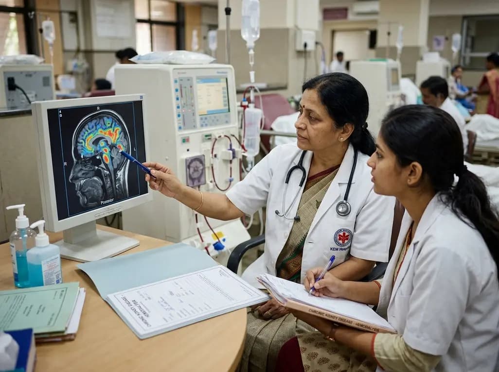
A retrospective clinical analysis reveals that hemodialysis-associated cognitive dysfunction arises from synergistic pathological mechanisms, including severe chronic anemia, worsening renal clearance, abnormal regional brain iron deposition, and an imbalance between neuroinflammatory and neurotrophic factors.
Today

A multiparametric MRI approach combining quantitative T2 shading, apparent diffusion coefficient (ADC) measurements, and morphological signs provides superior diagnostic accuracy over subjective evaluation to differentiate ovarian endometriomas from benign hemorrhagic cysts.
Today

Discover a novel laparoscopic approach for iatrogenic diaphragmatic hernia repair following pedicled omentoplasty. This modified Sugarbaker technique integrates dorsal pedicle lateralization and round ligament reinforcement to secure the diaphragmatic defect while preserving vital omental vascularity.
Yesterday
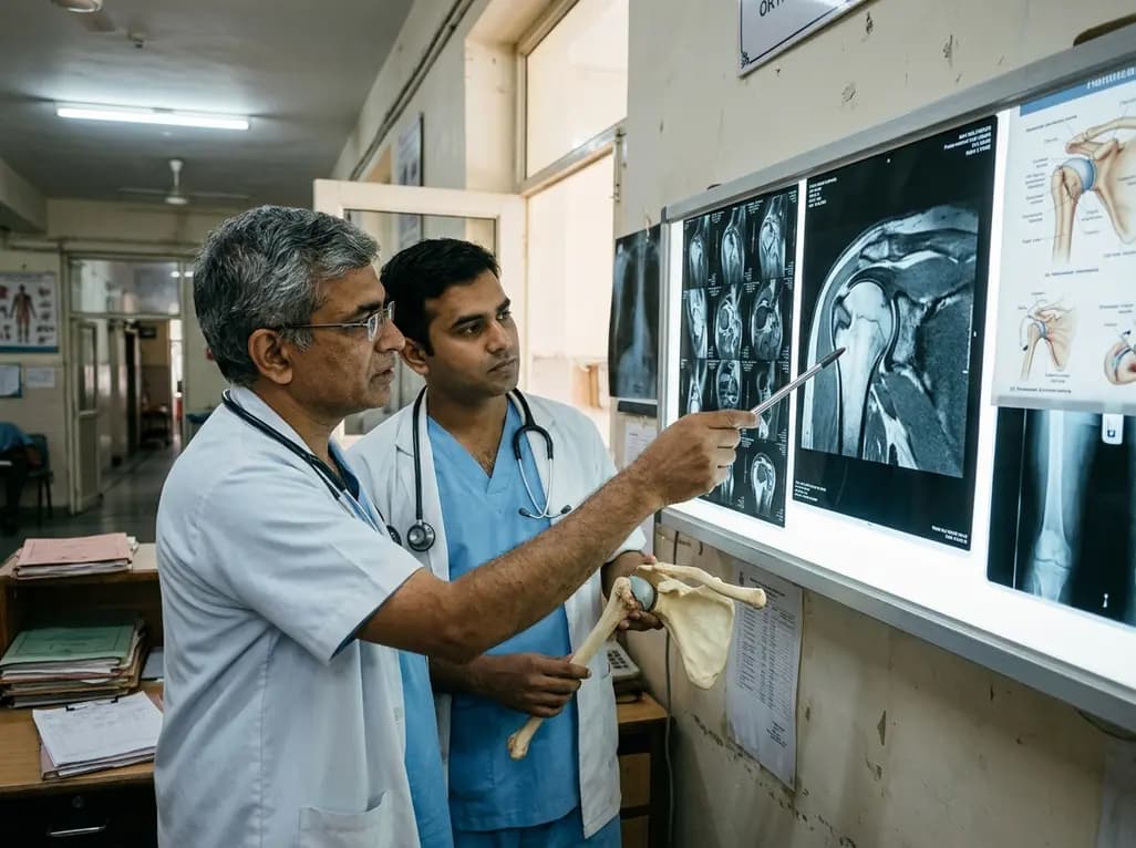
Evaluating Hill-Sachs lesion size and the distance to dislocation is critical in recurrent shoulder instability. As bone loss concepts shift from binary on-track classifications to continuous risk assessment, clinicians must identify when isolated Bankart repair is insufficient and supplementary procedures are needed.
Today

New research reveals that M1 macrophage-derived exosomes aggravate diabetic nephropathy by transferring WTAP to stabilize S1PR2 mRNA. Silencing WTAP in these vesicles attenuates endothelial injury and renal fibrosis, pointing toward innovative nanomedicine therapies.
Today
