.webp&w=384&q=75)

Loading, please wait...
Loading, please wait...

Pediatric hydrocephalus represents a challenging neurosurgical condition characterized by the abnormal accumulation of cerebrospinal fluid within the cerebral ventricles. Clinicians frequently encounter diagnostic and monitoring hurdles when managing these vulnerable patients. Routine evaluations traditionally depend on repeated computed tomography scans or high-field magnetic resonance imaging. However, both modalities impose distinct clinical burdens, including repeated ionizing radiation exposure and the logistical complexity of hospital transfers. In recent neurosurgical research, the advent of portable MRI pediatric hydrocephalus assessment protocols offers a promising paradigm shift. This emerging point-of-care technology delivers rapid, accessible neuroimaging directly at the bedside, fundamentally transforming how clinicians evaluate ventricular caliber and identify progressive ventriculomegaly.
Furthermore, standard high-field neuroimaging suites often require deep sedation or general anesthesia in young children to prevent motion artifacts during scans. Transporting critically ill pediatric patients from intensive care units also introduces hemodynamic and airway risks. Consequently, clinicians urgently require non-invasive, radiation-free neuroimaging alternatives that operate seamlessly at the point of care. Ultra-low-field magnetic resonance systems successfully address these persistent limitations. These compact scanners operate safely in ordinary clinical environments without specialized radiofrequency shielding. Therefore, investigating their diagnostic concordance against standard imaging remains essential for pediatric neurosurgery.
To evaluate whether low-field imaging can accurately monitor ventricular dimensions, investigators initiated a prospective, single-blind trial across three tertiary academic medical centers. The study enrolled 153 pediatric patients who presented with suspected or confirmed ventriculomegaly. The cohort demonstrated a balanced sex distribution, comprising 53.59% male and 46.4% female participants, with an average age of 9.55 ± 6.39 years. Researchers systematically recruited patients from pediatric neurology clinics, neurosurgery services, and emergency departments to reflect real-world clinical variation across care settings.
Additionally, the trial analyzed a diverse spectrum of underlying hydrocephalus etiologies commonly encountered in pediatric practice. The primary etiologies included posthemorrhagic hydrocephalus in 27.45% of cases, myelomeningocele in 15.03%, and aqueductal stenosis in 15.03%. Each participant underwent bedside neuroimaging using an ultra-low-field 0.064-Tesla magnetic resonance device alongside standard high-field scans. Strict ethical protocols and privacy standards protected all participant data throughout the multi-institutional study. By comparing standardized axial projections, neuroradiologists rigorously examined anatomical landmarks without knowing which scanner acquired the comparative image sets. Consequently, this multi-institutional trial provides crucial validation data regarding point-of-care scanner deployment.
The primary scientific objective centered on whether ultra-low-field images provide quantitative concordance with conventional neuroimaging modalities. Specifically, investigators calculated the Evans index and the frontal-occipital horn ratio across both modalities. These linear indices serve as universally recognized radiological standards for tracking ventricular expansion and monitoring shunt function in hydrocephalus. Statistical analyses using Bland-Altman plots revealed near congruent agreement between the point-of-care portable scans and standard-of-care imaging.
Moreover, the researchers evaluated Lin's concordance correlation coefficient to establish statistical reliability across imaging platforms. The concordance correlation coefficient reached 0.922 for the Evans index, with a tight 95% confidence interval spanning from 0.8941 to 0.9428. Similarly, the frontal-occipital horn ratio demonstrated exceptional concordance at 0.9419, with a confidence interval between 0.9206 and 0.9576. These robust metrics clearly show that ultra-low-field imaging reliably detects ventricular caliber changes. Hence, pediatric clinicians can depend on portable measurements to gauge ventricular size without subjecting pediatric patients to high-field scan delays or transport complications.
Children suffering from chronic hydrocephalus often undergo dozens of surveillance scans throughout their developmental years. Historically, clinicians relied on head computed tomography because of its rapid acquisition time and widespread accessibility in emergency departments. Nevertheless, cumulative ionizing radiation from repeated computed tomography scans significantly elevates lifetime risks of secondary malignancies and radiation-induced cognitive morbidity. In contrast, ultra-low-field portable systems utilize non-ionizing radiofrequency pulses, thereby eliminating radiation risks entirely.
Additionally, performing imaging directly at the bedside eliminates the substantial dangers associated with intrahospital patient transport. Critically ill children connected to mechanical ventilation, external ventricular drains, or intravenous infusions face risks of accidental extubation and line displacement during transit. Portable magnetic resonance scanners roll directly into intensive care rooms, completely avoiding these transit hazards. Furthermore, the 0.064-Tesla magnetic field does not disrupt external programmable shunt valves. Consequently, medical teams do not need to reprogram magnetic shunt valves after every scan, significantly streamlining clinical workflow.
Although the multicenter trial demonstrated excellent agreement for linear ventricular indices, investigators highlighted critical diagnostic boundaries. Ultra-low-field scanners are not designed to serve as standalone diagnostic platforms for complete intracranial evaluation. Because ultra-low-field scanners operate at lower signal-to-noise ratios, they cannot replicate the fine parenchymal resolution of 1.5-Tesla or 3-Tesla scanners. Subtle structural abnormalities, including small intracranial tumors, subtle cortical malformations, or microvascular pathologies, may escape detection on low-field images.
Therefore, neurosurgeons must view portable systems as supplementary surveillance tools rather than complete replacements for comprehensive diagnostic scans. Initial disease characterization, surgical pathway planning, and fine vascular assessments still demand conventional high-field neuroimaging. Nevertheless, point-of-care imaging excels at routine monitoring, shunt malfunction screenings, and rapid emergency evaluations. Furthermore, because these portable devices run on standard electrical wall outlets, they present immense potential for resource-constrained regions and district hospitals lacking conventional scanner infrastructure.
Ultra-low-field portable MRI demonstrates excellent quantitative concordance with standard neuroimaging modalities for measuring ventricular dimensions. Clinical studies show concordance correlation coefficients exceeding 0.92 for the Evans index and 0.94 for the frontal-occipital horn ratio. Consequently, bedside scanners reliably track ventricular caliber and identify progressive ventriculomegaly in pediatric patients.
No, portable MRI cannot entirely replace conventional high-field neuroimaging systems. While portable scanners excel at assessing ventricular size and detecting gross ventriculomegaly, they lack the spatial resolution required to evaluate subtle parenchymal pathologies or intracranial tumors. Therefore, clinicians must utilize portable MRI as a supplementary surveillance tool alongside standard diagnostic scans.
Bedside portable MRI eliminates harmful ionizing radiation exposure associated with serial computed tomography scans. Furthermore, point-of-care imaging prevents hazardous intrahospital transport for critically ill pediatric patients. The ultra-low-field strength also avoids unintentional reprogramming of magnetic programmable shunt valves, significantly improving patient comfort, diagnostic efficiency, and overall clinical safety during routine follow-up examinations.
Disclaimer: This content is for informational and educational purposes only. It is not intended to provide medical advice, diagnosis, or treatment. Always seek the advice of your physician or other qualified health provider with any questions you may have regarding a medical condition. Clinicians should use their clinical judgement and correlate findings clinically. Refer to the latest local and national guidelines for clinical practice.
References

Read summarized clinical updates, watch expert medical content, and earn CME certifications right from your smartphone.
Open your smartphone camera to scan and install.





A multicenter feasibility study demonstrates that ultra-low-field portable MRI offers substantial concordance with standard imaging for measuring ventricular size in pediatric hydrocephalus. Point-of-care MRI reduces radiation risks and transport hazards, serving as a reliable complementary surveillance tool.
Today

The CARE study protocol introduces a multilingual, RAG-powered conversational interface designed by IIT Bombay to support tobacco cessation in India. Evaluating feasibility and behavioral outcomes across three cessation centers, this digital health initiative aims to enhance quitting support for diverse users.
Today

A large UK cohort study of 1.4 million women shows that pregnancy complications, including gestational diabetes, hypertension, and preterm birth, substantially raise long-term risks for hypertension, chronic kidney disease, type 2 diabetes, and cardiovascular events, highlighting early postpartum intervention needs.
Today
A cross-sectional study reveals that hypertension knowledge, illness perceptions, depression, and substance use significantly predict self-care behaviors. Addressing mental health and cognitive beliefs in primary care is crucial for optimal blood pressure control and cardiovascular disease prevention.
Today
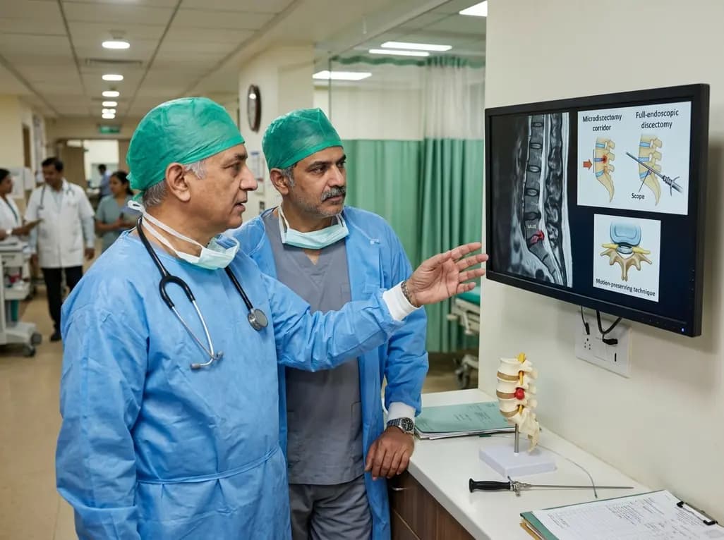
This clinical overview examines recent advancements in lumbar disc herniation surgery, comparing microscopic and full-endoscopic discectomy while highlighting biportal techniques and emerging motion-preserving innovations for spine specialists.
Today

Small interfering RNA (siRNA) therapeutics harness RNA interference to degrade disease-causing messenger RNA. By delivering hepatocyte-targeted, durable gene silencing, agents targeting PCSK9, LPA, and angiotensinogen offer potent cardiovascular risk reduction with infrequent, biannual dosing schedules.
Today
