.webp&w=384&q=75)

Loading, please wait...
Loading, please wait...

Rapid neuroimaging remains the cornerstone of modern acute ischemic stroke evaluation. However, conventional high-field magnetic resonance imaging systems face substantial logistical bottlenecks, high maintenance costs, and strict architectural shielding requirements. To address these barriers, portable low-field MRI has emerged as a disruptive bedside diagnostic modality. Operating at field strengths typically below 0.1 Tesla, these point-of-care devices bring neuroimaging directly to intensive care units, emergency bays, and remote healthcare facilities. Nevertheless, physical constraints often compromise native image resolution and quantitative precision. Recent clinical breakthroughs demonstrate that artificial intelligence can bridge this diagnostic divide, transforming portable neuroimaging into a robust quantitative tool.
Conventional 1.5-Tesla and 3.0-Tesla MRI scanners deliver superb anatomic detail and high spatial resolution. However, transporting critically unstable stroke patients to distant radiology suites introduces hazardous clinical delays. Furthermore, conventional scanners demand specialized infrastructure, including cryogenic helium cooling and radiofrequency shielding cages, which substantially escalate capital expenditures. In contrast, portable low-field MRI systems operate using standard wall electrical outlets and require minimal structural shielding. Consequently, clinicians can deploy these lightweight scanners directly beside the patient, drastically minimizing time-to-scan intervals.
Despite these operational strengths, low-field systems inherently generate weaker magnetic resonance signals. Therefore, native scans exhibit lower signal-to-noise ratios and coarse spatial resolution compared to high-field counterparts. Consequently, subtle ischemic changes, punctate lacunar infarcts, and posterior circulation strokes may escape rapid visual detection. Clinicians urgently require diagnostic parity with conventional scanners before integrating bedside systems into acute revascularization decision pipelines. Advanced super-resolution machine learning addresses this limitation directly. By reconstructing low-resolution acquisitions into synthetic high-resolution volumes, algorithmic post-processing restores fine anatomical boundaries. Thus, portable low-field systems can expand timely neuroimaging access across emergency departments, resource-limited regional hospitals, and rural healthcare centers.
To overcome native hardware constraints, investigators developed deep learning-based synthetic super-resolution magnetic resonance imaging, termed SynthMRI. Researchers deployed a sophisticated neural network architecture designated as SCUNet (Swin-Conv-UNet). This framework uniquely synergizes Swin Transformer blocks with convolutional neural network layers, effectively capturing both global contextual relationships and local textural features within brain parenchyma. Initially, researchers pretrained the SCUNet model on massive, diverse open-source neuroimaging repositories. Subsequently, they fine-tuned the model using paired clinical datasets comprising stroke patients and healthy controls who underwent both portable low-field and high-field examinations.
During processing, the neural network ingests low-field acquisitions and reconstructs high-fidelity volumetric representations that closely simulate high-field image contrast. Importantly, the algorithm preserves essential diffusion dynamics and structural geometry rather than merely smoothing pixel artifacts. Furthermore, the pipeline integrates quantitative apparent diffusion coefficient mapping, ensuring accurate tissue characterization. Consequently, SynthMRI generates synthetic images that exhibit superior contrast-to-noise ratios, sharpened lesion margins, and suppressed background noise. This computational reconstruction allows clinicians to visualize subtle cytotoxic edema patterns that standard low-field sequences frequently obscure, thereby elevating diagnostic confidence during critical clinical evaluations.
The clinical validation of SynthMRI demonstrates profound diagnostic improvements over unprocessed low-field acquisitions. In rigorous testing sets, native low-field scans achieved a sensitivity of 77.1% and a specificity of 89.2% for ischemic lesion detection. In sharp contrast, SynthMRI boosted sensitivity to 89.0% and increased specificity to 93.8%. Consequently, artificial intelligence enhancement significantly reduces false-negative interpretations, particularly for small-volume infarcts and early ischemic presentations. Furthermore, SynthMRI markedly improves the delineation of lesion borders across both cortical and subcortical territories.
Clinicians frequently struggle to differentiate salvageable tissue from completed core when reviewing low-resolution bedside scans. However, SynthMRI provides clear gray-white matter differentiation and clarifies cytotoxic edema boundaries with high reproducibility. Moreover, inter-reader agreement among neuroradiologists increases substantially when evaluating SynthMRI datasets compared to native acquisitions. Readers identify acute territorial infractions more rapidly and exhibit fewer equivocal assessments. Therefore, synthetic super-resolution successfully mitigates the diagnostic ambiguity historically associated with point-of-care magnetic resonance systems. As a result, emergency physicians and neurointensivists can reliably assess acute parenchymal injury without relying exclusively on fixed, centralized high-field radiology facilities.
Accurate topographic scoring is paramount when determining patient eligibility for intravenous thrombolysis and endovascular thrombectomy. The study evaluated agreement between portable MRI and high-field reference standards using the Alberta Stroke Program Early CT Score (ASPECTS) for both anterior and posterior circulations. Native low-field imaging exhibited moderate correlation with high-field ground truth. However, SynthMRI demonstrated exceptionally high intraclass correlation coefficients for both diffusion-weighted ASPECTS and posterior circulation ASPECTS. Consequently, clinicians can accurately map regional ischemic topography directly at the bedside.
Beyond regional scoring, precise volumetric quantification of the ischemic core dictates clinical outcomes. The investigative team evaluated lesion volume agreement and mean apparent diffusion coefficient (ADC) values within infarcted zones. SynthMRI exhibited outstanding intraclass correlation coefficients and Pearson correlation coefficients when benchmarked against high-field MRI. Moreover, quantitative ADC values within reconstructed lesions aligned tightly with true physiological diffusion restriction. Therefore, SynthMRI does not merely enhance cosmetic visual appearance; it preserves quantitative biophysical metrics essential for hemodynamic and tissue viability analysis. Thus, clinicians gain an objective, mathematically validated assessment of tissue fate to guide critical emergency therapeutic pathways.
The democratization of neuroimaging holds profound clinical implications for healthcare systems worldwide, particularly across developing nations like India. In many community hospitals, secondary healthcare facilities, and rural clinics, high-field MRI installations remain completely inaccessible due to exorbitant infrastructure and operating expenses. Consequently, acute stroke victims often endure lengthy inter-facility transfers to obtain confirmatory imaging, losing precious therapeutic time windows. Deploying portable low-field systems equipped with synthetic super-resolution software can decentralize neuroimaging delivery, establishing immediate diagnostic capabilities at primary triage sites.
Furthermore, intensive care units frequently manage critically ill, mechanically ventilated stroke patients whose transfer to distant radiology suites poses severe hemodynamic and airway risks. Bedside portable MRI eliminates transport-related adverse events, enabling serial neuroimaging directly within intensive care cubicles. Emergency physicians can rapidly distinguish acute ischemic infarction from intracerebral hemorrhage or non-ischemic stroke mimics. Moreover, bedside quantitative assessment facilitates timely administration of thrombolytic therapy in community settings where round-the-clock specialized radiology staff might be unavailable. Therefore, integrating super-resolution artificial intelligence with affordable portable hardware bridges fundamental healthcare disparities and ensures standardized, high-quality stroke care across diverse clinical environments.
Deep learning models like SCUNet utilize advanced convolutional and transformer architectures pretrained on extensive imaging datasets. The algorithm effectively removes noise, enhances spatial resolution, and sharpens tissue contrast from portable low-field acquisitions. Consequently, it reconstructs synthetic high-resolution images that closely match high-field scans while preserving quantitative diffusion metrics.
Portable low-field MRI serves as a complementary, highly accessible bedside screening modality rather than a complete replacement for high-field systems. While deep learning dramatically improves lesion sensitivity and quantitative agreement, high-field MRI remains the gold standard for intricate microvascular pathology, subtle intracranial hemorrhage evaluation, and definitive advanced neurovascular characterization.
Portable systems operate on standard electrical power without requiring cryogenic helium cooling or specialized radiofrequency cages. Consequently, they significantly lower capital and operational costs. Furthermore, bedside deployment accelerates stroke triage in community hospitals, remote clinics, and crowded intensive care units where access to conventional high-field infrastructure remains severely restricted.
Disclaimer: This content is for informational and educational purposes only and should not be considered medical advice. Always consult a qualified healthcare professional regarding clinical decisions or treatments. Refer to the latest local and national guidelines for clinical practice.
References

Read summarized clinical updates, watch expert medical content, and earn CME certifications right from your smartphone.
Open your smartphone camera to scan and install.





Deep learning super-resolution significantly improves the diagnostic performance of portable low-field MRI in stroke. The SCUNet-based SynthMRI framework elevates lesion sensitivity to 89% and closely matches high-field MRI for volumetric and quantitative diffusion assessment, expanding bedside neuroimaging access.
Today

The Maharashtra FDA's recent licence suspension at Fortis Hospital Mulund highlights the urgent clinical need for strict culinary hygiene in healthcare facilities. Preventing cross-contamination, fungal infestation, and pest activity is essential to protect vulnerable inpatients from life-threatening infections.
Today

A Belgian study highlights maternal desires and healthcare provider perspectives on breastfeeding in women living with HIV. While viral suppression reduces transmission risk below 1%, significant clinical training and policy gaps remain, highlighting the urgent need for multidisciplinary counseling protocols.
Yesterday
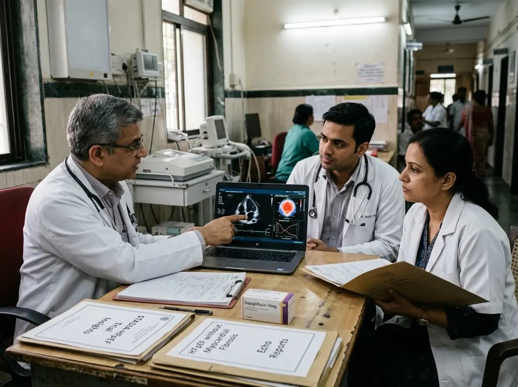
The STADIA-HFpEF trial evaluated whether dapagliflozin improves left ventricular distensibility in phenotypically stratified HFpEF patients without significant myocardial fibrosis. While primary diastolic endpoints were neutral, exploratory improvements in left atrial conduit strain and biomarkers emerged.
Today

A 2-center cohort study demonstrates that insurance status significantly influences diagnostic and surgical timing in acute Achilles tendon rupture repair. Addressing administrative hurdles and expediting orthopedic referrals can optimize patient outcomes and reduce wound complications.
Today

A landmark multi-ethnic study of 40,132 participants demonstrates that risk-weighted apoB (RW-apoB) captures coronary risk beyond measured apoB. By integrating the atherogenic potency of triglyceride-rich lipoproteins and Lp(a), RW-apoB improves cardiovascular risk discrimination across diverse populations.
Today
