.webp&w=384&q=75)

Loading, please wait...
Loading, please wait...
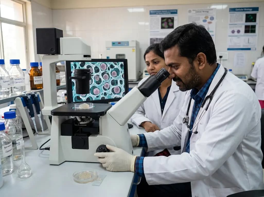
Articular cartilage lesions present an enduring clinical hurdle in orthopedic practice and regenerative medicine. Because native articular tissue lacks vascularity and neural networks, spontaneous self-repair remains severely restricted. Consequently, investigators have extensively evaluated cell-based scaffolds to treat osteochondral defects and degenerative joint disease. Cartilage tissue engineering holds immense promise for restoring functional hyaline articular surfaces. However, preserving the native chondrogenic phenotype during ex vivo manipulation poses major scientific challenges. Traditional three-dimensional matrices frequently distribute cells as isolated individual units. This dispersion disrupts natural intercellular communication pathways. Recent advances demonstrate that multicellular aggregation fundamentally alters cell signaling and biosynthetic activity. A groundbreaking study published in Advanced Healthcare Materials highlights an innovative biomaterial platform. The investigators utilized porous alginate hydrogels to systematically compare individual cell distribution against multicellular spheroids. Their findings establish that spatial cell arrangement critically determines chondrocyte viability and matrix production.
Clinicians managing chondral pathology frequently observe the limitations of conventional autologous chondrocyte implantation. When chondrocytes undergo standard monolayer expansion, they rapidly lose their characteristic spherical morphology. Therefore, dedifferentiation into fibroblastic phenotypes occurs, accelerating the synthesis of type I collagen rather than robust type II collagen and aggrecan. Tissue engineers design 3D hydrogel scaffolds to prevent this maladaptive shift. Hydrogels mimic the water-rich extracellular microenvironment of native cartilage. Nevertheless, standard bulk hydrogels encapsulate cells in an isolated state. This physical isolation prevents direct cadherin-mediated cell-cell junctions. As a result, critical juxtacrine signaling networks remain dormant. Cellular behavior depends heavily on spatial organization and neighbor proximity. Multicellular aggregates promote complex biological dialogues through tight junctions, gap junctions, and localized paracrine gradients. By investigating how single-cell dispersion transitions into multicellular spheroids within identical biomaterials, researchers can identify optimal structural microenvironments. Ultimately, these structural insights allow clinicians and bioengineers to develop functional cellular grafts that withstand biomechanical joint stress.
To investigate spatial cellular distribution without confounding material variables, researchers synthesized a tunable biomimetic scaffold platform. They formulated porous alginate hydrogels using an elegant droplet-templating technique. Specifically, the investigators introduced calcium chloride solution droplets into an uncrosslinked sodium alginate precursor. This controlled droplet introduction yielded uniform micropores within the surrounding crosslinked alginate matrix. The researchers created cell-loaded micropores within cell-free hydrogel matrices or dispersed individual chondrocytes directly inside the hydrogel mesh. Bovine articular chondrocytes served as the primary cell model. Within the confined geometry of the micropores, chondrocytes spontaneously aggregated into dense, three-dimensional spheroids. Conversely, chondrocytes embedded in the surrounding alginate network remained physically isolated as single cells. This hydrogel system allowed direct head-to-head comparison within an identical chemical substrate. Alginate provides excellent biocompatibility, minimal immunogenicity, and predictable ionic crosslinking. Furthermore, the porous architecture ensures efficient diffusion of nutrients, oxygen, and metabolic waste products. Therefore, this platform effectively eliminates diffusion barriers while maintaining distinct spatial configurations.
The spatial distribution of articular chondrocytes induced profound differences in transcriptomic profiles and biosynthetic activity. Chondrocytes organized as multicellular spheroids inside the micropores demonstrated robust upregulation of essential cartilaginous genes compared to single-cell counterparts. In particular, expression levels of SOX9, type II collagen (COL2A1), and aggrecan (ACAN) increased dramatically in spheroids. SOX9 serves as the master chondrogenic transcription factor, regulating downstream matrix structural genes. Enhanced SOX9 transcription indicates sustained phenotype preservation and active resistance against dedifferentiation. Furthermore, the aggregation of chondrocytes promoted cell proliferation rates within the porous cavities. Dense homotypic cell contacts trigger intracellular cascades, including N-cadherin signaling, which modulate cytoskeletal tension and metabolic survival pathways. Conversely, solitary chondrocytes in the hydrogel matrix exhibited suppressed proliferative capacity and lower cartilaginous marker expression. In addition, the close proximity of cells within spheroids established concentrated microdomains of autocrine and paracrine growth factors. Consequently, the coordinated cellular microenvironment reinforced chondrocytic identity and elevated biosynthetic throughput far beyond isolated cells.
Functional restoration of load-bearing articular surfaces requires extensive deposition of high-quality extracellular matrix. Histological and biochemical evaluations revealed that chondrocyte spheroids secreted significantly higher quantities of sulfated glycosaminoglycans and type II collagen. The newly synthesized matrix accumulated densely within the micropores, gradually remodeling the surrounding hydrogel architecture. Moreover, spheroids demonstrated superior extracellular matrix distribution, forming continuous cartilaginous tissue patches over extended culture durations. Direct comparison within the same biomaterial construct proved that spheroid-mediated cell-cell interactions drive superior matrix synthesis. Individual chondrocytes in the alginate matrix synthesized baseline extracellular components, but their physical isolation restricted matrix interconnectivity. In contrast, spheroids actively bridged intra-pore spaces to produce cohesive neocartilage units. Mechanical integrity of engineered cartilage depends entirely on this dense, interconnected proteoglycan and collagen network. Thus, stimulating initial spheroid formation within porous hydrogels accelerates neo-tissue maturation. These biochemical outcomes emphasize that spatial cell architecture acts as a potent regulator of functional cartilage regeneration.
These compelling biomaterial discoveries provide vital translational lessons for orthopedic surgeons, rheumatologists, and regenerative medicine specialists. Current clinical interventions for joint restoration often yield fibrocartilage, which lacks the long-term durability of native hyaline cartilage. By integrating multicellular spheroids into porous hydrogels, clinicians may soon deploy advanced therapies with superior regenerative fidelity. Additionally, the droplet-templating alginate platform offers high scalability and cell compatibility, making it attractive for translational biomanufacturing. This porous hydrogel paradigm can easily merge with three-dimensional bioprinting technologies to construct anatomically tailored osteochondral implants. Furthermore, this approach opens promising avenues for combining chondrocytes with mesenchymal stem cells or immune-modulatory biofactors. Such multi-component systems could mitigate chronic synovitis in osteoarthritic joints while actively rebuilding articulating surfaces. Clinicians must follow emerging clinical trials and scaffold safety evaluations closely. Adopting bioengineered scaffolds that harness homotypic cell interactions will transform focal defect repair and improve patient-reported joint mobility.
Multicellular spheroid formation maintains intense homotypic cell-cell contacts and activates essential cadherin-mediated signaling pathways. These close interactions upregulate key chondrogenic transcription factors such as SOX9 while repressing fibroblastic gene expression. Furthermore, spheroids create concentrated microenvironments for autocrine growth factors that protect the rounded cellular morphology. Consequently, chondrocytes preserved in spheroids resist dedifferentiation and consistently produce hyaline-specific type II collagen and aggrecan rather than inferior fibrocartilage components.
Porous alginate hydrogels offer outstanding biocompatibility, gentle ionic crosslinking under physiological conditions, and low baseline immunogenicity. In addition, the porous architecture provides spacious cavities that accommodate multicellular spheroids without imposing excessive mechanical confinement. The interconnected micropores also facilitate the unhindered diffusion of nutrients, oxygen, and cellular metabolites throughout the scaffold. Therefore, these hydrogels maintain optimal cell viability while supporting robust extracellular matrix deposition and structural remodeling.
Traditional cell therapies, such as standard autologous chondrocyte implantation, often struggle with cellular leakage, dedifferentiation, and poor biomechanical integration. In contrast, spheroid-laden porous hydrogels deliver pre-aggregated, highly functional cellular clusters capable of immediate extracellular matrix synthesis. This strategy enhances graft retention at the defect site and accelerates functional tissue maturation. Consequently, patients may experience superior joint biomechanics, more durable hyaline-like repair, and reduced risks of premature graft failure.
Disclaimer: This content is for informational and educational purposes only and does not constitute medical advice. Healthcare professionals should evaluate individual patient circumstances and refer to the latest local and national guidelines for clinical practice.
References

Read summarized clinical updates, watch expert medical content, and earn CME certifications right from your smartphone.
Open your smartphone camera to scan and install.





Recent research reveals that porous alginate hydrogels with spheroid spatial cell distribution significantly boost chondrocyte proliferation, cartilaginous gene expression, and extracellular matrix deposition, offering major breakthroughs for cartilage tissue engineering and clinical joint repair.
last month

Modern renal nutrition is shifting toward additive-aware dietary guidance. This paradigm differentiates between intrinsic nutrients in whole foods and hyper-bioavailable inorganic additives, offering practical strategies to mitigate additive stacking and toxicity without restricting nutritious whole foods.
Today

A multiparametric MRI approach combining quantitative T2 shading, apparent diffusion coefficient (ADC) measurements, and morphological signs provides superior diagnostic accuracy over subjective evaluation to differentiate ovarian endometriomas from benign hemorrhagic cysts.
Today

A landmark case report reveals a donor heart beating for a century in a 63-year-old recipient, challenging conventional age limits in cardiac transplantation and offering critical lessons on organ longevity, donor selection, and long-term graft survival.
Today
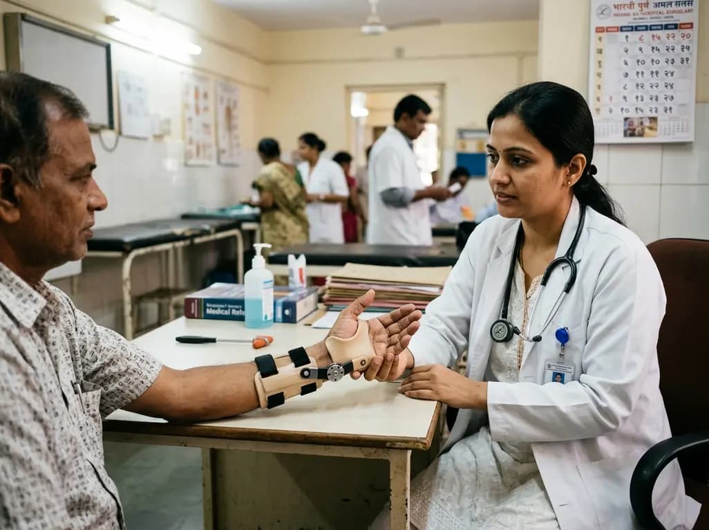
A new randomized crossover trial reveals that a hinged ulnar gutter orthosis significantly improves hand function and weight-bearing tolerance compared to standard volar splints in patients with degenerative triangular fibrocartilage complex lesions, supporting functionally permissive orthotic designs.
Today

New research reveals that M1 macrophage-derived exosomes aggravate diabetic nephropathy by transferring WTAP to stabilize S1PR2 mRNA. Silencing WTAP in these vesicles attenuates endothelial injury and renal fibrosis, pointing toward innovative nanomedicine therapies.
Yesterday
