

Loading, please wait...
Loading, please wait...

Preeclampsia remains one of the most formidable hypertensive complications encountered during pregnancy, contributing substantially to maternal and perinatal morbidity and mortality across the globe. Characterized primarily by new-onset hypertension and end-organ dysfunction after 20 weeks of gestation, the primary pathophysiologic origin lies within defective trophoblast invasion and impaired spiral artery remodeling. Consequently, clinicians have continuously searched for non-invasive imaging techniques that provide earlier and more precise structural and functional insights into placental pathology. In this context, placental shear wave elastography combined with vascular Doppler velocimetry represents a meaningful leap forward in characterizing placental health, opening new horizons for clinical risk stratification.
Normal placentation entails extensive remodeling of maternal spiral arteries by extravillous cytotrophoblasts, converting narrow muscular vessels into high-flow, low-resistance conduits. However, in preeclampsia, incomplete trophoblastic invasion leaves spiral arteries constricted and hyper-reactive. This primary vascular defect generates chronic placental ischemia, oxidative stress, and widespread maternal systemic endothelial inflammation. The persistent ischemic insult promotes substantial histopathological alterations within the chorionic villi, including syncytiotrophoblast knotting, accelerated villous maturation, parenchymal fibrin deposition, and progressive intervillous thrombosis. Therefore, maternal-fetal units face heightened risks of severe fetal growth restriction and oligohydramnios.
As these microstructural remodeling processes intensify, tissue elasticity progressively declines while interstitial fibrosis accumulates. Furthermore, maternal systemic vascular resistance escalates concurrently, creating a detrimental hemodynamics loop that conventional two-dimensional B-mode ultrasound struggles to quantify directly. Although gray-scale sonography can identify gross morphological lesions such as retroplacental hematomas or substantial infarctions, it lacks sensitivity for diffuse microscopic fibroelastic changes. Consequently, integrating quantitative stiffness mapping with maternal-fetal hemodynamics provides a more comprehensive physiological overview.
Clinicians have long relied on conventional Doppler ultrasonography of the maternal uterine arteries and fetal umbilical artery to evaluate downstream hemodynamic resistance. Elevation of the uterine artery pulsatility index and persistence of early diastolic notching serve as established markers of defective spiral artery transformation. However, Doppler parameters primarily quantify macrovascular blood velocity profiles rather than the intrinsic parenchymal biomechanical properties of the organ itself.
To overcome this diagnostic gap, placental shear wave elastography offers a non-invasive acoustic technique that generates acoustic radiation force impulses into tissue. By tracking the lateral propagation velocity of the resulting shear waves, the ultrasound software directly derives parenchymal stiffness values, expressed quantitatively in kilopascals or meters per second. When clinicians combine uterine and umbilical artery Doppler velocimetry with shear wave imaging, they capture both the macrovascular flow impedance and the microvascular structural stiffness. Thus, this multi-parametric ultrasound evaluation bridges the missing link between vascular supply and organ tissue remodeling.
A recent prospective observational comparative study rigorously examined sixty pregnant women, comprising thirty patients diagnosed with preeclampsia and thirty normotensive healthy controls. The investigative team performed bilateral uterine artery and umbilical artery Doppler ultrasonography, capturing peak systolic velocities, pulsatility indices, and resistance indices. Simultaneously, examiners performed shear wave elastography across multiple discrete anatomic sites, measuring the right peripheral, central, and left peripheral zones across both fetal and maternal placental surfaces.
The study demonstrated marked hemodynamic differences between the two cohorts. Specifically, patients with preeclampsia exhibited significantly elevated uterine artery pulsatility indices compared to healthy pregnant women. The mean right uterine artery pulsatility index measured 1.19 ± 0.65 in preeclamptic pregnancies compared to 0.74 ± 0.24 in healthy controls (p = 0.003). Similarly, the left uterine artery pulsatility index reached 1.35 ± 0.57 in the preeclampsia cohort versus 0.84 ± 0.26 in controls (p < 0.001). These robust statistical elevations confirm severe maternal vascular malperfusion and incomplete spiral artery remodeling.
Importantly, the placental elastography measurements revealed striking differences across parenchymal layers. Preeclamptic placentas exhibited significantly higher shear wave velocity and tissue stiffness values than control placentas across maternal and central regions. Furthermore, investigators observed that elevated parenchymal stiffness correlated directly with adverse perinatal outcomes, including earlier gestational age at delivery, lower five-minute Apgar scores, and compromised umbilical cord blood gas parameters. Hence, tissue stiffness reliably mirrors clinical disease severity.
Understanding how shear wave technology functions helps clinicians interpret numerical elastography readouts. Unlike legacy strain elastography, which relied on variable manual compression by the sonographer, shear wave systems deliver standardized acoustic impulses from the transducer. Because shear waves propagate significantly faster through rigid, fibrous structures than through soft, hydrated matrices, faster wave speeds (meters per second) and higher Young's modulus values (kilopascals) indicate advanced tissue stiffness.
In healthy normotensive pregnancies, the placenta remains a compliant, vascular sponge optimized for low-pressure blood pools and rapid nutrient exchange. Conversely, in preeclamptic placentas, extensive perivillous fibrinoid deposits, infarction, and interstitial edema create rigid acoustic barriers that dramatically accelerate shear wave velocity. Moreover, regional mapping shows that the maternal surface and central cotyledonary regions consistently display the highest stiffness increments. By standardizing these regional kilopascal thresholds, clinicians gain a reliable, operator-independent tool that objectively tracks ongoing microvascular damage.
The translation of these imaging discoveries into daily obstetric practice holds remarkable promise for refining patient management algorithms. When obstetricians identify elevated resistance indices on routine second-trimester Doppler screenings, immediately incorporating elastography can clarify borderline clinical scenarios. If a pregnant patient exhibits borderline uterine artery velocimetry alongside markedly elevated placental stiffness, clinicians can escalate surveillance frequency, initiate timely fetal biophysical monitoring, and coordinate targeted serial growth scans.
Additionally, this multi-parametric framework enhances clinical decision-making regarding the timing and location of delivery. In tertiary referral centers, distinguishing between mild hypertensive disease and severe, rapidly decompensating preeclampsia is critical for preventing eclampsia and abruptio placentae. Placentas demonstrating diffuse stiffening and high maternal vascular resistance alert the obstetric team to prepare for potential neonatal resuscitation and neonatal intensive care unit admission. Thus, elastography shifts clinical practice from reactive intervention to proactive risk stratification.
Looking ahead, integrating artificial intelligence algorithms with multi-parametric ultrasound data will likely transform prenatal risk assessment. Researchers are currently evaluating machine learning pipelines that combine first-trimester angiogenic biomarkers, such as soluble fms-like tyrosine kinase-1 and placental growth factor, with maternal mean arterial pressure, uterine artery Doppler waveforms, and shear wave elastography maps. Such integrated predictive models could identify vulnerable pregnancies weeks before overt clinical hypertension emerges.
Nevertheless, widespread clinical implementation requires robust international standardization of elastographic measurement protocols. Differences in maternal body mass index, ultrasound probe frequencies, placental location, and gestational age can introduce subtle measurement variability. Consequently, academic bodies must establish normative reference nomograms across diverse ethnicities and trimesters. Once standardized, non-invasive elastography will undoubtedly secure a fundamental role alongside Doppler ultrasound in modern obstetric imaging.
Placental shear wave elastography is an advanced, non-invasive ultrasound technique that uses acoustic radiation impulses to measure tissue stiffness quantitatively. By tracking how rapidly shear waves propagate through the chorionic plate and placental parenchyma, the ultrasound system calculates tissue rigidity in kilopascals or meters per second, providing objective information regarding underlying microscopic fibrosis, vascular congestion, or cellular necrosis.
Preeclampsia alters placental stiffness primarily through defective trophoblastic invasion and microvascular ischemia. Chronic underperfusion leads to tissue hypoxia, accelerated villous maturation, extensive perivillous fibrinoid deposition, and parenchymal infarction. These pathological processes replace soft, vascular tissue with rigid fibrous tissue, markedly accelerating shear wave propagation and elevating tissue elasticity measurements compared to healthy, normotensive pregnancies.
No, shear wave elastography is designed to complement rather than replace conventional Doppler ultrasound. While Doppler velocimetry evaluates blood flow impedance and macrovascular resistance in the uterine and umbilical vessels, elastography assesses physical parenchymal compliance and microscopic tissue damage. Combining both modalities yields superior predictive accuracy and diagnostic depth for evaluating preeclampsia and associated fetal growth restriction.
Disclaimer: This content is for informational and educational purposes only and is intended solely for healthcare professionals. It does not constitute medical advice, diagnosis, or treatment recommendations. Clinical decisions should always be made by qualified medical practitioners based on comprehensive patient evaluations and individual clinical circumstances. Refer to the latest local and national guidelines for clinical practice.
References
1. Akalın B et al. Assessment of doppler ultrasonography and placental shear wave elastography findings in pregnancies complicated by preeclampsia: a prospective observational comparative study. J Matern Fetal Neonatal Med. 2026 Dec undefined. doi: 10.1080/14767058.2026.2743342. PMID: 42893936.
2. Cimsit C, Yoldemir T, Akpinar IN. Shear wave elastography in placental dysfunction: comparison of elasticity values in normal and preeclamptic pregnancies in the second trimester. J Ultrasound Med. 2015 Jan;34(1):151-159. doi: 10.7863/ultra.34.1.151. PMID: 25542951.
3. Tian F, Dou LF, Tang LW, Gao QM, Li BW. Predictive value of placental real-time shear wave elastography combined with 3-dimensional power Doppler index for preeclampsia. Medicine (Baltimore). 2024 Mar 8;103(10):e37372. doi: 10.1097/MD.0000000000037372. PMID: 38457566.

Read summarized clinical updates, watch expert medical content, and earn CME certifications right from your smartphone.
Open your smartphone camera to scan and install.





A prospective study demonstrates that combining placental shear wave elastography with Doppler ultrasonography detects increased parenchymal stiffness and impaired perfusion in preeclampsia, offering valuable non-invasive insights for clinical risk stratification and maternal-fetal surveillance.
Today

Kenya's confirmation of its eighth Ebola infection highlights the acute risk of secondary transmission in hospital environments. Clinicians must maintain high vigilance, enforce strict infection prevention protocols, and implement rapid triage mechanisms for viral hemorrhagic fevers across healthcare settings.
Today

A study reveals that the MYLK c.4079G>A, p.(Gly1360Asp) variant is linked to high-penetrance heritable thoracic aortic disease and early-onset aortic dissection, showing a founder effect in Finnish cohorts. Findings emphasize early cascade screening and vigilant surveillance.
Yesterday
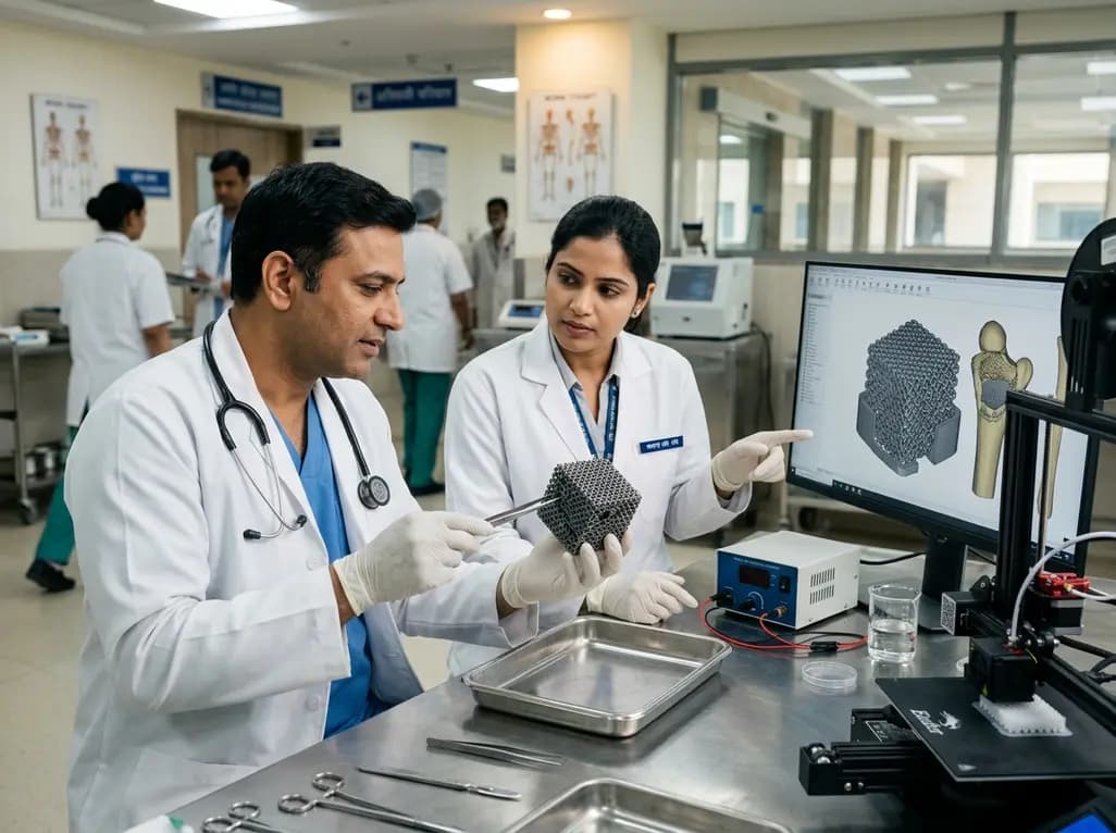
A novel sacrificial-template technique combines FDM polymer 3D printing and micro-arc oxidation to fabricate orientation-controlled porous titanium scaffolds. These bioactive constructs offer tailored stiffness, exceptional multidirectional load resistance, and robust osseointegration for bone defect repair.
Today
A novel label-free electrochemical biosensor utilizing magnetic nanocomposites enables ultrasensitive TERT C228T mutation detection (LOD: 0.67 pM) for rapid risk stratification in papillary thyroid cancer, offering promising point-of-care utility for endocrine oncology.
Today
