.webp&w=384&q=75)

Loading, please wait...
Loading, please wait...

Modern diagnostic imaging continually evolves through the integration of artificial intelligence and advanced biophotonic technologies. Specifically, photoacoustic computed tomography has emerged as a transformative modality that synergizes optical absorption contrast with high-resolution acoustic propagation. This hybrid technology offers substantial promise for non-invasive soft tissue assessment, particularly during breast cancer screening and diagnostic oncologic evaluations. However, clinicians and medical imaging researchers increasingly recognize that the rapid proliferation of deep learning algorithms requires rigorous, objective validation. Without standardized evaluation frameworks, reconstructed clinical images may harbor imperceptible artifacts that compromise diagnostic accuracy.
Photoacoustic computed tomography operates by delivering nanosecond laser pulses into biological tissue, inducing localized thermoelastic expansion. Consequently, this transient thermal event produces ultrasonic acoustic waves that travel outward to surrounding detector arrays. Dedicated inversion algorithms then reconstruct spatial maps of optical absorption, highlighting endogenous chromophores like hemoglobin. Therefore, this mechanism enables physicians to visualize functional microvasculature, tissue oxygenation, and tumor-associated angiogenesis with remarkable clarity. Furthermore, the optical contrast provides high biological specificity, whereas ultrasonic detection avoids the severe scattering typically encountered in pure optical imaging. Nevertheless, achieving high fidelity requires solving complex acoustic inverse problems that account for tissue heterogeneity and acoustic attenuation. Traditional analytical inversion methods, such as filtered backprojection, frequently demand exhaustive computational resources and dense sensor coverage. To overcome these operational constraints, investigators have increasingly deployed neural network architectures to reconstruct images from sparse or incomplete tomographic data. Although these deep learning approaches demonstrate exceptional computational acceleration, they present substantial challenges regarding generalization and diagnostic fidelity.
Historically, computational imaging researchers have relied on traditional image quality metrics, including peak signal-to-noise ratio and structural similarity index measure. However, these mathematical indices primarily quantify pixel-level intensity differences rather than clinically meaningful anatomical features. In clinical breast imaging, an algorithm might achieve impressive numerical scores while completely smoothing over subtle microcalcifications or architectural distortions. Furthermore, deep neural networks often minimize mean squared errors by generating perceptually pleasing but anatomically fabricated approximations. Consequently, an algorithm can easily hallucinate smooth parenchyma while obliterating tiny malignant lesions. This discrepancy poses critical risks in medical oncology, where early lesion detection directly dictates patient prognosis. Moreover, traditional metrics fail to evaluate whether a radiologist can reliably classify or delineate suspicious margins. Thus, reliance on non-clinical metrics has created an artificial sense of technical progress across photoacoustic computed tomography research. Radiologists require robust assurance that reconstruction algorithms preserve true pathology rather than optimizing cosmetic appearance.
To establish rigorous validation standards, researchers engineered a standardized open-source benchmarking framework for deep learning acoustic inversion. Specifically, the investigators generated over 10,000 two-dimensional stochastic breast numerical models reflecting anatomically plausible tissue variations. In addition, these numerical phantoms systematically incorporate clinically relevant lesions of varying sizes, shapes, and optical contrast levels. The team simulated photoacoustic wave propagation across multiple degrees of physical modeling complexity, capturing realistic acoustic reverberations and acoustic speed variations. By pairing known numerical ground truths with synthetic acoustic sensor measurements, this framework provides an unbiased testing platform. Furthermore, open-source access enables global development teams to train and benchmark diverse network architectures under identical conditions. Consequently, researchers can clearly distinguish genuine mathematical improvements from dataset-specific algorithmic artifacts. This rigorous standardization addresses longstanding concerns regarding reproducibility and algorithmic fragility in machine learning imaging studies.
The benchmarking initiative integrated both conventional fidelity measures and rigorous task-based image quality assessments to evaluate clinical utility. Specifically, task-based metrics directly examine how well a reconstruction preserves critical diagnostic information necessary for lesion detection and localization. Interestingly, the preliminary benchmarking study revealed alarming discrepancies between conventional metrics and true lesion recovery performance. While several deep learning models recorded superior structural similarity scores, they frequently failed to reconstruct subtle malignant lesions accurately. In contrast, certain physics-informed reconstruction methods preserved lesion margins much more reliably despite displaying slightly higher background noise. Consequently, these empirical findings demonstrate that high signal fidelity does not necessarily correlate with clinical efficacy. Therefore, adopting task-based evaluation protocols represents a necessary evolution in medical image reconstruction. Clinicians must prioritize algorithms that accurately maintain pathological margins, vascular boundaries, and microlesion conspicuity under challenging measurement conditions.
Ultimately, standardized benchmarking frameworks pave the way for successful clinical translation of photoacoustic computed tomography into hospital workflows. As healthcare institutions evaluate artificial intelligence tools for diagnostic breast imaging, verifiable algorithmic robustness becomes paramount. Furthermore, combining synthetic data benchmarking with task-based validation allows engineers to optimize transducer geometry, acoustic sampling rates, and laser parameters efficiently. Consequently, medical device developers can reduce costly hardware iterations while ensuring high diagnostic sensitivity. In addition, these computational methodologies could soon assist Indian diagnostic centers in screening younger women with dense breast tissue, where conventional mammography exhibits reduced sensitivity. However, multi-center clinical trials on diverse patient populations remain indispensable before widespread commercial adoption occurs. By bridging computational innovation with rigorous task-based validation, the medical community ensures that emerging imaging technologies genuinely elevate oncologic diagnostics and patient safety.
Traditional metrics like peak signal-to-noise ratio and structural similarity primarily quantify mathematical pixel variance across an entire image frame. Consequently, these metrics often reward algorithms that smooth out background noise while simultaneously erasing small pathological lesions. In contrast, clinical diagnosis requires accurate boundary preservation and lesion conspicuity. Therefore, traditional metrics can generate misleadingly high scores for algorithms that fail to detect clinically vital oncologic abnormalities during routine screening.
Synthetic datasets provide tens of thousands of precisely controlled, anatomically plausible numerical models with known ground truths. Consequently, investigators can systematically train neural networks across diverse tissue densities, variable acoustic speeds, and heterogeneous lesion morphologies. Furthermore, this standardized framework eliminates biological variability during early algorithmic development. As a result, developers can objectively benchmark different inversion architectures, detect subtle reconstruction artifacts, and enhance model generalization before initiating expensive human clinical trials.
Photoacoustic computed tomography combines optical absorption contrast with ultrasonic resolution, enabling functional visualization of tumor neoangiogenesis without ionizing radiation. Consequently, this modality offers significant clinical value for younger women presenting with dense breast tissue, where conventional mammography often exhibits decreased sensitivity. Furthermore, because malignant tumors stimulate extensive microvascular proliferation, photoacoustic imaging can assist oncologists in characterizing tumor margins, monitoring neoadjuvant chemotherapy response, and improving overall diagnostic specificity in clinical practice.
Disclaimer: This content is for informational and educational purposes only, and should not be construed as professional medical advice, diagnosis, or treatment. It does not replace clinical judgment or institutional protocols. Always consult an appropriate healthcare provider for medical decisions. While we endeavor to keep the information accurate and up to date, clinical knowledge changes rapidly. Refer to the latest local and national guidelines for clinical practice.
References

Read summarized clinical updates, watch expert medical content, and earn CME certifications right from your smartphone.
Open your smartphone camera to scan and install.





A study introduces a benchmarking framework using over 10,000 synthetic breast objects to evaluate deep learning image reconstruction in photoacoustic computed tomography, revealing critical diagnostic limitations of traditional image quality metrics.
Today

Novel biomimetic flexible polypyrrole sensors integrate mechanical hemodynamics with exhaled ammonia detection, offering noninvasive monitoring for chronic kidney disease and remote clinical care.
Today
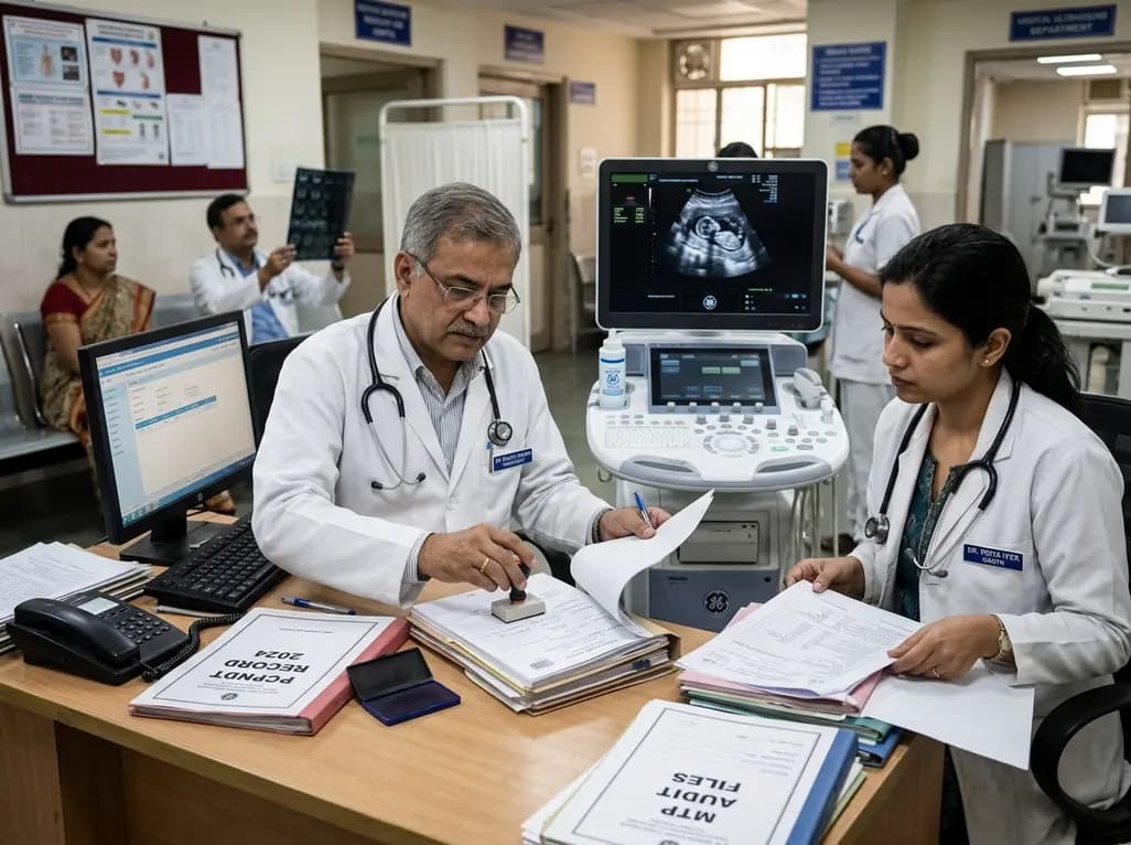
Maharashtra has initiated the appointment of two dedicated special public prosecutors in every district alongside a joint police-health SOP to fast-track PCPNDT and MTP cases. Medical imaging centers, radiologists, and obstetricians must strengthen statutory documentation and audit preparedness.
Today
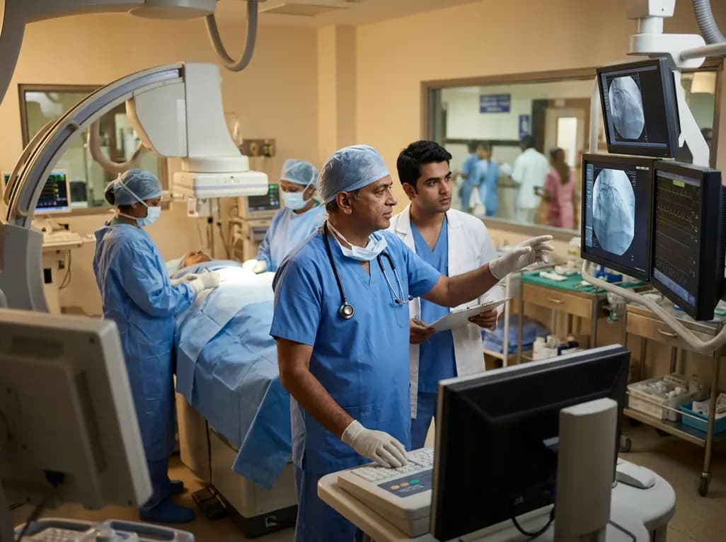
A prospective study evaluates percutaneous renal denervation in high-risk patients with resistant hypertension, demonstrating significant short-term blood pressure reduction alongside notable procedural considerations.
Today
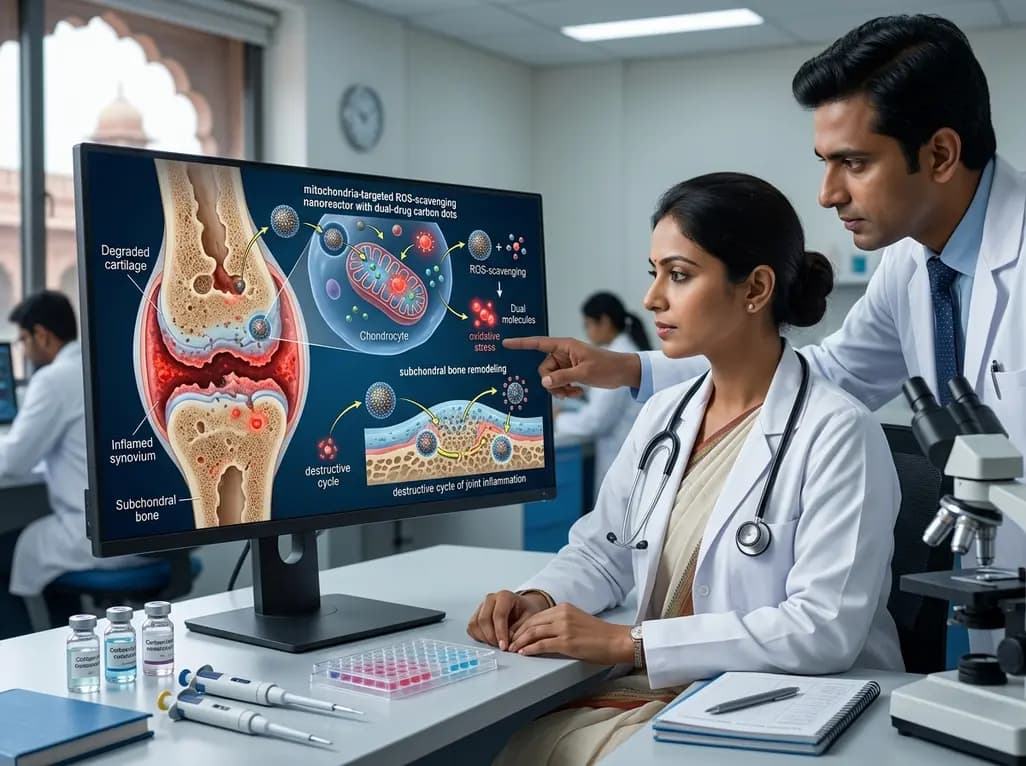
A novel mitochondria-targeted ROS-scavenging nanoreactor with dual-drug carbon dots provides a multi-mechanism strategy for osteoarthritis therapy. By neutralizing oxidative stress and remodeling subchondral bone, this platform breaks the destructive cycle of chronic joint inflammation and cartilage degeneration.
Today

A modified Delphi study identified crucial clinical and research priorities for older transgender adults. Key findings emphasize provider education, screening for social isolation, navigation of community resources, and long-term evaluation of gender-affirming hormone therapy safety during the aging process.
Today
