.webp&w=384&q=75)

Loading, please wait...
Loading, please wait...

Advanced molecular diagnostics have revolutionized modern neuro-oncology by refining tumor taxonomy and prognostic risk stratification. Over the past decade, high-throughput next-generation sequencing and DNA methylation profiling have become indispensable cornerstones in neuropathology. However, these established genomic technologies characterize static tumor identity rather than active cellular biochemistry. Functional phosphoproteomic profiling addresses this diagnostic gap by measuring real-time enzymatic signaling cascades in clinical tissue specimens. Central nervous system neoplasms display profound biological complexity that genomic sequencing cannot fully resolve. For example, glioblastomas frequently harbor epidermal growth factor receptor amplifications. Nevertheless, copy number alterations fail to predict therapeutic sensitivity to tyrosine kinase inhibitors reliably. Downstream signal transduction cascades undergo complex post-translational modifications that decouple enzymatic activity from genomic gene dosage. Consequently, measuring phosphorylation states provides an essential orthogonal dimension of biological information. Phosphoproteomics captures transient pathway dynamics and active kinase states within neoplastic cells. Furthermore, functional proteomic evaluations explain why tumors with identical genomic classifications often exhibit strikingly different clinical trajectories. Capturing this functional dimension therefore empowers oncologists to interpret tumor behavior with unprecedented molecular depth. Ultimately, integrating functional biochemistry with genomic diagnostics paves the way for precise therapeutic stratification.
Historically, comprehensive phosphoproteomic profiling required snap-frozen tissue to prevent rapid enzymatic dephosphorylation. However, fresh tissue preservation poses substantial logistical barriers in standard clinical practice across hospitals. In contrast, formalin-fixed, paraffin-embedded blocks represent the universal standard across pathology laboratories globally. Recent methodological advancements have successfully overcome the technical hurdles created by formalin cross-linking. Specifically, optimized heat-induced antigen retrieval and advanced detergent-compatible extraction protocols now preserve intact peptides efficiently. Additionally, ultra-sensitive mass spectrometry instruments achieve deep proteomic coverage from minute clinical specimens. A rigorous study demonstrated this capability by analyzing ten comprehensively characterized IDH-wildtype glioblastomas from routine archives. The researchers profiled five EGFR-amplified tumors alongside five non-amplified specimens. Remarkably, the analytical workflow quantified over seven thousand unique proteins per sample with outstanding reproducibility. Moreover, the platform identified more than thirteen thousand distinct phosphosites across the entire patient cohort. Comparable intensity distributions confirmed robust quantification across all analyzed specimens. These technical achievements conclusively show that routine pathology blocks retain highly stable, biologically coherent signaling information. Thus, neuropathologists can now evaluate archival cohorts without needing specialized liquid nitrogen infrastructure. Consequently, vast historical biobanks become immediately accessible for translational cancer research.
Global proteomic profiling revealed marked group-level elevations in EGFR abundance among amplified glioblastomas. However, substantial intertumoral overlap existed across individual specimens, highlighting the limitations of assessing protein expression alone. When investigators adjusted phosphoproteomic measurements for total protein abundance, they identified four hundred forty-three significantly altered phosphosites. Consequently, phosphoproteomic profiling revealed intricate signaling variations that standard genomic assays missed completely. Kinase-substrate enrichment analyses uncovered coordinated signaling cascades governed by EGFR and SRC-family kinases. Notably, individual tumors displayed striking variability in their downstream kinase engagement despite sharing identical genomic alterations. For instance, one amplified tumor exhibited pronounced SRC-family signaling, while another prioritized alternative downstream metabolic cascades. Therefore, phosphoproteomic profiling illuminates patient-specific functional circuitry rather than relying on assumed downstream effects. Furthermore, detecting activated secondary kinases uncovers critical bypass pathways that drive therapeutic resistance. Mapping these active enzymatic networks allows multidisciplinary tumor boards to select rational drug combinations tailored to individual patients. Ultimately, functional profiling transforms static diagnostic reports into dynamic blueprints of cellular enzymatic behavior. In addition, this granular functional insight explains why monotherapies frequently fail in clinical trials. As a result, clinicians gain actionable data to construct effective multi-targeted treatment regimens.
Glioblastoma represents one of the most heterogeneous primary brain neoplasms encountered in clinical oncology. Spatial variations in receptor tyrosine kinase expression frequently undermine the therapeutic efficacy of targeted monotherapies. For example, EGFR amplification often occurs in mosaic subclonal patterns throughout the resected tumor mass. Furthermore, alternative splicing events and secondary point mutations frequently alter downstream receptor signaling properties. Routine immunohistochemistry provides spatial localization but cannot determine whether the expressed receptor is catalytically active. In contrast, quantitative phosphoproteomics directly assesses receptor autophosphorylation and the activation status of downstream transducers. By measuring specific tyrosine phosphorylation sites, neuropathologists can distinguish active oncogenic drivers from inert passenger proteins. Additionally, phosphoproteomic profiling identifies compensatory feedback loops that tumors activate when exposed to therapeutic stress. Specifically, co-activation of parallel receptor tyrosine kinases often sustains downstream cell survival when clinicians inhibit EGFR. Understanding these intricate interactions helps neuro-oncologists anticipate treatment resistance early in the disease course. Therefore, comprehensive functional characterization provides an effective solution to the challenge of intratumoral signaling heterogeneity. Consequently, incorporating these functional measurements refines clinical decision-making significantly. Moreover, assessing functional dynamics enables clinicians to track biological evolution over time. Thus, oncologists can adapt treatment strategies proactively as tumors acquire secondary resistance mechanisms.
Integrating functional proteomics into clinical oncology workflows holds immense therapeutic potential for tertiary cancer centres across India. Currently, neuro-oncologists rely heavily on targeted next-generation sequencing panels to identify actionable alterations. However, many glioblastoma patients fail to achieve meaningful responses with genotype-matched targeted therapies. Incorporating phosphoproteomic profiling from archival FFPE specimens enables clinicians to confirm active target engagement before prescribing costly therapies. Furthermore, retrospective analyses of archived trial specimens can uncover functional biomarkers that explain past trial failures. This retrospective approach permits investigators to re-evaluate previously discontinued kinase inhibitors within specifically enriched patient subsets. Additionally, standardizing FFPE proteomics streamlines translational research by eliminating the logistical requirement of maintaining liquid nitrogen cryobanks. Pathologists can readily utilize excess tissue sections from standard diagnostic biopsies for deep multi-omic characterization. Consequently, this technology bridges the gap between academic discovery and daily clinical neuropathology practice. As analytical turnaround times decrease, routine functional profiling will guide individualized neuro-oncology treatment regimens with exceptional precision. In addition, expanding local testing infrastructure reduces diagnostic delays for critically ill patients. Therefore, Indian clinicians can implement timely, personalized interventions that improve patient outcomes. Ultimately, establishing standardized proteomic workflows will democratize advanced molecular diagnostics across regional cancer hospitals. Together, these advancements represent a major leap forward for oncology.
Recent protocol enhancements overcome formalin cross-linking through optimized heat decrosslinking and refined extraction buffers. Consequently, mass spectrometry can consistently identify thousands of intact peptides and regulatory phosphosites from archived pathology archives. This development confirms that routine clinical blocks reliably preserve functional phosphorylation states despite prolonged room-temperature storage.
Genomics and epigenomics establish tumor classification and static structural variations effectively. However, they cannot reveal real-time pathway activation or post-translational signaling changes. Phosphoproteomics measures active intracellular signaling cascades directly. Therefore, it bridges the gap between molecular categorization and actionable drug targets, providing a comprehensive view of active tumor biology.
Kinase-substrate enrichment analysis maps active kinase cascades, such as coordinated EGFR and SRC-family signaling networks. Consequently, oncologists can identify functional enzymatic drivers rather than relying solely on gene amplification status. This functional characterization supports rational multi-kinase inhibitor combinations, helping overcome common therapeutic resistance mechanisms in recurrent glioblastomas.
Disclaimer: This content is for informational and educational purposes only... Refer to the latest local and national guidelines for clinical practice.
References

Read summarized clinical updates, watch expert medical content, and earn CME certifications right from your smartphone.
Open your smartphone camera to scan and install.





A landmark study demonstrates that phosphoproteomic profiling of routine FFPE CNS tumor tissue captures functional signaling networks, complementing genomic and epigenomic classifications to advance precision neuro-oncology.
Today
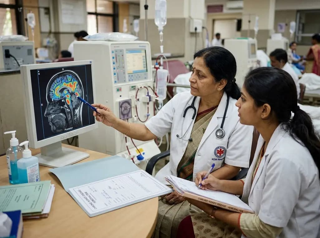
A retrospective clinical analysis reveals that hemodialysis-associated cognitive dysfunction arises from synergistic pathological mechanisms, including severe chronic anemia, worsening renal clearance, abnormal regional brain iron deposition, and an imbalance between neuroinflammatory and neurotrophic factors.
Today

A multiparametric MRI approach combining quantitative T2 shading, apparent diffusion coefficient (ADC) measurements, and morphological signs provides superior diagnostic accuracy over subjective evaluation to differentiate ovarian endometriomas from benign hemorrhagic cysts.
Today

Discover a novel laparoscopic approach for iatrogenic diaphragmatic hernia repair following pedicled omentoplasty. This modified Sugarbaker technique integrates dorsal pedicle lateralization and round ligament reinforcement to secure the diaphragmatic defect while preserving vital omental vascularity.
Today
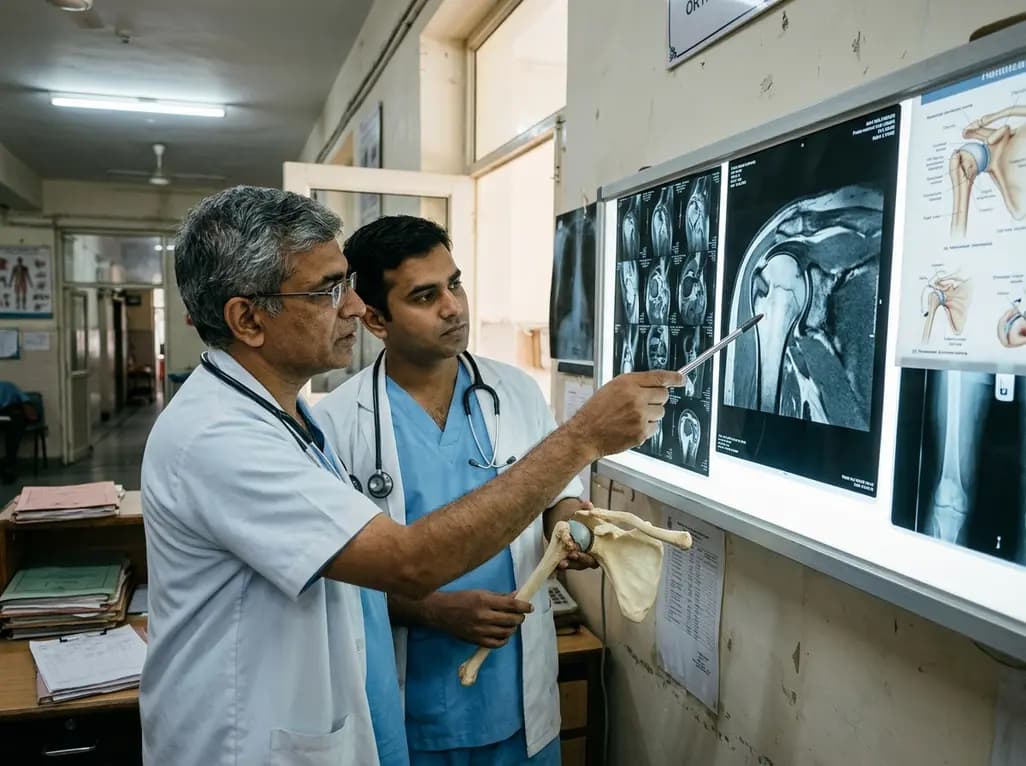
Evaluating Hill-Sachs lesion size and the distance to dislocation is critical in recurrent shoulder instability. As bone loss concepts shift from binary on-track classifications to continuous risk assessment, clinicians must identify when isolated Bankart repair is insufficient and supplementary procedures are needed.
Today

New research reveals that M1 macrophage-derived exosomes aggravate diabetic nephropathy by transferring WTAP to stabilize S1PR2 mRNA. Silencing WTAP in these vesicles attenuates endothelial injury and renal fibrosis, pointing toward innovative nanomedicine therapies.
Today
