

Loading, please wait...
Loading, please wait...
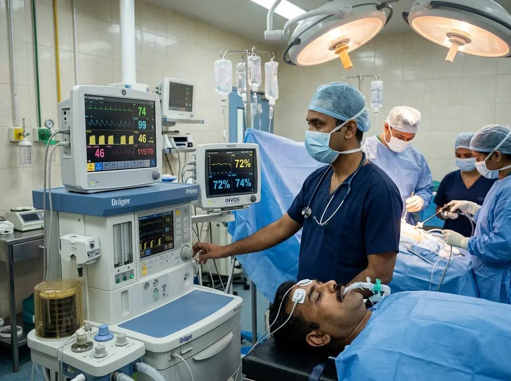
Intraoperative management of ventilation plays a crucial role in maintaining organ perfusion during major surgery. In recent years, permissive hypercapnia has emerged as a promising strategy to mitigate intraoperative cerebral hypoxia and secondary neurological injury. Emerging clinical evidence suggests that mild elevations in carbon dioxide tension may optimize microvascular cerebral perfusion. Consequently, clinicians are re-evaluating conventional normocapnic targets to determine whether controlled hypercapnia can improve outcomes.
Cerebral blood flow closely mirrors fluctuations in arterial carbon dioxide partial pressure. Specifically, carbon dioxide readily diffuses across the blood-brain barrier to alter perivascular extracellular pH. This local acidosis triggers vascular smooth muscle relaxation and induces profound cerebral arteriolar vasodilation. As a direct result, cerebral blood flow increases and regional oxygen delivery rises significantly. In contrast, standard normocapnia or accidental hyperventilation promotes vasoconstriction, which potentially exposes brain tissues to ischemic stress. Furthermore, mild hypercapnia shifts the oxyhemoglobin dissociation curve to the right through the classical Bohr effect. This physiologic shift facilitates oxygen unloading directly to metabolically active brain cells. Consequently, regional cerebral tissue saturation improves during prolonged anesthesia. In addition, experimental models indicate that moderate arterial carbon dioxide elevations can suppress systemic inflammatory cytokine cascades. Therefore, this ventilatory approach protects microvascular integrity and reduces cellular metabolic stress. These combined physiological actions establish a robust scientific rationale for maintaining mildly elevated carbon dioxide levels during high-risk surgical procedures.
A recent comprehensive systematic review and meta-analysis synthesized data from seven clinical trials comprising 824 adult surgical patients. Specifically, the investigators systematically compared permissive hypercapnia against conventional normocapnia across diverse operative settings. Most notably, the meta-analysis revealed that permissive hypercapnia significantly improved intraoperative regional cerebral oxygen saturation, showing a mean difference of 6.65 percent. Furthermore, this substantial improvement in cerebral tissue oxygenation remained consistent across various surgical models, including laparoscopic interventions and one-lung ventilation. In addition, the researchers evaluated critical secondary outcomes, such as postoperative cognitive dysfunction and postoperative delirium. Accordingly, patients managed with controlled hypercapnic targets demonstrated lower rates of neurocognitive decline during early recovery. Moreover, the analysis demonstrated that arterial blood gas parameters and pulmonary compliance remained favorable in the hypercapnia cohorts. Importantly, the meta-analysis found no increase in all-cause mortality or severe adverse hemodynamic complications. Thus, these findings provide compelling quantitative validation that maintaining mildly elevated arterial carbon dioxide safely enhances cerebral tissue oxygenation during major operations.
Postoperative cognitive impairment represents a severe complication that disproportionately affects elderly surgical candidates. Indeed, intraoperative cerebral hypoperfusion and occult tissue hypoxia act as primary drivers for both delirium and delayed cognitive dysfunction. When cerebral oxygen saturation drops below baseline thresholds, neuronal oxidative distress escalates rapidly. Consequently, neuroinflammation ensues and disrupts fragile synaptic networks. Specifically, the latest evidence demonstrates that preserving adequate regional oxygenation through tailored ventilation directly mitigates these neurocognitive risks. For instance, patients who maintain higher regional saturation scores during surgery exhibit superior performance on standardized cognitive assessments following extubation. Furthermore, researchers observe significantly lower circulating levels of central nervous system injury biomarkers, including S100-beta and neurofilament light chains. Therefore, elevating microvascular blood flow appears to shield vulnerable cortical and subcortical neurons from perioperative injury. In addition, avoiding cognitive decline shortens intensive care unit stays and decreases post-anesthesia care unit recovery times. Ultimately, these clinical improvements translate into enhanced patient independence and reduced long-term healthcare expenditure.
Implementing hypercapnic ventilation requires clinicians to carefully balance respiratory physiology and systemic hemodynamics. Although elevated carbon dioxide augments cardiac output through sympathetic activation, excessive hypercapnia can produce myocardial depression and severe arrhythmias. Fortunately, clinical trials show that maintaining carbon dioxide between 45 and 55 millimeters of mercury preserves hemodynamic stability without requiring high inotropic support. Furthermore, controlled hypercapnia facilitates lung-protective ventilation by permitting lower tidal volumes and reduced airway driving pressures. Consequently, this protective ventilatory strategy diminishes mechanical shear stress and reduces the incidence of postoperative pulmonary complications, such as atelectasis. However, anesthesiologists must continuously evaluate baseline right ventricular function and pulmonary vascular resistance before initiating this protocol. Because hypercapnic acidosis can elevate pulmonary arterial pressure, clinicians should exercise caution in patients with preexisting pulmonary hypertension. Therefore, diligent real-time monitoring of arterial blood gases and invasive arterial pressures remains mandatory. Through meticulous surveillance, healthcare teams can safely deliver the neuroprotective benefits of hypercapnic ventilation while preserving stable cardiovascular parameters.
To successfully incorporate permissive hypercapnia into routine practice, multidisciplinary surgical teams must establish structured patient selection criteria. Specifically, high-risk candidates undergoing prolonged laparoscopic procedures, steep Trendelenburg positioning, or thoracic one-lung ventilation derive substantial neurovascular benefits from this strategy. Accordingly, anesthesiologists should utilize near-infrared spectroscopy to monitor bilateral regional cerebral oxygen saturation continuously throughout the operative period. In addition, clinicians must titrate minute ventilation carefully to maintain end-tidal carbon dioxide within the predefined therapeutic window. Conversely, certain patient populations require strict exclusion from hypercapnic ventilation protocols. For instance, individuals with elevated intracranial pressure, acute traumatic brain injury, or severe cerebral edema must not undergo deliberate hypercapnia, as hypercapnic vasodilation can precipitate catastrophic intracranial hypertension. Similarly, patients presenting with severe uncompensated metabolic acidosis or severe chronic obstructive pulmonary disease necessitate individualized management. Therefore, structured institutional checklists and standardized operating room protocols ensure that eligible patients receive targeted neuroprotection without compromising safety. Ultimately, this tailored strategy fosters enhanced recovery pathways and improves surgical care.
Moving forward, intraoperative ventilatory management will increasingly rely on personalized physiological endpoints rather than rigid generic targets. Although recent meta-analytic data substantiate the cerebral oxygenation benefits of permissive hypercapnia, future randomized trials should determine optimal carbon dioxide thresholds across diverse subspecialties. Specifically, investigating long-term functional recovery, quality of life, and sustained neurocognitive outcomes beyond the immediate postoperative hospital stay will clarify enduring patient benefits. Furthermore, integrating multimodal neuromonitoring tools, such as processed electroencephalography alongside cerebral oximetry, will permit finer titration of ventilation to individual neurovascular autoregulation. In addition, evaluating the synergistic interaction between lung-protective mechanical ventilation and goal-directed fluid therapy will optimize microcirculatory perfusion throughout the perioperative continuum. Ongoing clinical trials will also establish clearer safety profiles for patients with borderline cardiovascular compromise or mild cerebrovascular disease. Therefore, continued clinical investigation into individualized hypercapnic targets promises to refine anesthesiology practice and elevate standard surgical care. Ultimately, adopting precision ventilation strategies will transform perioperative neuroprotection for high-risk surgical cohorts worldwide.
Permissive hypercapnia directly enhances cerebral tissue protection by raising arterial carbon dioxide levels, which prompts cerebral arteriolar vasodilation and augments cerebral blood flow. Furthermore, it shifts the hemoglobin-oxygen dissociation curve to the right, enabling easier oxygen release to brain tissues. Consequently, this technique elevates regional cerebral oxygen saturation, prevents covert intraoperative cerebral ischemia, and attenuates perioperative neuroinflammation. These combined mechanisms collectively lower the vulnerability of neurons during lengthy surgical interventions.
Clinicians must strictly avoid deliberate hypercapnia in patients diagnosed with intracranial hypertension, intracranial mass lesions, acute traumatic brain injury, or severe cerebral edema. In these clinical scenarios, hypercapnia-induced vasodilation significantly increases cerebral blood volume and dangerously accelerates intracranial pressure. Additionally, severe uncompensated metabolic acidosis, severe pulmonary hypertension, and unstable cardiac arrhythmias represent important relative contraindications. Therefore, thorough preoperative neurological and cardiovascular assessments remain essential prior to selecting patients for hypercapnic ventilation.
Anesthesiologists should utilize continuous near-infrared spectroscopy to track bilateral regional cerebral oxygen saturation in real time. Simultaneously, clinicians must monitor end-tidal carbon dioxide alongside regular arterial blood gas analyses to ensure arterial carbon dioxide remains within the intended target range. Continuous invasive arterial blood pressure monitoring and electrocardiography are also vital to promptly detect any hemodynamic instability. Furthermore, serial evaluations of ventilatory mechanics ensure lung-protective ventilation without creating excessive intrinsic positive end-expiratory pressure.
Disclaimer: This content is for informational and educational purposes only... Refer to the latest local and national guidelines for clinical practice.
References

Read summarized clinical updates, watch expert medical content, and earn CME certifications right from your smartphone.
Open your smartphone camera to scan and install.





A systematic review and meta-analysis reveals that intraoperative permissive hypercapnia significantly enhances regional cerebral oxygen saturation compared with normocapnia in adult surgical patients, offering neuroprotective benefits without compromising hemodynamic stability.
Today

A cross-sectional study reveals a 20.5% prevalence of GERD among medical students, identifying midnight snacking and postprandial exercise patterns as major independent determinants. Addressing these behavioral factors is vital for gastrointestinal health and academic performance.
Today

AIIMS researchers suggest yoga may alleviate gut dysbiosis in Polyendocrine Metabolic Ovarian Syndrome (PMOS) by modulating the gut-brain axis, lowering chronic inflammation, and enhancing parasympathetic tone to support metabolic and hormonal equilibrium.
Today

A prospective cohort study demonstrates that feeding tubes, formula exposure, aortic cross-clamp duration, and socioeconomic deprivation disrupt the neonatal gut microbiome in congenital heart disease, emphasizing the need for targeted nutritional and perioperative gut-protective strategies.
Today

Deep intramuscular vascular malformations in pediatric Klippel-Trenaunay syndrome can mimic Maffucci syndrome. Multimodality imaging with Doppler ultrasound and MRI, along with histopathology, ensures prompt differential diagnosis, targeted surgical debulking, and optimized conservative surveillance.
Today

A longitudinal feasibility pilot study highlights that combining polygenic risk score disclosure with in-person lifestyle counseling significantly enhances genomic literacy in individuals at risk for type 2 diabetes, overcoming educational disparities and reinforcing patient motivation.
Today
