.webp&w=384&q=75)

Loading, please wait...
Loading, please wait...
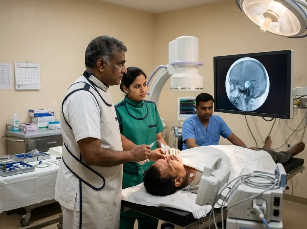
Percutaneous balloon compression serves as an essential, minimally invasive intervention for medically refractory trigeminal neuralgia, offering rapid pain relief to patients who cannot tolerate open microvascular decompression. For decades, neurosurgeons relied on standardized, commercially packaged kits to execute the procedure with predictable mechanical precision. However, the commercial withdrawal of the dedicated Mullan ganglion microcompression kit in 2016 created an unexpected global supply challenge. Consequently, surgical teams across tertiary centers had to adapt their workflows using alternative modular devices. Today, modern surgical teams successfully employ adaptable instrumentation frameworks to achieve excellent clinical outcomes. This comprehensive review examines current procedural adaptations, updated anatomical targeting strategies, and standardized protocols that ensure safety and procedural efficacy in contemporary trigeminal rhizotomy.
The withdrawal of the primary Food and Drug Administration-approved microcompression system fundamentally altered interventional pain practice. Previously, pre-packaged sets provided matched needles, stylets, and calibration tools that guaranteed standardized balloon positioning. In response to this market departure, neurosurgeons developed modular setups by combining readily available off-the-shelf equipment. Specifically, practitioners transitioned toward combining standard angiographic catheters, high-pressure stopcocks, and generic puncture needles. Consequently, surgical teams observed significant procedural variability across different health systems and regions. While some institutions developed proprietary bundled systems, others adopted technology-assisted workflows incorporating three-dimensional imaging or robotic guidance. Furthermore, international clinical registries confirmed that modular adaptations maintained therapeutic efficacy without increasing major complication rates. Therefore, procedural adaptability has successfully bridged the supply void, allowing centers worldwide to preserve this critical pain-relieving procedure for geriatric and medically fragile patients.
Accessing the trigeminal ganglion requires strict adherence to anatomical landmarks established by the classical Hartel percutaneous approach. Traditionally, the surgeon inserts the needle through the cheek, roughly 2.5 centimeters lateral to the oral commissure. The operator then directs the needle toward the intersection of two trajectories: one aimed at the ipsilateral pupil and another toward a point anterior to the external auditory meatus. Recent refinements emphasize modern fluoroscopic landmarks to improve target accuracy and minimize radiation exposure. In particular, clinicians frequently utilize the submental-vertex view and the fluoroscopic "H-figure" landmark to align the needle trajectory with the foramen ovale. Moreover, rotational C-arm cone-beam computed tomography provides real-time cross-sectional verification, thereby preventing accidental injury to adjacent vascular channels. As a result, modern operators cannulate the foramen ovale more efficiently, reducing tissue trauma, multiple needle passes, and procedural duration significantly.
Contemporary technical protocols rely primarily on a 13-gauge to 16-gauge puncture needle paired with a 3 Fr to 5 Fr Fogarty arterial embolectomy catheter. Because standardized commercial trocars are no longer readily accessible, surgeons frequently utilize bone marrow biopsy needles, Tuohy needles, or specialized spinal introducers. Therefore, careful assembly and inspection of the catheter-needle interface before skin puncture remain vital. The operator introduces the Fogarty catheter through the needle lumen once the needle tip engages the osseous boundary of the foramen ovale. Furthermore, clinicians must ensure that the catheter tip extends just beyond the needle bevel into the petrous apex. Using non-compliant connection tubing and low-friction stopcocks prevents inadvertent catheter recoil during advancing maneuvers. Consequently, these modular configurations replicate the mechanical performance of historical commercial sets while maintaining broad accessibility across varied clinical settings.
Achieving a true pear-shaped balloon configuration within Meckel's cave represents the technical hallmark of successful percutaneous balloon compression. Once the catheter reaches the trigeminal cistern, the surgeon slowly injects a non-ionic water-soluble contrast medium under continuous lateral fluoroscopic visualization. As the balloon expands, the tight dural aperture of the porus trigeminus constricts its proximal portion, creating the pathognomonic pear shape. If the balloon remains spherical or cylindrical, it indicates incorrect positioning in the subtemporal space or posterior fossa. Thus, the clinician must immediately deflate and reposition the catheter to avoid neurovascular complications. Most centers employ volume-controlled inflation, infusing approximately 0.5 to 1.0 milliliters of contrast agent for 60 to 90 seconds. Although real-time pressure transducers can measure intracavitary pressures, quantitative pressure monitoring remains limited to specialized academic protocols.
During balloon inflation, mechanical compression selectively injures large-diameter myelinated pain-mediating fibers while preserving smaller unmyelinated touch pathways. Consequently, patients experience immediate postoperative trigeminal analgesia with low rates of corneal anesthesia. However, surgeons must anticipate significant hemodynamic shifts during balloon inflation, particularly the trigeminocardiac reflex. This sudden autonomic surge often causes profound bradycardia, hypotension, or even transient asystole. Therefore, anesthesiologists must administer prophylactic anticholinergic agents, such as atropine or glycopyrrolate, before balloon expansion. Postoperative clinical assessments consistently demonstrate initial pain relief rates exceeding 90 percent across large institutional series. In addition, common minor adverse effects include transient masticatory muscle weakness and mild facial hypoesthesia, both of which typically resolve within three to six months.
The ongoing evolution of percutaneous balloon compression demonstrates that modularity and innovation can overcome major medical supply disruptions. Although the absence of universal commercial kits initially caused methodological fragmentation, contemporary literature reveals an emerging, highly consistent procedural consensus. Today, neurosurgeons routinely combine 4 Fr Fogarty catheters, 14-gauge introducers, biplane fluoroscopy, and volume-controlled contrast injection. Furthermore, the integration of advanced navigational aids and intraoperative three-dimensional imaging continues to lower the learning curve for early-career operators. In low-resource and high-volume centers, these cost-effective modular techniques provide sustainable solutions for treating medically refractory trigeminal neuralgia. Ultimately, standardizing institutional training protocols and sharing reproducible operative workflows will ensure that patients worldwide continue to receive safe, durable, and immediate relief from debilitating neuropathic facial pain.
The procedure works by mechanically compressing the Gasserian ganglion within Meckel's cave using an inflated balloon catheter. This targeted compression selectively disrupts medium and large myelinated nerve fibers responsible for transmitting lancinating paroxysmal pain signals. Consequently, it stops painful neuropathic discharges while sparing smaller unmyelinated fibers, thereby preserving basic corneal reflex function and sensory protective mechanisms across the affected facial dermatomes.
The characteristic pear shape develops when the inflated Fogarty balloon conforms to the rigid boundaries of Meckel's cave. The narrow neck of the pear corresponds to the porus trigeminus, where the dura restricts balloon expansion. Simultaneously, the broader body of the balloon expands into the trigeminal cistern. Fluoroscopic visualization of this morphology confirms accurate anatomical placement, guaranteeing adequate ganglion compression and optimal clinical outcomes.
The trigeminocardiac reflex arises from sudden mechanical stimulation of the trigeminal ganglion, causing acute vagal hyperactivation, profound bradycardia, and severe hypotension. Anesthesia teams mitigate this autonomic response by administering prophylactic anticholinergic medications, including atropine or glycopyrrolate, immediately before balloon inflation. Additionally, continuously monitoring hemodynamic parameters and briefly pausing inflation during severe heart rate drops ensure patient stability throughout the compression cycle.
Disclaimer: This content is for informational and educational purposes only and is not intended to serve as medical advice, diagnosis, or treatment. Healthcare professionals should rely on their clinical judgment and consult relevant medical literature and institutional protocols before making treatment decisions. Refer to the latest local and national guidelines for clinical practice.
References
Ranalli C et al. How Percutaneous Balloon Compression Is Performed Today: Technical Adaptations for Trigeminal Neuralgia in the Absence of Commercial Kits. Oper Neurosurg. 2026 Sep 01. doi: 10.1227/ons.0000000000002181. PMID: 42679315.
Mullan S, Lichtor T. Percutaneous microcompression of the trigeminal ganglion for trigeminal neuralgia. J Neurosurg. 1983;59(6):1007-1012.
Urculo E, Alfaro R, Arrazola M, et al. The transformation of the balloon shape in percutaneous balloon compression for trigeminal neuralgia. J Pain Res. 2021;14:3825-3834.

Read summarized clinical updates, watch expert medical content, and earn CME certifications right from your smartphone.
Open your smartphone camera to scan and install.





An evidence-based overview of percutaneous balloon compression for trigeminal neuralgia, reviewing post-kit technical adaptations, fluoroscopic landmarks, and modular toolkits.
4 weeks back

A nationwide Swedish cohort study shows oral glucocorticoid exposure over 1-2 years strongly predicts overall organ damage in SLE, while cardiovascular risk reflects cumulative doses across 3 years. These findings reinforce early steroid minimization and cautious long-term maintenance.
Today

A pharmacometric and machine learning study reveals that specific vaginal bacteria alter cervical antiretroviral exposure, highlighting the need for precision HIV PrEP strategies.
Today

New clinical evidence demonstrates a shift away from strict exercise restrictions in genetic heart disease. While patients with channelopathies engage in more vigorous activity than those with cardiomyopathies, shared decision-making and individualized risk assessments ensure safe, beneficial exercise participation.
Today
A scoping review highlights neural mobilization as a promising adjunct in managing cervicogenic headache. Integrating neurodynamics into multimodal physical therapy improves pain, range of motion, and posture, though standardized protocols and larger clinical trials are still needed.
Today

Recent research defines ferro-aging as a conserved iron-lipid axis driving organ decline across primates. Age-related iron dyshomeostasis stimulates ACSL4-mediated lipid peroxidation, promoting cellular senescence. Notably, vitamin C directly inhibits ACSL4, suppressing phospholipid oxidation and multi-organ decay.
Today
