.webp&w=384&q=75)

Loading, please wait...
Loading, please wait...
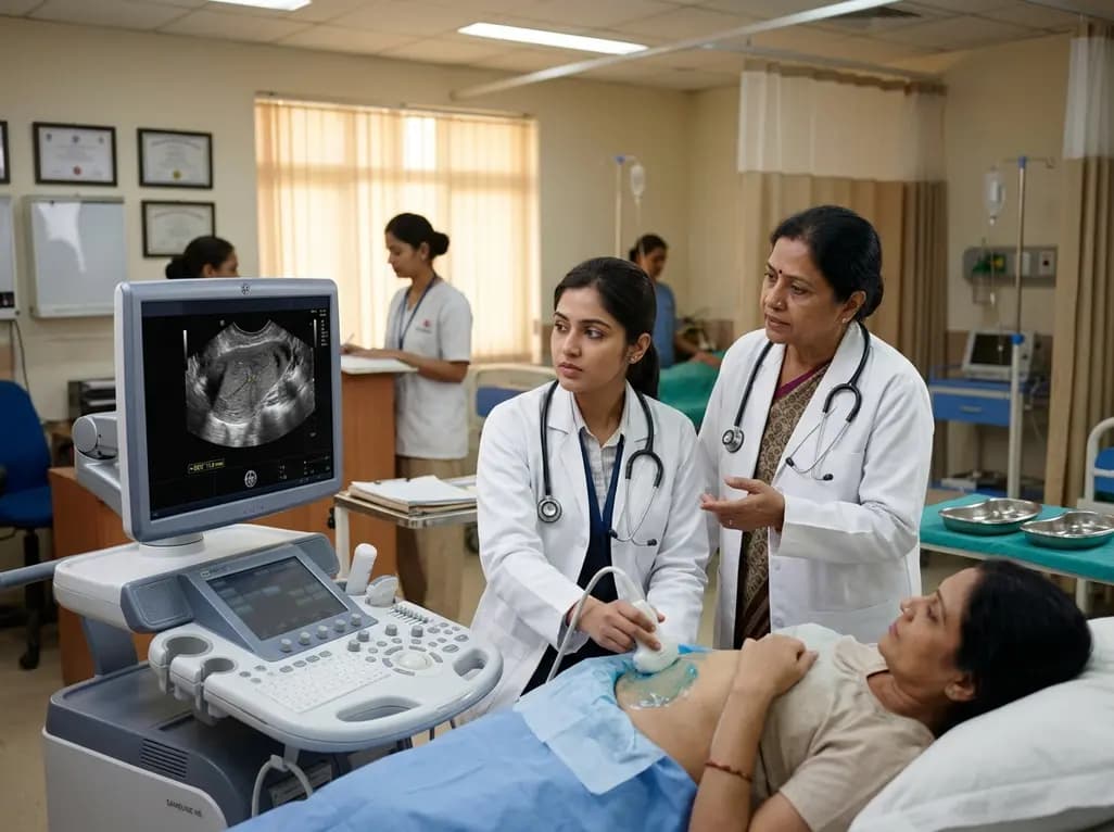
Abnormal uterine bleeding represents one of the most frequent gynecologic complaints in reproductive-aged individuals worldwide. Clinicians routinely utilize pelvic ultrasound as an initial diagnostic modality, yet debate persists regarding its predictiveness for premalignant lesions. Managing premenopausal abnormal uterine bleeding requires balancing timely oncologic detection against invasive procedures. A landmark study in the Journal of Ultrasound Medicine now provides critical evidence clarifying sonographic accuracy across bleeding etiologies.
Premenopausal abnormal bleeding encompasses diverse structural and nonstructural etiologies. Historically, clinicians have relied on postmenopausal endometrial thickness cutoffs to guide sampling. However, cyclical hormonal fluctuations in younger women introduce wide variations in normal endometrial stripe measurements. Consequently, establishing definitive sonographic thresholds has remained challenging.
Furthermore, medical consensus has questioned whether pelvic ultrasound reliably excludes malignancy in younger cohorts. Endometrial carcinoma occurs infrequently before menopause, but delayed recognition can cause devastating outcomes. Specifically, patients with chronic ovulatory dysfunction experience prolonged unopposed estrogen exposure, markedly increasing oncologic risk.
Therefore, practitioners frequently struggle with decisions regarding invasive tissue sampling versus conservative follow-up. While biopsy provides definitive histopathology, unnecessary procedures cause patient discomfort, anxiety, and substantial medical costs. In contrast, missing precursor lesions allows disease progression. Hence, clear evidence-based criteria are vital.
To resolve this diagnostic challenge, investigators conducted a cross-sectional study of 3,791 premenopausal patients presenting with abnormal bleeding between 2013 and 2021. Notably, all participants underwent pelvic ultrasound followed by endometrial sampling within 24 hours. This standardized timeline eliminated confounding effects from interval hormonal changes.
Researchers categorized bleeding etiologies using the FIGO PALM-COEIN classification system. Among the cohort, 23.6% had isolated anovulatory bleeding (AUB-O), 30.5% presented with AUB-O alongside other etiologies, and 45.9% had non-anovulatory causes. Additionally, histopathologists classified specimens into benign tissue, cancer precursors, and true endometrial neoplasia.
Moreover, multivariable logistic regression and predictive value analyses compared ultrasound parameters against pathology findings. By linking standardized imaging directly to immediate histology, the study established robust, real-world evidence regarding sonographic diagnostic accuracy across diverse clinical subsets.
The study demonstrated that pelvic ultrasound delivers excellent diagnostic accuracy for identifying endometrial pathology. Most importantly, researchers identified no cases of endometrial hyperplasia or carcinoma in patients with an endometrial thickness below 4 mm. Therefore, an endometrial stripe under 4 mm provides near-perfect negative predictive value.
Conversely, multiple sonographic abnormalities dramatically increased disease probability. When ultrasound demonstrated combined abnormalities, including severe thickening, heterogeneous architecture, and irregular margins, the probability of neoplasia or preneoplasia approached 1.0. Additionally, solitary abnormal findings correlated significantly with abnormal histopathology across all bleeding subtypes.
Furthermore, correlation between abnormal sonographic findings and malignant pathology was strongest in patients with isolated anovulatory bleeding. Because persistent anovulation drives unopposed proliferation, ultrasound signs in this subgroup proved exceptionally discriminative. Thus, sonography effectively distinguishes benign endometrium from high-risk proliferative changes.
These findings provide a clear risk stratification pathway for daily clinical practice. Clinicians can confidently use a thin endometrial measurement to defer invasive biopsy in low-risk premenopausal individuals. Specifically, when transvaginal ultrasound reveals a uniform endometrial stripe thinner than 4 mm, underlying neoplastic risk is negligible.
However, clinicians must evaluate sonography alongside patient-specific risk profiles. Conditions such as polycystic ovary syndrome, obesity, and prolonged amenorrhea substantially elevate baseline oncologic vulnerability. Therefore, when patients with chronic anovulation exhibit sonographic abnormalities, clinicians should promptly obtain endometrial tissue.
Additionally, sonographic evaluation must assess structural contour and intracavitary masses rather than thickness alone. Focal thickening and polypoid lesions require tissue sampling even in younger patients. Consequently, combining morphology with clinical history prevents both unnecessary procedures and missed diagnoses.
In outpatient settings, particularly across India, pelvic ultrasound remains the primary, cost-effective imaging modality. Many women presenting with abnormal bleeding initially visit primary health centres or community clinics where advanced diagnostic equipment is unavailable.
Consequently, confirming that an endometrial thickness under 4 mm safely excludes neoplasia offers immense clinical utility. Healthcare providers in resource-limited environments can reassure patients with thin stripes while prioritizing tissue sampling for suspicious imaging. Moreover, the high prevalence of polycystic ovary syndrome and metabolic dysfunction in South Asian populations underscores the need for structured triage.
To maximize diagnostic precision, sonographers should schedule transvaginal ultrasound during the early follicular phase whenever possible. During this window, normal endometrium appears thinnest and most homogeneous, minimizing false-positive suspicion. Additionally, operators must measure maximal double-layer thickness on a strict midsagittal view.
Furthermore, when evaluating intracavitary lesions, saline infusion sonohysterography or colour Doppler provides valuable architectural details. Clinicians should systematically document echogenicity, margins, and vascularity. If pelvic visualization remains limited due to large leiomyomas, magnetic resonance imaging provides helpful adjunctive clarity.
Ultimately, sonographic findings should drive patient counseling and collaborative management. Clinicians must clearly explain imaging results, risk factors, and monitoring plans. Integrating high-quality ultrasound with targeted histopathological sampling ensures superior diagnostic safety and patient outcomes.
Yes, pelvic ultrasound demonstrates strong negative predictive value for ruling out endometrial cancer in premenopausal women. Recent large-scale clinical evidence indicates that an endometrial thickness below 4 mm carries virtually zero risk of endometrial hyperplasia or carcinoma. Therefore, clinicians can safely avoid immediate endometrial sampling in asymptomatic or low-risk premenopausal patients presenting with normal, thin sonographic findings and absence of focal intracavitary lesions.
Premenopausal individuals require endometrial sampling when ultrasound demonstrates abnormal findings, such as an endometrial thickness exceeding normal thresholds, heterogeneous architecture, or focal masses. Furthermore, clinicians must promptly biopsy patients presenting with chronic anovulatory bleeding, obesity, polycystic ovary syndrome, or therapy-resistant abnormal uterine bleeding. When multiple sonographic abnormalities coexist with chronic anovulation, the probability of underlying premalignant or malignant pathology approaches nearly one hundred percent.
Ovulatory dysfunction significantly enhances the predictiveness of pelvic ultrasound for endometrial neoplasia. In patients with isolated anovulatory bleeding, persistent unopposed estrogen exposure causes chronic endometrial stimulation, increasing neoplastic risk. Consequently, sonographic abnormalities in this group correlate most strongly with premalignant and malignant histopathology. Therefore, identifying abnormal endometrial thickness or architectural disruption in anovulatory patients warrants immediate, high-priority tissue sampling and definitive histological evaluation.
Disclaimer: This content is for informational and educational purposes only and does not constitute medical advice. Refer to the latest local and national guidelines for clinical practice.
References

Read summarized clinical updates, watch expert medical content, and earn CME certifications right from your smartphone.
Open your smartphone camera to scan and install.





A study of 3,791 patients shows pelvic ultrasound reliably predicts endometrial neoplasia in premenopausal abnormal uterine bleeding, with EMT <4 mm effectively ruling out cancer.
last month

A systematic review of 41 preclinical studies reveals that baicalin dampens systemic inflammation, inhibits NF-κB and NLRP3 pathways, and improves organ preservation in sepsis models. However, substantial study heterogeneity and a complete lack of human clinical trials highlight the need for further translational research.
Today
Discover the crucial distinctions between mesonephric and mesonephric-like proliferations of the female genital tract. Learn the key histomorphologic, immunohistochemical, and molecular differences essential for accurate diagnosis and clinical management.
Today

A large UK Biobank study reveals that plasma proteomic signatures capture preclinical organ damage and improve multiorgan risk prediction across early CKM syndrome stages 0 to 2 beyond traditional PREVENT clinical models.
Today
A narrative review investigates whether the articularis genus muscle functions as an independent anatomical entity or blends with the vastus intermedius. We evaluate its morphology, role in retracting the suprapatellar bursa, and direct clinical significance in anterior knee pain and arthroplasty.
Today

A Bayesian multilevel meta-analysis reveals that aerobic training combined with moderate carbohydrate restriction modestly lowers HbA1c in type 2 diabetes. However, sparse data and very low certainty leave incremental benefits over exercise or diet alone unproven, highlighting the need for individualized care.
Today
