

Loading, please wait...
Loading, please wait...
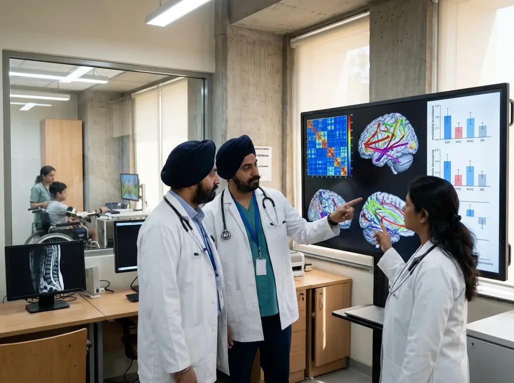
Traumatic spinal damage during childhood fundamentally disrupts critical developmental pathways. Consequently, clinical management of pediatric spinal cord injury demands an understanding of supraspinal neural adaptations beyond focal column injury. Children with complete trauma frequently experience profound sensorimotor loss alongside emergent cognitive and emotional struggles. However, clinicians often overlook these cerebral reorganizations during routine physical care. Therefore, advanced functional neuroimaging provides crucial insight into whole-brain remodeling.
Resting-state functional magnetic resonance imaging provides an exceptional non-invasive window into neural network architecture. In this pediatric investigation, researchers examined 34 children with complete spinal cord injuries alongside 36 age-matched healthy peers. Specifically, the entire cohort encompassed young patients aged 6 to 12 years. The investigators applied graph theoretical analysis to whole-brain resting-state scans based on the Automated Anatomical Labeling 90 atlas. As a result, they generated comprehensive functional correlation matrices for all participants.
Furthermore, graph theory allows clinicians to evaluate both global topological organization and localized nodal characteristics. By measuring specialized topological indices, the study mapped how neural communication segregates and integrates across the developing brain. In addition, the researchers quantified nodal centrality, modular structure, and path length metrics. Notably, this framework isolates distinct functional communities and measures inter-module communication efficiency. Consequently, clinicians gain an objective metric for brain plasticity. Thus, connectome analysis decodes functional network alterations following severe spinal disruption.
Traumatic disruption of ascending sensory signals profoundly alters subcortical processing hubs. In pediatric patients, the study observed significant reductions in nodal metrics across subcortical structures. Most notably, the bilateral thalamus exhibited marked decreases in nodal degree and network efficiency. Because the thalamus acts as a primary sensory relay station, peripheral deafferentation clearly dampens its resting communication capacity.
Furthermore, this subcortical attenuation reverberates through interconnected cortical and limbic loops. Healthy children maintain strong functional bridges between the thalamic complex and motor execution centers. In contrast, pediatric spinal trauma disconnects these pathways, impairing sensorimotor feedback synchronization. Therefore, lost sensory feedback drives progressive supraspinal disuse and network uncoupling. In addition, this subcortical depression disrupts affective processing loops that depend heavily on thalamostriatal integration. Clinicians must recognize that spinal trauma reshapes deep cerebral hubs during critical developmental windows. Consequently, early multimodal rehabilitation should actively engage preserved subcortical pathways.
Beyond subcortical deficits, the research identified profound shifts in core hub distribution. Typically, core hubs sustain efficient whole-brain communication and resource allocation. However, pediatric patients exhibited five newly added core nodes predominantly located within the default mode network. This network governs self-referential thought, emotional introspection, and episodic memory retrieval.
Additionally, patients presented heightened functional connectivity within the default mode network itself. Similarly, connectivity increased between the default mode network and the dorsal attention network. This hyperconnectivity suggests a compensatory neural adaptation to persistent physical sensory deprivation. When external sensory interaction declines, the developing brain redirects its functional architecture inward toward introspective thought processes. Unfortunately, excessive default mode engagement frequently correlates with depressive rumination and chronic anxiety. Moreover, reduced connectivity between default mode and visual modules hampers external spatial awareness. Hence, these topological shifts explain why pediatric patients often experience profound neuropsychological and emotional challenges alongside physical paralysis.
Healthy human brains maintain high modularity, organizing functionally specialized areas into distinct, well-segregated clusters. In contrast, children with complete spinal trauma demonstrated significantly lower overall modularity. This finding reveals compromised functional boundaries between distinct neural modules. Specifically, normal modular segregation deteriorated substantially across the pediatric patient cohort.
Furthermore, modular interactions exhibited stark decoupling between sensorimotor domains and associative systems. The study demonstrated markedly reduced connectivity between the default mode network and the vision network. In addition, communication between the attention network and subcortical networks dropped precipitously. Consequently, the injured brain loses its balanced dynamic orchestration between sensory input and cognitive execution. Children rely on robust modular boundaries to develop refined motor schemas and spatial cognition. Therefore, this systemic breakdown contributes directly to motor planning deficits and executive processing difficulties. By understanding these disrupted modular boundaries, rehabilitation teams can tailor specific neuromodulatory interventions.
One of the study's most actionable discoveries involves the left temporal pole. Specifically, the participation coefficient of the left middle temporal gyrus positively correlated with motor examination scores. In graph theoretical analysis, the participation coefficient measures how evenly a node distributes its connections across diverse modules. Therefore, higher temporal participation reflects superior functional communication across widely distributed cortical domains.
Furthermore, the temporal pole integrates multi-sensory information, semantic cognition, and socio-emotional processing. When this critical region maintains flexible inter-modular connections, patients exhibit better motor preservation and adaptive responsiveness. Consequently, the participation coefficient of the left temporal pole serves as a promising imaging biomarker. Pediatric neurologists can utilize this objective parameter to evaluate individual functional status and monitor longitudinal progress. In addition, mapping preserved functional hubs provides precise anatomical targets for future non-invasive brain stimulation. Thus, functional connectomics bridges the gap between neuroimaging data and bedside motor recovery.
These topological alterations highlight urgent targets for clinical rehabilitation strategies. Because spinal injury severely disconnects sensory-motor channels, alternative sensory systems must compensate for lost afferent pathways. Notably, the study revealed significant decoupling between visual and attention networks. Therefore, structured visual-feedback rehabilitation provides an ideal pathway to restore functional connectivity.
Moreover, virtual reality and interactive visual biofeedback engage intact cortical circuits during physical therapy. By presenting real-time visual representations of movement, therapists stimulate dormant sensorimotor representations. In addition, addressing the psychocognitive burden remains essential for pediatric recovery. Because default mode hyperconnectivity correlates with depressive symptoms, comprehensive care must integrate dedicated mental health interventions. Clinicians should implement routine psychological assessments and supportive cognitive therapies alongside motor training. Ultimately, targeting both cognitive-emotional wellbeing and visual-motor integration optimizes long-term outcomes in young patients.
Pediatric complete spinal trauma significantly reduces overall brain modularity and disrupts functional boundaries between specialized neural networks. This structural degradation impairs communication between subcortical, attentional, and sensory domains. Consequently, the brain struggles to segregate localized cognitive processes while integrating complex multi-sensory tasks during daily physical and cognitive activities.
Following complete spinal disconnection, severe peripheral sensory loss prompts the developing brain to shift its neural resources inward. As a consequence, the default mode network increases internal connectivity and alters its interaction with attention systems. This maladaptive introspective hyperconnectivity frequently predisposes pediatric patients to persistent emotional distress and depressive rumination.
The participation coefficient of the left middle temporal pole correlates positively with motor scores in pediatric patients. Because this region coordinates multi-sensory and associative information across modules, its connectivity reflects neural integration capacity. Clinicians can utilize this metric as a prognostic biomarker and potential target for non-invasive neuromodulation.
Disclaimer: This content is for informational and educational purposes only. It is not intended to be a substitute for professional medical advice, diagnosis, or treatment. Always seek the advice of your physician or other qualified health provider with any questions you may have regarding a medical condition. Never disregard professional medical advice or delay in seeking it because of something you have read here. The authors and publishers are not responsible for any adverse effects resulting from the use of the information presented. Refer to the latest local and national guidelines for clinical practice.
References

Read summarized clinical updates, watch expert medical content, and earn CME certifications right from your smartphone.
Open your smartphone camera to scan and install.





A resting-state fMRI study reveals significant brain network reorganization in pediatric complete spinal cord injury, including subcortical thalamic decoupling, default mode network reorganization, and modularity breakdown, highlighting the role of visual-feedback and cognitive neurorehabilitation.
Today

A pioneering clinical trial across 200 general practices in England evaluates cutting-edge blood tests to detect vague abdominal malignancies earlier. By combining artificial intelligence with protein biomarker profiling, clinicians aim to triage high-risk patients rapidly and improve long-term survival.
Today

A preclinical study reveals that cinnamic acid counters D-galactose-induced premature ovarian failure in mice. By enhancing total antioxidant capacity and upregulating protective genes like Nrf2 and Foxo3a, cinnamic acid improved estrous cyclicity and follicular reserve, offering new insights into ovarian health.
Today

A systematic review of 18 studies reveals very low certainty evidence for cardiac autonomic modulation following intermittent normobaric hypoxia, showing insufficient data to confirm reproducible changes in heart rate variability or baroreflex sensitivity.
Today

An 8-week functional restoration program provides sustained psychological benefits at six-month follow-up for workers' compensation patients. The multidisciplinary intervention significantly improved pain self-efficacy, reduced anxiety and depression, and progressively resolved chronic insomnia severity.
Today

A multinational study of 2,573 medullary thyroid cancer patients refines AJCC TNM-8 staging, proposing N1c for level VII nodes and a new I-IVB schema for better survival stratification.
Today
