.webp&w=384&q=75)

Loading, please wait...
Loading, please wait...

Pediatric Gaucher disease is a rare, autosomal recessive lysosomal storage disorder that presents profound diagnostic dilemmas in resource-limited healthcare environments. Caused by a deficiency of the lysosomal enzyme acid beta-glucosidase, this condition leads to progressive multisystem pathology. Clinicians in South Asia frequently encounter atypical or delayed presentations due to low baseline clinical awareness and limited access to specialized metabolic laboratories. Recognizing subtle clinical signals early is essential to prevent permanent morbidity and reduce early childhood mortality.
Pediatric Gaucher disease arises from pathogenic mutations within the GBA1 gene, which encodes the lysosomal enzyme beta-glucocerebrosidase. Consequently, deficient enzyme activity impairs the physiological degradation of glucosylceramide into glucose and ceramide. As a result, undegraded glycolipids accumulate progressively within the mononuclear phagocyte system, generating characteristic lipid-laden macrophages known as Gaucher cells. These abnormal cells infiltrate key reticuloendothelial tissues, primarily the spleen, liver, bone marrow, and occasionally the lungs or nervous system.
Furthermore, this widespread infiltration provokes chronic inflammation, progressive organ enlargement, and bone marrow displacement. Clinicians classify the disorder into three distinct clinical types based on the presence and rate of neurological progression. Type 1 represents the non-neuronopathic form, whereas Type 2 and Type 3 involve acute and chronic neurovisceral deterioration, respectively. Because clinical severity varies widely across genotypes, early identification remains critical for initiating organ-preserving therapies and preventing irreversible skeletal or hematologic complications.
The clinical spectrum of pediatric lysosomal storage disorders often overlaps with common pediatric ailments, making initial identification difficult. Children typically present during infancy or early childhood with progressive abdominal distension caused by massive hepatosplenomegaly. In addition, patients frequently exhibit profound failure to thrive, systemic fatigue, recurrent fever, and chronic chest infections. Because these signs mirror endemic pediatric conditions, clinicians may initially suspect tuberculosis, visceral leishmaniasis, malnutrition, or primary hematologic malignancies.
Moreover, hematologic manifestations represent a hallmark of the disease. Infiltration of the marrow microenvironment alongside splenic sequestration leads to progressive cytopenias, including persistent anemia and thrombocytopenia. Consequently, affected children frequently suffer from recurrent epistaxis, easy bruising, petechiae, and secondary infections. Clinicians must also remain highly vigilant for family history red flags. In regions with elevated rates of consanguinity, a history of unexplained infant sibling deaths, neonatal jaundice, or early-onset visceral enlargement strongly indicates an underlying inherited metabolic disease.
Establishing a definitive diagnosis requires a structured laboratory algorithm. In low-resource settings, clinicians frequently perform a diagnostic bone marrow aspiration when investigating unexplained pancytopenia and organomegaly. Bone marrow cytology often reveals pathognomonic Gaucher cells, which appear as large, eccentric macrophages with characteristic 'wrinkled tissue paper' cytoplasm. Although identifying these cells provides strong presumptive evidence, cytologic evaluation is invasive and cannot serve as the sole confirmatory diagnostic modality.
Therefore, international and national guidelines mandate measuring beta-glucosidase enzymatic activity in peripheral blood leukocytes or dried blood spot samples to confirm the diagnosis. A profound reduction in enzyme activity firmly establishes the disorder. Additionally, molecular genetic analysis of the GBA1 gene identifies specific pathogenic variants, aiding phenotypic prediction and familial risk assessment. Clinicians should also measure sensitive plasma biomarkers, such as glucosylsphingosine (Lyso-Gb1) or chitotriosidase, to gauge baseline disease severity and monitor ongoing therapeutic response.
Healthcare practitioners in South Asia face substantial systemic hurdles when diagnosing rare metabolic disorders. Because specialized biochemical and genetic testing facilities remain concentrated within tertiary academic institutions, rural and semi-urban patients experience long diagnostic journeys. Consequently, misdiagnoses and prolonged delays are common, during which children suffer irreversible skeletal deformities, growth failure, or fatal hemorrhagic episodes. Limited clinician awareness regarding atypical lysosomal storage presentations further exacerbates these delays.
To overcome these challenges, regional healthcare systems must integrate simplified screening protocols into standard pediatric care pathways. For instance, dried blood spot testing provides a robust, cost-effective, and easily transportable method for field-level enzyme analysis. Furthermore, establishing dedicated regional referral networks between primary care centers and pediatric genetic centers accelerates diagnostic confirmation. Increasing clinical suspicion among general practitioners and hematologists ensures that symptomatic children receive specialized evaluations before irreversible organ dysfunction occurs.
The therapeutic paradigm for Gaucher disease has advanced substantially with the development of targeted systemic interventions. Enzyme replacement therapy, administered via regular intravenous infusions, represents the cornerstone of management for pediatric patients with Type 1 and Type 3 disease. Exogenous recombinant enzymes effectively clear accumulated glycolipids from macrophages, thereby reversing splenomegaly, restoring hematologic parameters, and promoting normal linear growth and bone mineral density.
Nevertheless, access to continuous enzyme therapy remains financially challenging in many developing nations. Consequently, comprehensive supportive management is essential. Clinicians must provide packed red blood cell or platelet transfusions for severe cytopenias, aggressive antibiotic coverage for secondary respiratory infections, and optimized nutritional support to counter cachexia. In addition, routine skeletal surveillance using magnetic resonance imaging allows early detection of osteopenia, avascular necrosis, or Erlenmeyer flask deformities. Multidisciplinary coordination between pediatricians, hematologists, orthopedicians, and geneticists ensures optimal long-term quality of life.
Because Gaucher disease follows an autosomal recessive pattern of inheritance, comprehensive genetic counseling forms an indispensable component of clinical management. When a proband receives a confirmed diagnosis, clinicians must educate parents regarding the 25 percent recurrence risk in each subsequent pregnancy. Furthermore, extended family pedigree analysis is vital, particularly in communities where consanguineous marriages are culturally prevalent, as carrier frequencies may be elevated.
Accordingly, clinicians should actively offer cascade screening to identify asymptomatic or mildly affected siblings. Early biochemical screening of at-risk relatives enables timely therapeutic intervention before irreversible clinical manifestations develop. Furthermore, offering prenatal diagnosis through chorionic villus sampling or amniocentesis provides informed reproductive choices for affected couples. Integrating accessible genetic counseling into tertiary care centers fosters community awareness and mitigates the long-term familial burden of rare inherited disorders.
Definitive diagnosis requires demonstrating deficient beta-glucocerebrosidase enzyme activity in peripheral blood leukocytes or dried blood spot samples. Although bone marrow aspiration frequently demonstrates characteristic lipid-laden Gaucher cells with wrinkled tissue paper cytoplasm, enzymatic assays and molecular genetic analysis of the GBA1 gene provide the essential confirmatory standard.
Clinicians initiate enzyme replacement therapy in symptomatic pediatric patients demonstrating severe cytopenias, debilitating hepatosplenomegaly, growth failure, or progressive skeletal pathology. Timely initiation effectively reverses visceral enlargement, restores normal hematopoiesis, prevents chronic bone crises, and supports appropriate pediatric physical development.
Gaucher disease is transmitted in an autosomal recessive manner, requiring two copies of a mutated GBA1 gene to cause disease. Consanguineous marriages significantly increase the likelihood that both parents carry identical ancestral pathogenic mutations, substantially elevating the incidence of homozygous affected offspring.
Disclaimer: This content is for informational and educational purposes only and should not be considered medical advice. Clinical decisions must be made by qualified healthcare professionals based on individual patient presentation and diagnostic evaluation. Refer to the latest local and national guidelines for clinical practice.
References
Waqas M et al. Diagnostic Challenge of Pediatric Gaucher Disease in a Low-Resource South Asian Setting: A Case Report. Clin Case Rep. 2026 Aug undefined. doi: 10.1002/ccr3.73346. PMID: 42609480.
Shemesh E, Deroma L, Bembi B, et al. Revised recommendations for the management of Gaucher disease in children. Eur J Pediatr. 2015;174(1):37-47.
Kapoor S, Jones SA, Bhattacharya K, et al. Diagnosis and Management of Gaucher Disease in India - Consensus Guidelines of the Gaucher Disease Task Force of the Society for Indian Academy of Medical Genetics and the Indian Academy of Pediatrics. Indian Pediatr. 2018;55(2):143-153.

Read summarized clinical updates, watch expert medical content, and earn CME certifications right from your smartphone.
Open your smartphone camera to scan and install.





Pediatric Gaucher disease presents critical diagnostic challenges in South Asia due to overlapping clinical features and limited diagnostic access. Early recognition, enzymatic assays, and timely interventions improve outcomes.
last month

Health authorities in the Democratic Republic of the Congo report unprecedented mortality from the Bundibugyo virus epidemic. Clinicians worldwide must understand the virological profile, atypical hemorrhagic manifestations, early diagnostic protocols, and containment strategies for this evolving health threat.
Today

A nationwide South Korean study shows underreporting of smoking among women dropped from 57.4% to 36.4% between 2008 and 2021. However, over one-third of female smokers still conceal their habit, highlighting the critical role of cotinine verification in clinical risk assessment.
Today
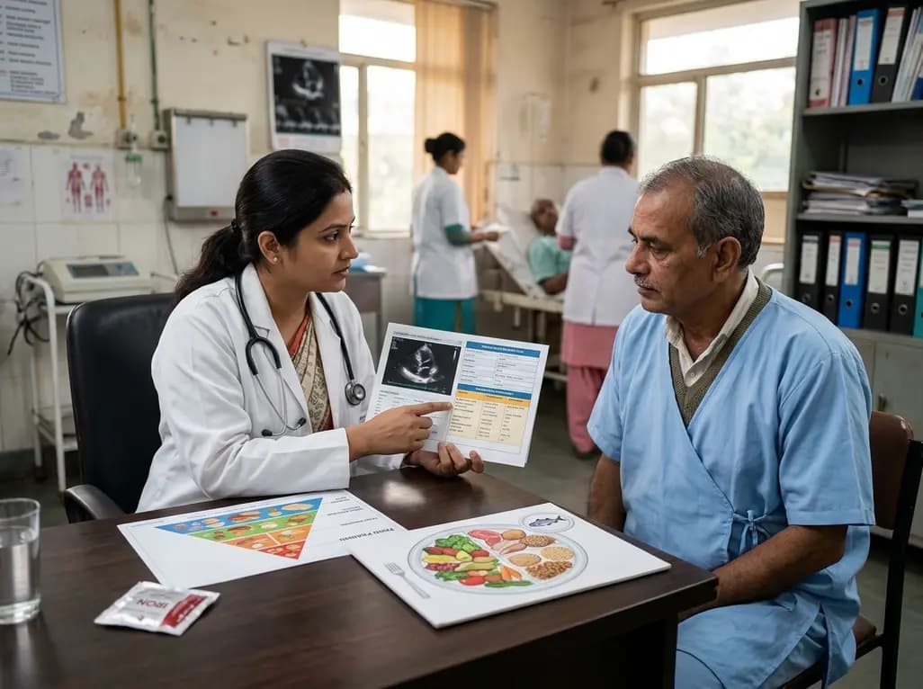
A scientific statement highlights the crucial role of nutrition in managing chronic heart failure. It emphasizes GLIM-based screening, individualized dietary patterns like the Mediterranean diet, appropriate protein and energy intake, avoiding unnecessary fluid restriction, and targeted iron supplementation.
Today

A new study evaluates the rheological stability of decellularized peripheral nerve matrix hydrogels under varied storage conditions. Findings show -80°C storage and freeze-drying preserve gelation kinetics and mechanical stiffness, offering key insights for shipping and translational clinical applications.
Today

A qualitative meta-synthesis highlights the complex interplay between gender dysphoria, minority stress, and eating disorders in transgender and gender-diverse individuals. Clinicians must recognize these distinct drivers to offer supportive, gender-affirming, and interdisciplinary eating disorder care.
Today
