

Loading, please wait...
Loading, please wait...

Paediatric valvular heart disease represents an immense challenge across global healthcare settings, particularly within low- and middle-income countries. Conventional prostheses cannot accommodate somatic growth in young children, which inevitably necessitates repeated surgical revisions. Recent surgical breakthroughs introduce partial heart transplantation as a transformative therapeutic paradigm. This technique implants viable donor valve roots that preserve cellular turnover and functional remodeling. Consequently, cardiothoracic teams can provide durable hemodynamic restoration without subjecting growing children to cyclical open-heart reoperations.
Young children with complex congenital heart defects require prompt, durable valvular interventions. Currently, standard surgical options include mechanical prostheses, xenograft conduits, and cryopreserved homografts. However, none of these standard replacements can adapt to a child's ongoing cardiovascular growth. Mechanical valves necessitate lifelong anticoagulation regimens, which introduce substantial hemorrhagic and thromboembolic hazards in active children. Meanwhile, bioprosthetic substitutes and cryopreserved tissues inevitably undergo accelerated calcification, immune-mediated degradation, and structural failure. As a result, paediatric patients frequently face multiple open-heart operations before reaching adulthood. Each subsequent sternotomy carries escalating surgical risks, dense adhesions, bleeding complications, and increased perioperative mortality. Furthermore, families in developing nations often encounter catastrophic out-of-pocket expenses for successive cardiac operations. In addition, rural communities lack pediatric cardiac surgical centers capable of handling complex redo operations safely. Therefore, congenital cardiac surgeons have long sought a durable biological valve substitute that retains cellular vitality and expands alongside normal somatic growth. Partial heart transplantation directly addresses these persistent clinical limitations.
Partial heart transplantation provides a living biological solution by harvesting intact semilunar valve roots from human donors. Unlike acellular or fixed bioprosthetic grafts, this technique preserves living donor interstitial cells and functional endothelial surfaces. These viable cells actively maintain extracellular matrix homeostasis, synthesize fresh collagen, and remodel under changing pressures. Early clinical evidence confirms that implanted allografts display proportional annular enlargement and leaflet elongation over serial evaluations. Consequently, the implanted living valves match the child’s growth without creating progressive stenosis or regurgitation. Furthermore, preserving cellular architecture substantially diminishes the catastrophic structural deterioration observed in acellular homografts. Because surgeons transplant only the valvular apparatus, they preserve the recipient's native ventricular myocardium entirely. Thus, the recipient avoids ventricular denervation, global ischemia-reperfusion trauma, and donor-specific ventricular pump dysfunction. This selective anatomical preservation secures immediate hemodynamic stability following cardiopulmonary bypass. In addition, maintaining native ventricular architecture prevents long-term heart failure risks, ensuring superior postoperative recovery in fragile infants.
When evaluated alongside orthotopic heart transplantation, partial heart transplantation exhibits remarkable physiological, surgical, and logistical benefits. Orthotopic transplantation demands stringent donor-recipient size matching, rapid transit, and aggressive triple-drug immunosuppression. In contrast, partial valve grafts demonstrate significant tolerance toward prolonged cold graft ischemia times. Because isolated valve leaflets maintain minimal metabolic activity compared to dense myocardium, grafts retain functional viability after lengthy transit periods. In addition, surgical teams can harvest pristine semilunar valves from donor hearts deemed completely unsuitable for orthotopic transplantation. For instance, hearts displaying severe left ventricular dysfunction often retain pristine aortic and pulmonary valves. This dual sourcing dramatically expands the severely restricted pediatric donor registry. Moreover, recipients typically achieve graft stability with low-dose tacrolimus monotherapy alone. Avoiding aggressive antimetabolite and corticosteroid regimens significantly diminishes the cumulative hazards of opportunistic infections and renal impairment. Therefore, this targeted surgical strategy minimizes pharmacological toxicities while maximizing donor organ utilization across resource-constrained healthcare environments.
Despite profound clinical advantages, establishing partial heart transplantation in low- and middle-income regions introduces considerable systemic hurdles. Foremost among these challenges is the acute scarcity of coordinated organ procurement organizations. Many resource-limited jurisdictions lack centralized organ allocation frameworks and clear legal mechanisms for pediatric brain-death declaration. Furthermore, therapeutic drug monitoring remains inconsistently accessible across regional healthcare centers. Because maintenance tacrolimus therapy mandates tight blood concentration surveillance, unreliable laboratory infrastructure introduces severe risks of graft rejection or nephrotoxicity. Additionally, socioeconomic disparities cause high rates of patient loss to follow-up after hospital discharge. Many impoverished families cannot maintain continuous access to specialized cardiology clinics or afford lifelong medications. Without reliable transportation networks, digital registries, and financial support, post-discharge outcomes deteriorate rapidly. Moreover, persistent supply chain disruptions frequently interrupt the distribution of essential maintenance pharmaceuticals across remote districts. Finally, regional operating suites must maintain modern cardiopulmonary bypass equipment and surgical teams trained in complex root reconstructions.
Implementing partial heart transplantation in resource-limited nations requires a carefully structured, progressive institutional roadmap. Initially, national health ministries should anchor partial transplant programs within established tertiary hospitals that already operate seasoned pediatric cardiac surgery services. These premier regional referral centers maintain critical cardiopulmonary support infrastructure, specialized pediatric intensive care units, and experienced perfusionists. Furthermore, healthcare teams can optimize cost efficiency by utilizing widely available generic immunosuppressive agents. Establishing regional immunoassay drug monitoring laboratories will optimize pharmacokinetic surveillance at affordable costs. Meanwhile, international academic partnerships can facilitate vital clinical knowledge exchange, virtual surgical proctoring, and structured recipient selection registries. Concurrently, public health authorities must formulate clear regulatory guidelines that govern deceased organ donation and ensure equitable graft distribution. Additionally, establishing decentralized follow-up satellite clinics will help monitor rural patients and prevent therapy discontinuation. By combining existing surgical foundations with standardized post-discharge community monitoring, middle-income healthcare systems can establish safe, sustainable programs for disadvantaged children facing terminal valvular failure.
Partial heart transplantation delivers viable donor heart valves containing living endothelial and interstitial cells, unlike conventional mechanical or bioprosthetic replacements. Standard synthetic or cryopreserved implants cannot grow and progressively suffer from structural calcification or thromboembolism. In contrast, living transplanted valve roots expand proportionally with pediatric somatic growth, significantly reducing the cumulative requirement for repeated open-heart surgeries over a child's lifetime.
Partial heart transplantation replaces only isolated semilunar valve structures rather than the entire ventricular myocardium. Because heart valves possess low cellular mass and limited vascularity compared to thick myocardial tissue, they evoke a less aggressive immune response. Consequently, clinical protocols primarily utilize low-dose tacrolimus monotherapy, safely avoiding the multi-drug regimens, high-dose steroids, and toxic immunosuppressive burdens mandatory in orthotopic heart transplants.
Yes, partial heart transplantation can successfully harvest functional semilunar valves from donor hearts deemed unsuitable for orthotopic transplantation. For instance, donor hearts affected by severe ventricular systolic failure, prolonged downtime, or anatomical ventricular anomalies often retain pristine aortic and pulmonary valve apparatuses. Utilizing these alternative grafts markedly broadens the pediatric donor pool and maximizes precious organ donation utility in resource-constrained environments.
Disclaimer: This content is for informational and educational purposes only... Refer to the latest local and national guidelines for clinical practice.
References

Read summarized clinical updates, watch expert medical content, and earn CME certifications right from your smartphone.
Open your smartphone camera to scan and install.





Partial heart transplantation offers living, growth-capable cardiac valve replacements for children. While orthotopic heart transplantation faces severe logistical barriers in low-resource settings, partial transplantation presents lower graft ischemia sensitivity and simplified immunosuppression protocols.
Today
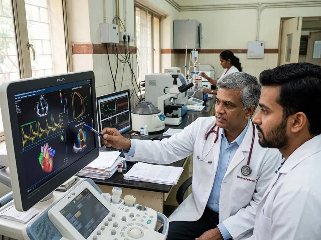
A study evaluates a cardiometabolic HFpEF model combining metabolic stress with chronic mineralocorticoid excess. The findings reveal profound diastolic stiffness, endothelial dysfunction, and pulmonary hypertension, illuminating key pathophysiological mechanisms for targeted cardiopulmonary therapies.
Today
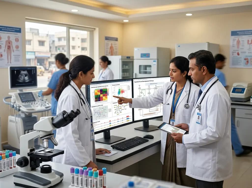
The Human Reproduction and Development Database (HRD) consolidates bulk and single-cell transcriptomics, epigenomics, metabolomics, proteomics, and clinical trial records into a unified platform to advance reproductive health research.
Today
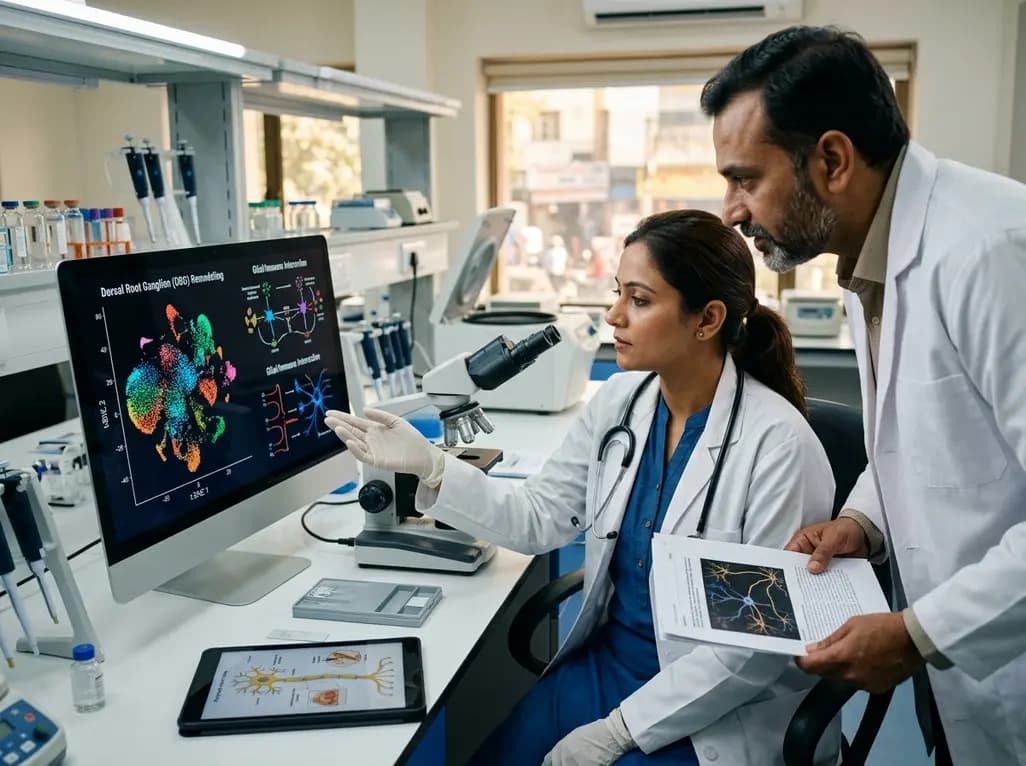
A single-cell transcriptomic atlas reveals how peripheral nerve injuries cause dorsal root ganglion remodeling. By evaluating proximal nerve crush and resection, researchers identify distinct glial and immune programs that resolve neuropathic pain and promote regeneration.
Today
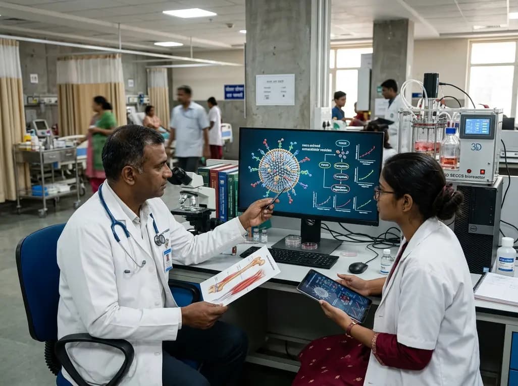
A breakthrough study demonstrates that fullerol-primed extracellular vesicles generated in a 3D SECCS bioreactor attenuate ferroptosis and restore myogenic regeneration in diabetes via the FOXO3 pathway, offering a cell-free therapeutic strategy for diabetic musculoskeletal degeneration.
Today
