

Loading, please wait...
Loading, please wait...

Ischemic stroke remains one of the leading global causes of long-term adult disability and motor impairment. Although acute reperfusion therapies save lives, thousands of survivors live with severe, persistent arm weakness. Recently, paired vagus nerve stimulation emerged as an innovative bioelectronic strategy designed to overcome chronic neurological plateaus. By synchronizing electrical stimulation of the vagus nerve with intensive physical rehabilitation, clinicians can drive profound neuroplastic changes. Historically, pivotal clinical trials primarily enrolled individuals recovering from supratentorial hemispheric strokes. However, exciting new clinical observations show that this targeted neuromodulatory technique also delivers meaningful benefits for patients who suffered debilitating infratentorial brainstem or cerebellar vascular injuries.
The approval of vagal neurostimulation by regulatory authorities marked a pivotal paradigm shift in restorative neurology. For decades, physical medicine teams believed motor recovery invariably stalled after six to twelve months. Consequently, patients with chronic deficits had few therapeutic alternatives beyond compensatory adaptations. Paired vagus nerve stimulation fundamentally challenges this dogma by demonstrating that dormant neural networks can reorganize years after an initial insult. In this approach, a neurosurgeon implants a small pulse generator within the chest wall. During active upper limb rehabilitation, an occupational therapist delivers short electrical pulses to the left cervical vagus nerve. Specifically, each burst coincides directly with successful task completion, reinforcing specific movement patterns. Furthermore, extensive preclinical research confirms that paired stimulation induces lasting reorganization in the primary motor cortex. As a result, patients regain functional control over movements they thought were lost forever. In addition, real-world registry data demonstrates robust patient satisfaction and durable functional recovery across diverse rehabilitation environments.
Posterior fossa ischemic strokes account for approximately twenty percent of all ischemic cerebrovascular events. Unfortunately, infratentorial infarcts involving the brainstem, pons, and cerebellum produce complex, devastating motor syndromes. In addition to hemiparesis, patients frequently experience debilitating ataxia, dysmetria, and bilateral coordination deficits. Because critical descending corticospinal tracts and ascending sensory pathways tightly converge within the brainstem, even tiny vascular lesions can cause extensive motor impairment. Furthermore, conventional stroke rehabilitation protocols often yield diminished returns in this cohort due to concurrent cerebellar dysfunction. Historically, clinical investigators excluded posterior circulation strokes from major restorative device trials to maintain homogenous study populations. Consequently, stroke specialists lacked clear evidence regarding whether neuromodulation could alleviate chronic deficits originating from infratentorial brain injury. Therefore, chronic posterior fossa stroke survivors remained medically underserved. Clinicians often assumed that damage to brainstem nuclei or cerebellar circuitry would prevent the synaptic plasticity required for stimulation-induced motor gains. Nevertheless, emerging clinical reports are actively dismantling these conservative assumptions.
To understand how paired vagus nerve stimulation benefits infratentorial stroke survivors, one must examine its neurochemical cascade. When electrical pulses stimulate the left vagus nerve, ascending afferent signals rapidly travel through the solitary tract. Subsequently, these impulses trigger the simultaneous activation of two major subcortical nuclei: the locus coeruleus and the nucleus basalis of Meynert. As a result, these nuclei release bursts of norepinephrine and acetylcholine throughout the cerebral cortex. This targeted neurotransmitter surge opens a transient window of heightened neuroplasticity. Furthermore, pairing this biochemical surge with goal-directed physical tasks strengthens active synapses via Hebbian learning principles. Even when an ischemic injury damages the brainstem or cerebellum, intact cortical motor areas can undergo compensatory reorganization. Thus, repeated paired trials enable cortical pathways to recruit alternative downstream tracts. Moreover, recent neuroimaging investigations suggest that paired neuromodulation alters functional connectivity across both cerebral hemispheres. Therefore, the brain constructs robust new neural networks around the damaged posterior fossa territory.
A landmark case series recently demonstrated the tangible clinical impact of paired vagus nerve stimulation in posterior circulation stroke survivors. Investigators evaluated four patients presenting with persistent, disabling upper extremity deficits following remote posterior fossa infarcts. Notably, all four participants had sustained their strokes months to years prior to device implantation. Despite having previously plateaued in traditional physical therapy, every patient achieved significant functional recovery following paired stimulation protocols. Clinicians documented substantial improvements on standardized motor rating scales, including the Fugl-Meyer Assessment for Upper Extremity. In addition, patients demonstrated meaningful gains on the timed Wolf Motor Function Test. Importantly, these statistical gains translated into transformative real-world improvements in daily activities. Patients reported greater ease with feeding, grooming, and household tasks. Furthermore, participants experienced notable improvements in overall quality of life and subjective limb confidence. These remarkable outcomes indicate that the therapeutic window for posterior stroke recovery extends well beyond traditional rehabilitation timeframes.
Implementing paired vagus nerve stimulation requires seamless collaboration between stroke neurologists, neurosurgeons, and specialized rehabilitation therapists. The surgical procedure involves implanting a miniature pulse generator subcutaneously in the left chest wall. Meanwhile, the surgeon tunnels a bipolar helical lead subcutaneously to the neck and wraps it around the left vagus nerve. Surgeons avoid the right vagus nerve because right-sided stimulation carries a higher risk of cardiac bradyarrhythmias. Postoperative recovery proceeds quickly, allowing patients to begin paired outpatient rehabilitation within several weeks. Typically, patients attend intensive clinic sessions three times per week alongside independent home exercises. Looking forward, clinicians must conduct larger prospective clinical trials to define ideal patient selection criteria for posterior circulation strokes. In addition, future studies should evaluate whether noninvasive transcutaneous vagus nerve stimulation can replicate these clinical results. Ultimately, these groundbreaking observations offer renewed hope to chronic stroke survivors, proving that meaningful neurological recovery remains possible years after injury.
Paired vagus nerve stimulation stimulates the cervical vagus nerve precisely during task-oriented physical rehabilitation exercises. Consequently, this targeted electrical discharge activates subcortical nuclei, including the locus coeruleus and nucleus basalis. These centers immediately release key neuromodulators such as acetylcholine and norepinephrine into the cerebral cortex. Therefore, the brain reorganizes surviving motor networks around the infarction, enhancing synaptic plasticity and accelerating motor relearning in hemiparetic stroke survivors.
Ideal candidates include adult ischemic stroke survivors suffering from moderate to severe chronic upper extremity hemiparesis. Typically, patients must be at least six months post-stroke and maintain plateaued progress despite conventional physical therapy. Furthermore, candidates must retain minimal residual distal arm movement, such as slight wrist extension or finger flexion. However, severe contractures, complete flaccid plegia, active medical instability, and prior bilateral vagotomy represent major contraindications to receiving surgical implantation.
Surgical implantation of the vagus nerve stimulator generally presents a favorable safety profile similar to epilepsy procedures. However, potential surgical complications include transient vocal cord paresis, hoarseness, cough, and localized neck pain during active stimulation. In addition, rare risks involve wound infection, hematoma formation, or lead migration requiring revision. Neurosurgeons mitigate these surgical risks through precise anatomical dissection within the carotid sheath and careful bipolar nerve stimulation during intraoperative testing.
Disclaimer: This content is for informational and educational purposes only... Refer to the latest local and national guidelines for clinical practice.
References

Read summarized clinical updates, watch expert medical content, and earn CME certifications right from your smartphone.
Open your smartphone camera to scan and install.





Recent clinical evidence highlights the utility of paired vagus nerve stimulation for chronic posterior circulation stroke. By pairing vagal neuromodulation with upper extremity physical therapy, clinicians achieved significant motor improvements and enhanced daily functioning years after the cerebrovascular event.
Today
Explore the three-tier harness framework for clinical LLM agents. Learn how orchestration, clinical adaptation, and failure-driven evaluation surpass raw model capability to deliver safe, reliable healthcare AI integration.
Today
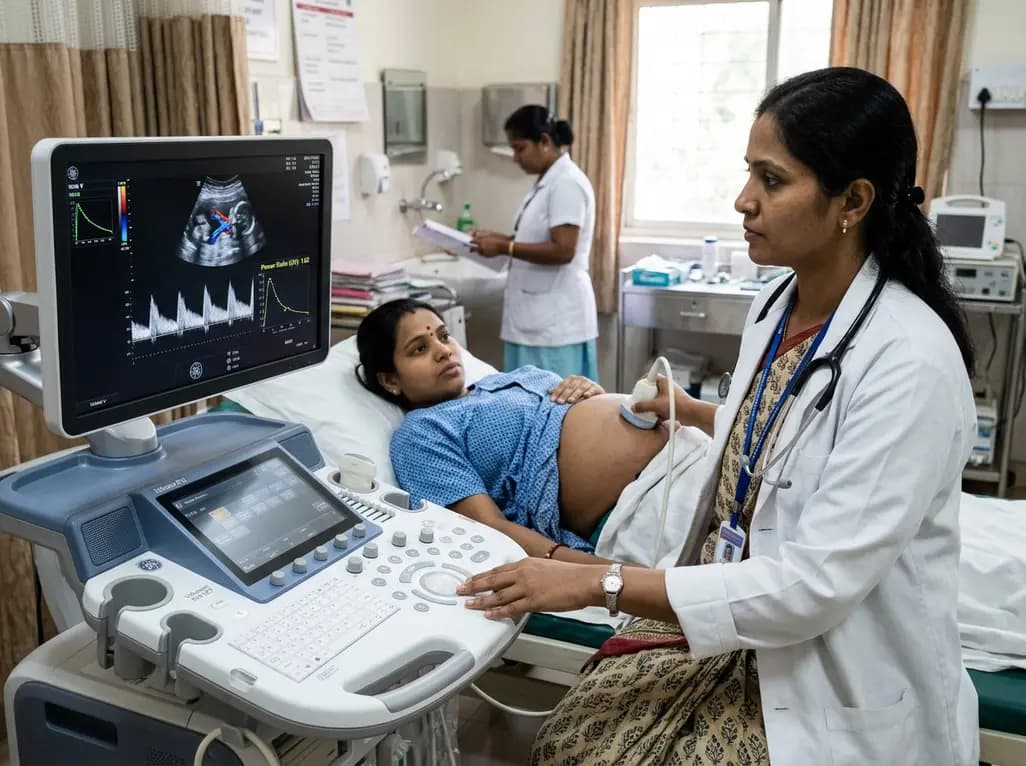
Adaptive spectral analysis of umbilical venous Doppler recordings reveals that an elevated second-trimester maternal-fetal power ratio can distinguish pregnancies at risk of preeclampsia, offering a novel biophysical approach to enhance early vascular risk stratification.
Today

A real-world cohort study of 6,537 patients from the FOUNTAIN platform demonstrates that initiating finerenone significantly reduces albuminuria by 40.7% with minimal eGFR decline and manageable hyperkalemia risk, confirming its practical effectiveness and safety in diabetic kidney disease.
Today

A comprehensive analysis of MAP therapy resistance in osteosarcoma, examining canonical and non-canonical mechanisms across damage avoidance, enhanced DNA repair, and replication stress tolerance to identify novel vulnerabilities.
Today
Near-infrared autofluorescence provides real-time label-free optical guidance during endoscopic transsphenoidal pituitary surgery. By highlighting stark signal contrasts between normal pituitary gland and adenomas, this technique aids gross-total tumor resection while safeguarding critical endocrine function.
Today
